3077
Combining HOSVD-based joint reconstruction with shifted acquisition sampling pattern enables liver DWI in a single breath-hold1ShanghaiTech University, Shanghai, China, 2United Imaging Healthcare, Shanghai, China
Synopsis
Keywords: Image Reconstruction, Diffusion/other diffusion imaging techniques
Fast liver DWI scan has significant clinical relevance, however, motion artifacts, long scan time and low SNR remain as major technical challenges for liver DWI. In this study, we proposed a joint recon method based on global and local high order tensor SVD (HOSVD), combining with shifted acquisition sampling pattern across diffusion directions. The proposed method improves DWI image quality and enables fast liver DWI scan within a single breath-hold.Introduction
Diffusion-weighted imaging (DWI) plays an important role in the clinical and research liver imaging applications. Single-shot echo-planar imaging (SS-EPI) is widely used in liver diffusion studies due to short scanning time and thus relatively insensitivity to motion. However, the liver EPI-DWI is still faced with significant technical challenges, including the residual motion artifacts due to organ physiological motion, low signal-to-noise ratio (SNR), etc. Many approaches have being developed to accelerate the acquisition of DWI, including conventional GRAPPA/SENSE, compressed sensing based reconstruction methods1-3. In recent years, low rank matrix based recon methods like RAC-LORAKs has been applied in EPI-DWI and shown very promising results4. To further improve the image quality of liver DWI, we proposed a joint recon method, combining the global and local High Order Tensor SVD (HOSVD) approach5-7 with shifted acquisition sampling pattern across diffusion directions, resulting in 3-folded under-sampling DWI data acquisition in a single breath-hold and improved liver DWI image quality.Methods
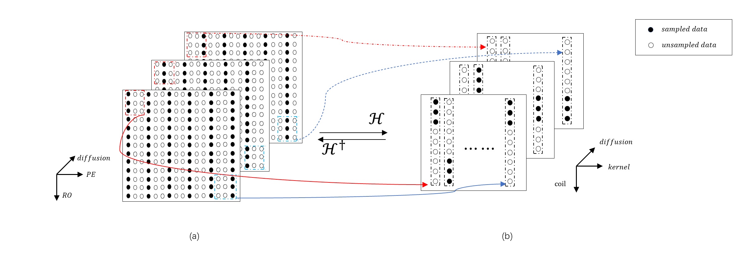
Data acquisition: Images were acquired on three healthy volunteers on an uMR880 3.0T scanner (United Imaging Healthcare, Shanghai, China), using 24ch Rx Body coil and Spine coil. All experiments were performed under the written consent of the volunteer. As shown in Figure 1a, the shifted k-space view sampling pattern among diffusion directions, when combined, corresponded to a full k-space encoding and thus provided complementary information during joint image recon. The key acquisition parameters were: FOV=400mm×360mm, 20 slices with thickness=5mm, matrix=144×128, b-values=50 and 500s/mm2, 3 orthogonal diffusion directions, TE/TR = 62.6/2102ms, uniform under-sampling rate=3, total scan time=17s.Reconstruct methods: The formulation of the proposed recon method is expressed as:
$$\hat k = argmin ‖Fk-d‖_2^2+λ‖C‖_*^2 $$
where F is the under-sampled mask, k is the synthesized data, d is the measured data, $$$||.||_*$$$ denotes the nuclear norm, and C is the low rank tensor constructed jointly by data from all diffusion directions. The way to construct the low rank tensor was shown in Figure 1b. The block-wise Hankel matrix was first formed by organizing the multichannel data for each diffusion direction, and then concatenated along the diffusion direction into the high order tensor.
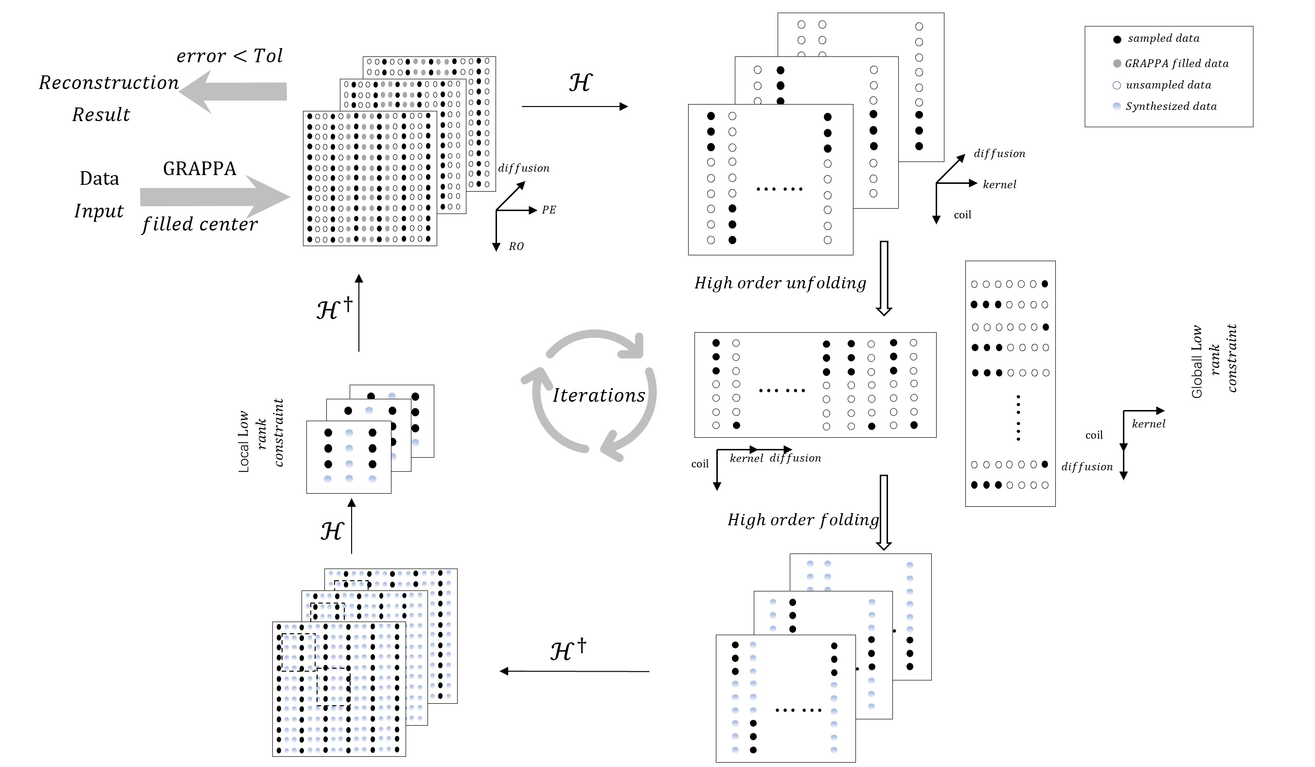
The optimization problem (equation above) was solved iteratively using the higher-order singular value decomposition, which analyzed the multilinear subspace of the tensor. The flowchart of the proposed recon method was shown in Figure 2. The center region of k-space was first filled using traditional GRAPPA method, which provided the initial reconstruction and accelerated the convergence of the iterative algorithm. During each iteration, four steps were carried out in the algorithm. (1) a large high order tensor was generated using k-space data from all diffusion directions; (2) global HOSVD recon was then performed to synthesis the missing data in the tensor by unfolding the diffusion dimension to the kernel and coil dimensions separately; (3) the global tensor was transformed back to k-space, and local high order tensors were constructed by clustering locally similar cubes in k-space into stacks and forming small size tensors, followed by the local HOSVD recon process; (4) the resulting tensors were transformed back to k-space.
Results
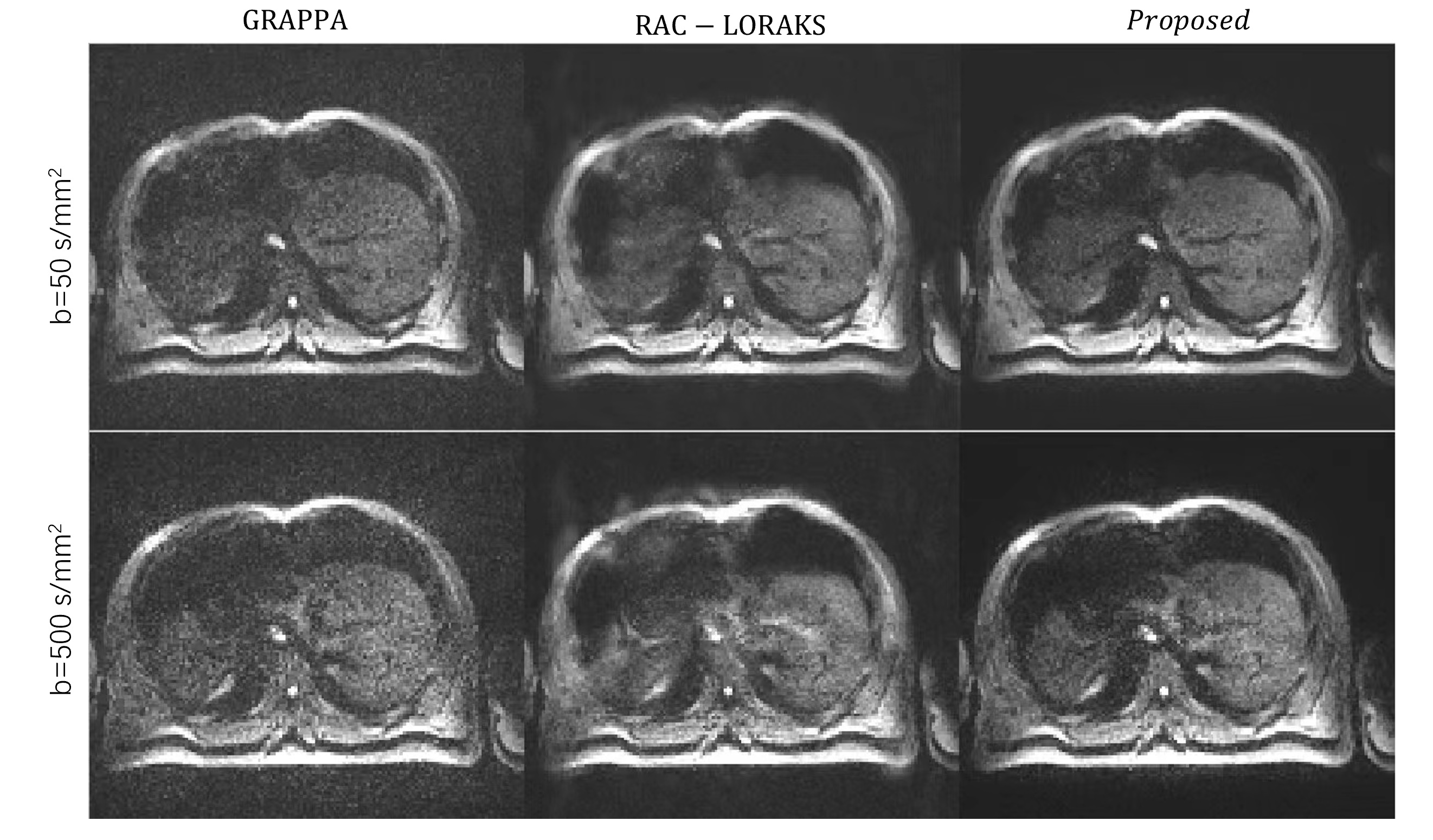
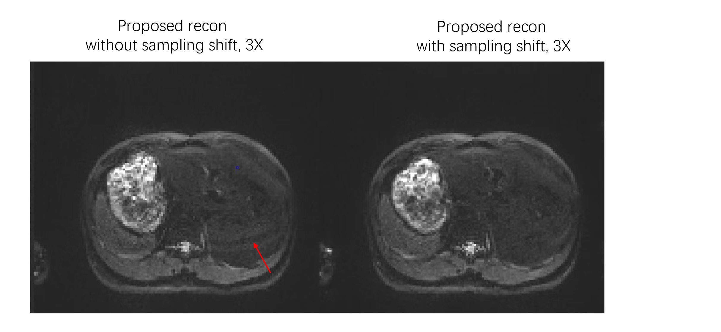
Figure 3 shows the example images of liver DWI reconstructed by conventional GRAPPA, RAC-LORAKs and the proposed method in one volunteer slice. In general, both RAC-LORAKs and the proposed method significantly improve image SNR comparing to GRAPPA, while the proposed method produces images with less alias artifacts comparing to RAC-LORAKs. Figure 4 shows the result images reconstructed by proposed method with and without the shifted view ordering in 3X under-sampling acquisitions. The improved image quality was found for images using proposed recon method with sampling pattern shifting, as alias artifacts can be seen in the images without using sampling pattern shifting.Discussion
A methodology was proposed for liver DWI scan in a single breath-hold. The resulting image quality was superior to conventional GRAPPA and state-of-art RAC-LORAKs method. The joint recon using low rank tensor constrain exploits common spatial support and coil sensitivity information shared among different virtual k-space kernels and diffusion directions, while the shifted sampling scheme provides the complementary information among different diffusion directions, leading to improved SNR and more effective elimination of alias artifacts.Although the proposed method can achieve outstanding results for liver DWI, the recon process takes too long and prevents its clinical use. Future work will include deep learning model in the recon process to overcome this limitation.
Conclusion
The recon method based on low rank tensor with high-order SVD combined shifted sampling scheme significantly improve the image quality of liver DWI with 3X under-sampling rate, which enable the liver DWI scan in single breath-hold.Acknowledgements
NoneReferences
1. Griswold, M.A., et al., Generalized autocalibrating partially parallel acquisitions (GRAPPA). MRM, 2002. 47(6): p. 1202-1210.
2. Pruessmann, K.P., et al., SENSE: sensitivity encoding for fast MRI. MRM, 1999. 42(5): p. 952-962.
3. Knoll, F., et al., Deep-Learning Methods for Parallel Magnetic Resonance Imaging Reconstruction: A Survey of the Current Approaches, Trends, and Issues. IEEE Signal Process Mag, 2020. 37(1): p. 128-140.
4. Lobos, R.A., et al., Robust autocalibrated structured low-rank EPI ghost correction. MRM, 2021. 85(6): p. 3403-3419.
5. Yi, Z., et al., Joint calibrationless reconstruction of highly undersampled multicontrast MR datasets using a low‐rank Hankel tensor completion framework. MRM, 2021. 85(6): p. 3256-3271
6. Vannieuwenhoven, N., et al., A new truncation strategy for the higher-order singular value decomposition. SIAM Journal on Scientific Computing, 2012. 34(2): p. A1027-A1052
7. Bilgic, B., et al., Improving parallel imaging by jointly reconstructing multi‐contrast data. MRM, 2018. 80(2): p. 619-632
Figures