3072
The application of Quantitative Perfusion Analysis of GRASP for assessing pathologic prognostic factors in rectal cancer1Department of Radiology, Sichuan Provincial People's Hospital, chengdu, China, 2Department of MR Scientific Marketing, Siemens Healthcare, Shanghai, China
Synopsis
Keywords: Data Analysis, Perfusion, rectum
Colorectal cancer’s pathological prognostic factors directly affect the patient's prognosis. Golden-angle RAdial Sparse Parallel (GRASP) imaging was invented to calculate the accurate perfusion parameters including influx forward volume transfer constant (Ktrans), rate constant (Kep), and plasma volume fraction (Ve). We found GRASP parameters showed significant differences in many prognostic factors, including histology type, extramural venous invasion (EMVI), lymphovascular invasion (LVI), tumor deposit (TD) and lymph node metastasis (LNM), and were independent factors for histology type, LNM, LVI and EMVI. GRASP parameters were strongly associated with preoperative prognostic factors in rectal cancer, providing useful information for treatment and follow-up protocol.Introduction
Colorectal cancer is the third most common cancer, its prognosis depends on many factors, such as differentiation grade, T classification, tumor deposit (TD), extramural venous invasion (EMVI)and lymphovascular invasion (LVI). Previous studies have demonstrated the role of Dynamic contrast-enhanced MRI (DCE-MRI) in the preoperative diagnosis of rectal cancer, and the quantitative parameters can be used to predict and evaluate tumor grading and the response of patients with rectal cancer after neoadjuvant chemotherapy and radiation therapy1. Conventional DCE-MRI is known to be sensitive to motion and requires breath-holding during scan, resulting in poor temporal and spatial resolution. To overcome this limitation, a novel DCE-MRI technique known as Golden-angle RAdial Sparse Parallel (GRASP) imaging was invented to achieve an acquisition with much higher temporal resolution without breath-holding, thus increasing patient comfort while allowing higher spatial resolution images to be acquired and more accurate perfusion to be quantified2. The purpose of this study was to explore the diagnostic value of the influx forward volume transfer constant (Ktrans), rate constant (Kep) , and plasma volume fraction (Ve) from GRASP MRI for the assessment of prognostic factors in resectable rectal cancer.Material and Methods
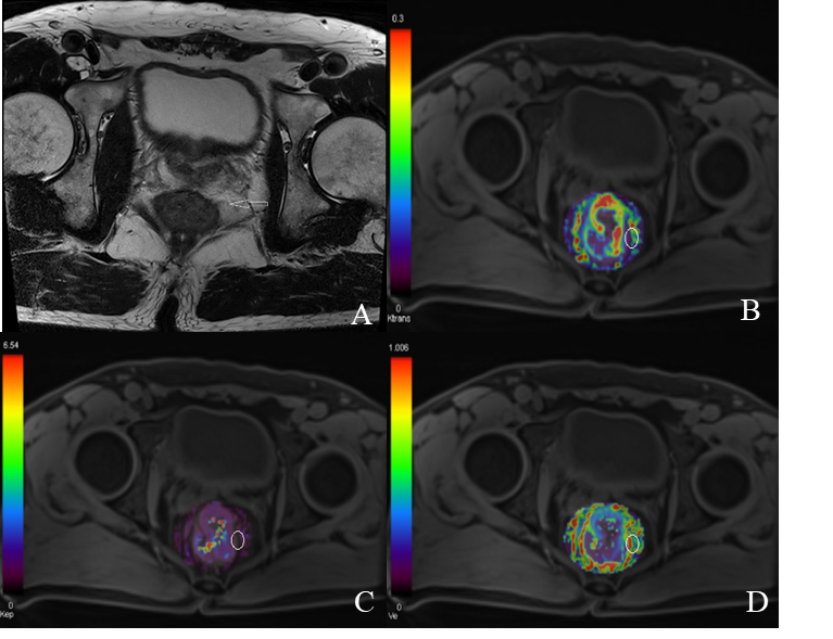
A total of 103 patients (78 males and 25 females, 25-82 years old, with a mean age of 62±13.1 years old) with rectal cancer were enrolled (from December 2021 to August 2022), who had undergone GRASP MRI and conventional sequences by using 3.0 T MR system with 30-channels coil (MAGNETOM VIDA, Siemens Healthcare, Erlangen, Germany). The details of parameters are shown in Table 1. Image processing was performed by TOFTS model’s software (MR Tissue4D; Siemens Healthineers), including Ktrans, Kep, and Ve. Two radiologists independently placed freehand region of interest (ROI) on perfusion maps on the largest tumor cross-section (Figure 1). Histopathologic findings including histology type, T classification, lymph node metastasis (LNM), EMVI, TD, and LVI were reported. Independent samples t-test or Mann-Whitney U test were used to compare the statistical difference between different groups. Univariate and multivariate logistic regression analyses identified the independent risk factors. The receiver operating characteristic (ROC) curve was adopted to determine the performance for GRASP parameters in discrimination of clinicopathologic characteristics of rectal cancer, indicated by the area under the curve (AUC). The statistical analyses were performed using SPSS version 26 (IBM Corporation).Results
The results of correlations between prognostic factors and GRASP parameters are shown in Table 2. The Ktrans was larger in the poor differentiation, LNM-positive, TD-positive, LVI-positive, and EMVI-positive groups (p <0.05). The Kep was higher in the poor differentiation, and LNM-positive, EMVI-positive groups (p <0.05). The Ve was larger in EMVI-positive groups (p=0.026). The univariate and multivariate logistic regression results for screening out the independent risk factors for prognosis are summarized in Table 3. The Ktrans and Kep were independent factors for histology type (OR= 227.318, 11.303, p<0.001, =0.044). The Ktrans and Kep were independent factors for LNM (OR= 31.365, 7.19, p=0.003, =0.026). The Ktrans was an independent factor for LVI (OR=727.139, p <0.001) and EMVI (OR= 35.202, p =0.024). As illustrated in Table 4, diagnostic performance of GRASP parameters for assessment of clinicopathological characteristics. Combine of Ktrans and Kep Can significantly improve the diagnostic efficacy of clinicopathological characteristics.Discussion
There were significant difference in Ktrans of differentiation grade, LNM, LVI, and EMVI. Ktrans reflects the ability of the contrast agent to be transported from the blood vessels to the interstitial space and reflects the tissue capillary permeability and blood perfusion. The higher the degree of malignancy, the more capillaries leading to the higher the Ktrans value3. The Kep of poor differentiation, LNM positive, and EMVI positive group were significantly higher than the contrast group. Similarly, a higher Kep value represents greater blood return to the vasculature. EMVI positive may lead to incompleteness and leakage of vascular endothelial cells due to the excessive growth of vien. Therefore, a higher Kep value indicates more leakage of the contrast medium. In addition, Kep is only affected by the contrast concentration and fractional volumes in the tumor extravascular extracellular space (EES) and might thus more accurately reflect the tumor capillary permeability4. In addition, the Ve of the EMVI positive group was significantly higher than the contrast group. There were more significant venous invasion in EMVI positive group, leading to more contrast agent leaking from the intravascular to the extravascular, and higher ratio of the extravascular space to the extravascular space increases. As far as we know, there were no studies about the correlation between GRASP parameters and pathological prognostic factors of rectal cancer. Our results showed that the Ktrans could be used to predict differentiation grade, LNM, LVI, and EMVI, but the specificity of assessing LNM was relatively low, which may requires additional caution for clinical use5.Conclusion
Quantitative parameters of GRASP pharmacokinetic model can be used to predict pathological prognostic factors preoperatively, which could assist the determination of treatment strategy and follow-up protocols in rectal cancer.Acknowledgements
No acknowledgement found.References
1. Ciolina M, Caruso D, De Santis D, et al. Dynamic contrast-enhanced magnetic resonance imaging in locally advanced rectal cancer: role of perfusion parameters in the assessment of response to treatment. Radiol Med. 2019;124:331-338.
2. Riffel P, Zoellner FG, Budjan J, et al. "One-Stop Shop": Free-Breathing Dynamic Contrast-Enhanced Magnetic Resonance Imaging of the Kidney Using Iterative Reconstruction and Continuous Golden-Angle Radial Sampling. Invest Radiol. 2016;51:714-719.
3. Zhu Y, Zhou Y, Zhang W, et al. Value of quantitative dynamic contrast-enhanced and diffusion-weighted magnetic resonance imaging in predicting extramural venous invasion in locally advanced gastric cancer and prognostic significance. Quant Imag Med Surg . 2021;11:328-340.
4. Koo HR, Cho N, Song IC, et al. Correlation of perfusion parameters on dynamic contrast-enhanced MRI with prognostic factors and subtypes of breast cancers. J Magn Reson Imaging. 2012;36:145-151.
5. Yeo DM, Oh N, Jung CK, et al. Correlation of dynamic contrast-enhanced MRI perfusion parameters with angiogenesis and biologic aggressiveness of rectal cancer: Preliminary results. J Magn Reson Imaging. 2015;41:474-480.
Figures