3023
Vendor-neutrality and upgrade immunity: Post-upgrade assessment of vendor-neutral qMRI from two perspectives1NeuroPoly Lab, Polytechnique Montreal, Montreal, QC, Canada, 2Montreal Heart Institute, University of Montreal, Montreal, QC, Canada, 3Center for Advanced Interdisciplinary Research, Ss. Cyril and Methodius University, Skopje, Macedonia
Synopsis
Keywords: White Matter, Quantitative Imaging, vendor-neutral, post-upgrade, T1, MTR, MTsat
We scanned the ISMRM/NIST system phantom and one healthy participant to compare vendor-neutral and vendor-native T1, MTR and MTsat maps pre- and post-upgrade. Our findings indicate a systematic T1 bias in the phantom (up to 18.5%) that is linked to post-upgrade prescan calibrations. In-vivo, MTR remains stable, whereas the T1 bias (up to 9%) affects MTsat more. On the other hand, end-to-end consistency of the vendor-neutral workflow is immune against the software upgrade (e.g., data was consistently exported in the BIDS format). To disentangle the effect of upgrades on longitudinal stability, qMRI would benefit from transparent vendor-neutral prescan calibrations.Introduction
Inter-scanner bias and scanner hardware/software upgrades are often unavoidable. The impact of upgrades on the reliability of longitudinal MRI analyses has been reported for a variety of applications, e.g., conventional imaging1, voxel-based morphometry2, diffusion imaging3, and relaxometry4,5. Recently, we showed a significant reduction in inter-vendor differences for three quantitative MRI (qMRI) metrics by developing a vendor-neutral 3D SPGR sequence coupled with end-to-end transparent workflows6 (VENUS). However, it is not well known whether vendor-neutrality can provide immunity against major system upgrades.In this work, we evaluate the impact of a major software upgrade on vendor-native and vendor-neutral qMRI workflows. We scanned the system phantom and one participant to compare vendor-neutral and vendor-native T1, MTR and MTsat maps pre- and post-upgrade. Post-upgrade changes are evaluated from the perspective of an engineer (longitudinal stability of the quantitative measurements) and from the perspective of an MRI technologist (end-to-end consistency of the operational procedures).
Methods
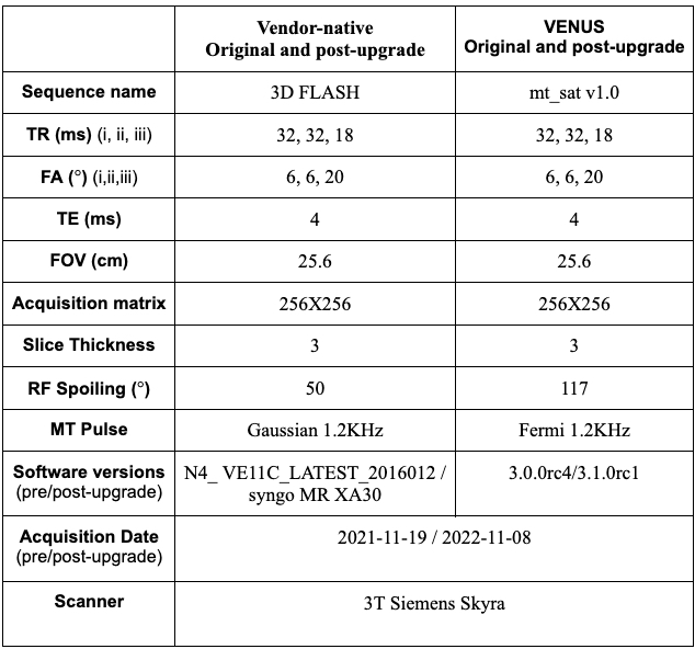
The original vendor-native and VENUS dataset was imported in the qMRI-BIDS7 format from the reference repository5 for the healthy participant and the system phantom. Following the upgrade, the same participant and phantom were scanned by the same technologist in a test-retest setting using the vendor-native and VENUS protocols (Fig. 1).Transmitter reference voltage (Vref) determines the RF radiation that produces a 90° flip in the center of the FOV. Therefore, there is 1-to-1 mapping between Vref and B1+. For the in-vivo acquisition using the same head coil, post-upgrade Vref (327.5V) was higher than the original (322.5V). The increase (5V) was comparable to the pre-upgrade variation of the Vref for the same participant at the same scanner and coil (319.3 ± 4.48V).
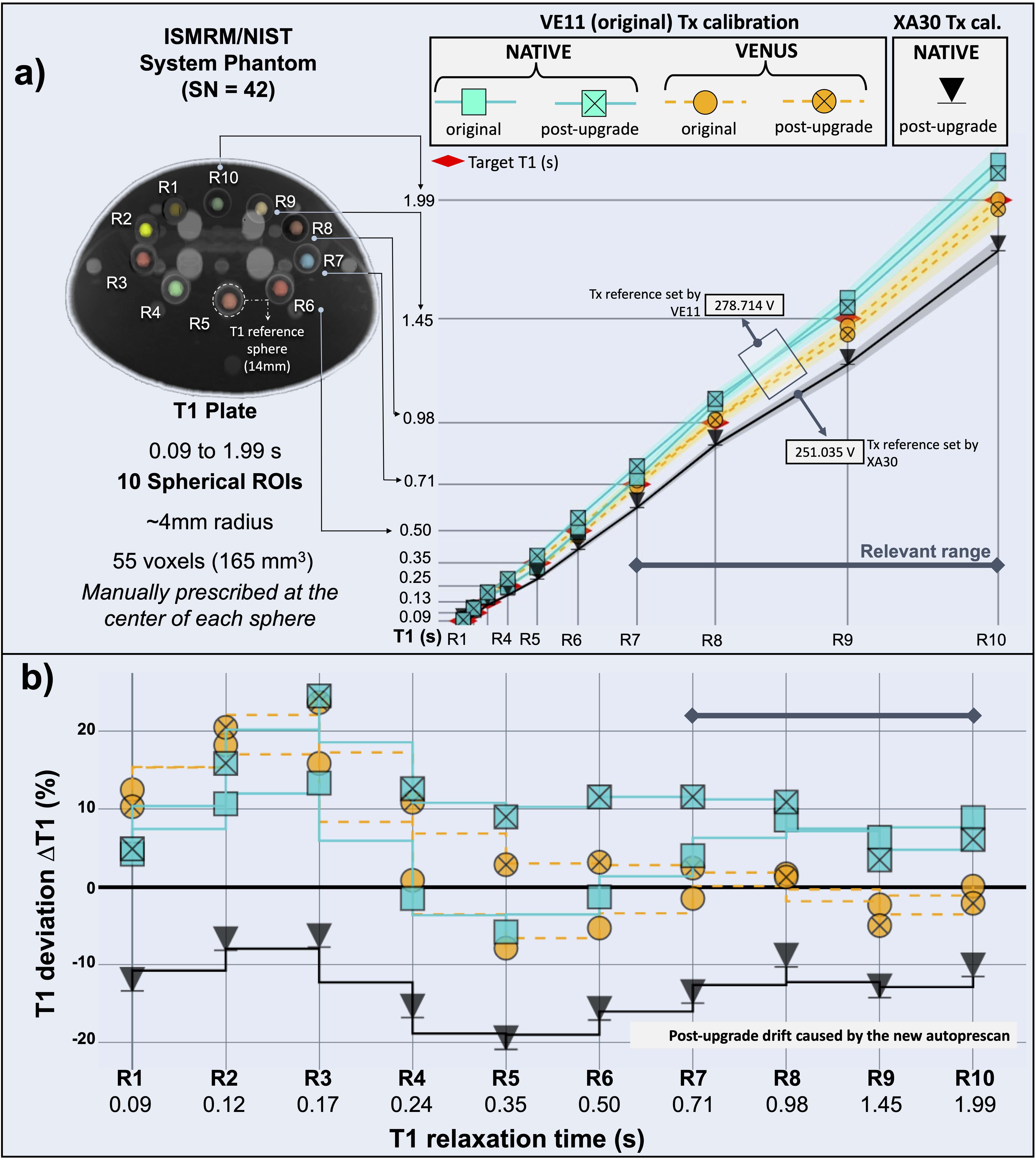
However, for the phantom acquisition, the original (278.7V) vs post-upgrade (251.0V) Vref difference was much larger (27.8V), exceeding the pre-upgrade variation (278.4±1.6V) by an order of magnitude. Because of this drastic change, we also performed an additional scan (phantom only) by manually setting the Vref to that of the original dataset.
Data processing and statistical analyses were performed as described in6 by running the same Nextflow pipeline published previously (https://github.com/qMRLab/VENUS). The code and BIDS-formatted data associated with this abstract are at https://github.com/qmrlab/ismrm23.
Results
Longitudinal stabilityPost-upgrade phantom T1 values closely follow the original measurements when the XA30 system uses the original transmit calibrations (Fig. 2a). As this holds true for both VENUS and vendor-native protocols, the accuracy improvement of VENUS persists.
Phantom test-retest post-upgrade measurements are stable across R1-10 reference spheres within two-decimal precision. However, they notably underestimate the ground truth. For example, the T1 drop in physiologically relevant values ranges from 0.2 to 0.5s, corresponding to a post-upgrade T1 drift up to 18.5% (Fig. 2a). Fig. 2b indicates that the upgrade increased the vendor-native deviation from the ground truth (blue squares vs black triangles).
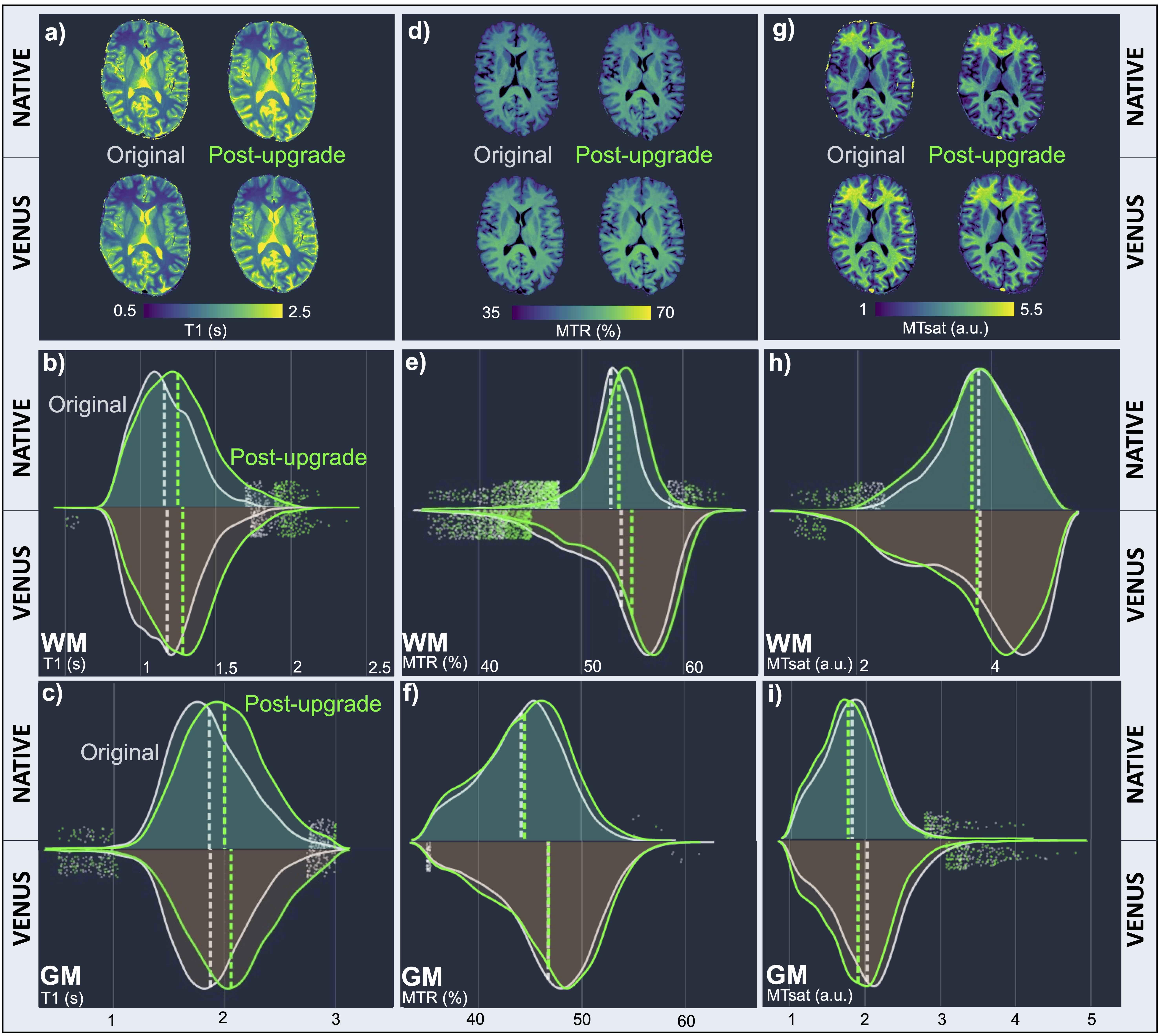
In-vivo T1 values show an overall post-upgrade increase, both with the vendor-neutral and the VENUS acquisitions. Fig. 3a-c reveals the systematic nature of the T1 bias, with a median increase of 8%. MTR remains mostly stable against the upgrade (Fig. 3d-f), whereas the T1 bias affects MTsat more (Fig. 3g-i).
End-to-end consistency
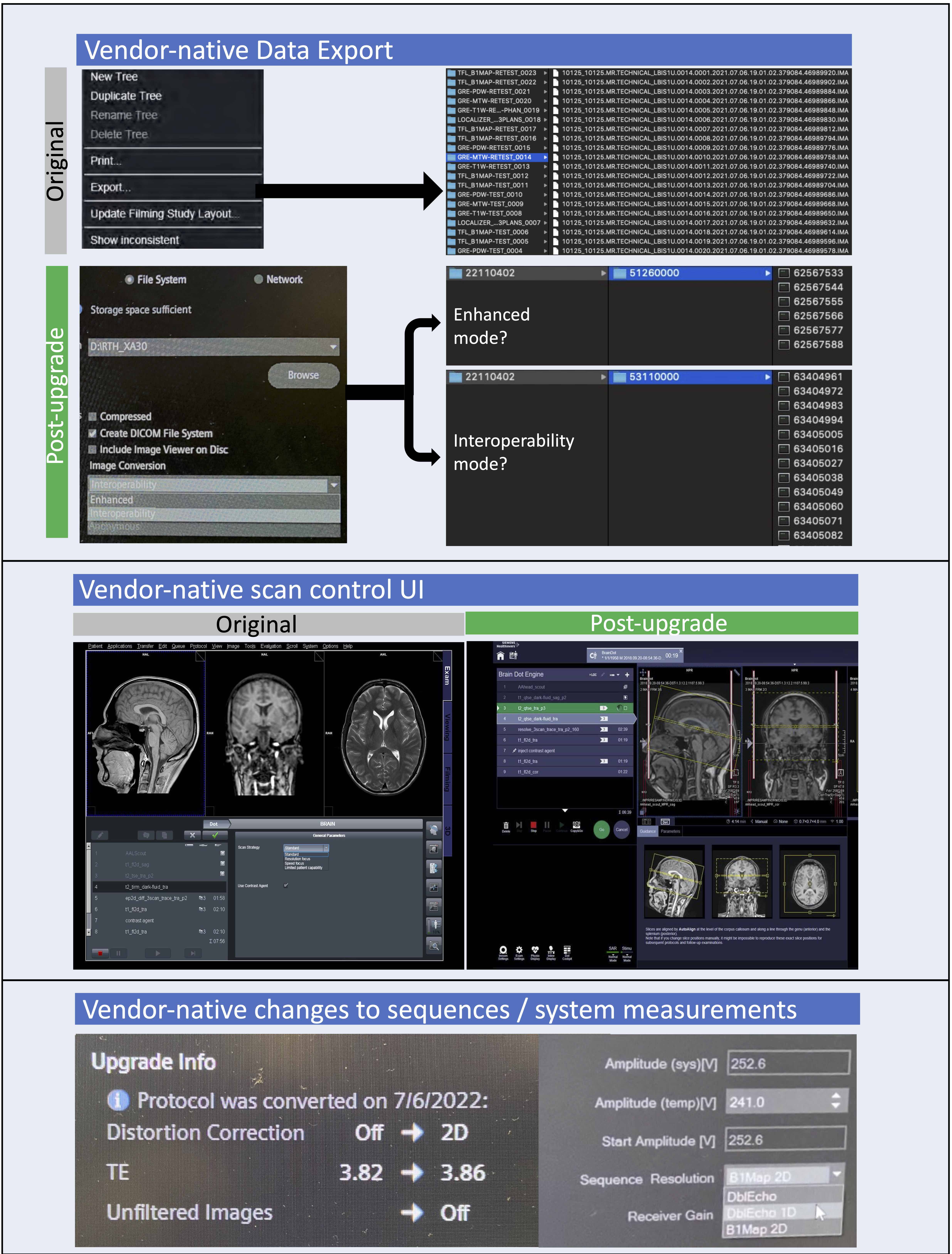
The new vendor-native data export module was notably different from the original one (Fig. 4). This change appears to cause a series of major problems for MRI researchers (see https://tinyurl.com/z5vkyrxt). Other challenges include a new scanner console and inadequate information on the changes made to the acquisition protocols (Fig. 4). Such difficulties place extra burden on the technologists in ensuring data consistency for research projects.
As for the practical aspects of the qMRI workflow, VENUS remained the same. For example, the VENUS data export module conformed with the qMRI-BIDS pre- and post-upgrade, and the scan control interface did not change.
Discussion
Engineering perspectivePrevious qMRI work that explored a scanner upgrade reported a dramatic (up to 30%) post-upgrade change in phantom T1 on a vendor-native system4. The changes were entirely attributed to contributions from B1+. Similarly, our findings indicate a directional relationship from decreased Vref to decreased T1 values in the phantom, and from increased Vref to increased T1 values, in-vivo.
Standardizing the acquisition and workflows using VENUS supports the hypothesis that the differences can be explained by B1+. Manually correcting for Vref in prescan recovers the T1 accuracy in both VENUS and vendor-neutral measurements (Fig. 2a-b). Addressing this correction at the root (before images are acquired) should be considered in addition to B1+ mapping.
Technologist perspective
We would like to conclude this abstract with a verbatim quote from a technologist: “Acquiring good research data is often much more complicated than performing routine clinical scans. After 28 years working as a technologist, I am yet to see one upgrade that makes MRI research easier.” This study shows that VENUS can guard against major convention changes that complicate research.
The proprietary nature of the post-upgrade prescan changes makes it impossible to tease out the exact origin of the drift in the transmit reference voltage. To disentangle these effects, qMRI would benefit from transparent vendor-neutral prescan calibrations.
Conclusion
Vendor-native software upgrades may lead to significant bias in qMRI experiments. To preserve the fidelity of these measurements, future work should develop vendor-neutral prescan protocols under version control.Acknowledgements
Canadian Foundation For Innovation, Grant/Award Number: Leader's Fund; Canadian Network for Research and Innovation in Machining Technology, Natural Sciences and Engineering Research Council of Canada, Grant/Award Numbers: 2016-06774; RGPIN-2019-07244; RGPIN-2022-05308; Canadian Open Neuroscience Platform, Grant/Award Number: Research scholarship; Fonds de Recherche du Québec - Nature et Technologies, Grant/Award Number: 2015-PR-182754; Fonds de Recherche du Québec - Santé, Grant/Award Numbers: 28826; FRSQ-35250; FRSQ-36759; Réseau en Bio-Imagerie du Quebec, Grant/Award Numbers: 5886; 8436-0501; TransMedTech Institute, Grant/Award Number: Excellence scholarshipReferences
1. Potvin, O. et al. Measurement Variability Following MRI System Upgrade. Front. Neurol. 10, 726 (2019).
2. Gunter, J. L., Shiung, M. M., Manduca, A. & Jack, C. R., Jr. Methodological considerations for measuring rates of brain atrophy. J. Magn. Reson. Imaging 18, 16–24 (2003).
3. Takao, H., Hayashi, N. & Ohtomo, K. Effect of scanner in asymmetry studies using diffusion tensor imaging. Neuroimage 54, 1053–1062 (2011).
4. Keenan, K. E. et al. Multi-site, multi-platform comparison of MRI T1 measurement using the system phantom. PLoS One 16, e0252966 (2021).
5. Lee, Y., Callaghan, M. F., Acosta-Cabronero, J., Lutti, A. & Nagy, Z. Establishing intra- and inter-vendor reproducibility of T1 relaxation time measurements with 3T MRI. Magn. Reson. Med. 81, 454–465 (2019).
6. Karakuzu, A., Biswas, L., Cohen-Adad, J. & Stikov, N. Vendor-neutral sequences and fully transparent workflows improve inter-vendor reproducibility of quantitative MRI. Magn. Reson. Med. 88, 1212–1228 (2022).
7. Karakuzu, A. et al. qMRI-BIDS: An extension to the brain imaging data structure for quantitative magnetic resonance imaging data. Sci Data 9, 517 (2022).
Figures