2974
Magnetic Resonance Imaging Based Radiomics of Axial and Sagittal Orientation in Suspected Placenta Accreta Spectrum Pregnancies1Radiology, UT Southwestern Medical Center, Dallas, TX, United States, 2Obstetrics & Gynecology, UT Southwestern Medical Center, Dallas, TX, United States, 3Parkland Health and Hospital System, Dallas, TX, United States, 4Advanced Imaging Research Center, UT Southwestern Medical Center, Dallas, TX, United States, 5Center for Imaging and Surgical Innovation, UT Dallas, Richardson, TX, United States, 6Bioengineering and Computer Science, UT Dallas, Richardson, TX, United States, 7Population and Data Sciences, UT Southwestern Medical Center, Dallas, TX, United States
Synopsis
Keywords: Placenta, Radiomics, placenta accreta spectrum, MR images, hysterectomy
Placenta accreta spectrum (PAS) is associated with significant morbidity and mortality. We applied ROI-based radiomics analysis on axial and sagittal plane MR images in women with placenta previa and a history of previous cesarean delivery. Our goal was to compare MR textural features extracted from axial and sagittal imaging planes and compare findings to surgical outcomes. Radiomics features were significant in both anatomical planes in the MRIs of women who underwent hysterectomy.Introduction
Placenta accreta spectrum (PAS) or pathologic adherence of the placenta in women with previous cesarean has become increasingly prevalent, affecting up to 1 in 272 pregnancies. Depending on PAS severity, patient management may involve a hysterectomy following cesarean delivery. Magnetic resonance (MR) imaging provides visualization of the entire placenta and is performed as an adjunct to aid in the diagnosis of PAS, enabling detailed operative planning for delivery. Publications of MR findings of PAS have often used images in sagittal and axial planes to demonstrate PAS MR-characteristics such as placental heterogeneity, low-attenuation T2 bands and placental-uterine interface(1,2). Radiomics analysis on placental ROI from PAS-suspected MR images has been carried out with either on axial(3) or sagittal plane(4-7). In this study, we sought to compare and correlate radiomics analysis extracted from axial vs. sagittal plane for the prediction of hysterectomy at the time of cesarean. This characterization will be valuable in our understanding of the placental spatial heterogeneity and the selection of effective radiomic features in the context of imaging plane.Methods
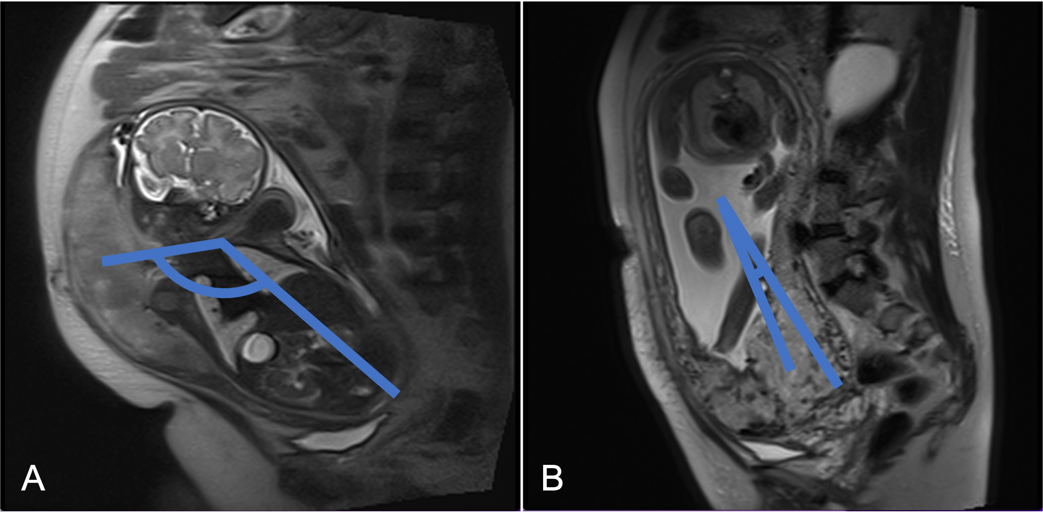
We performed an IRB approved retrospective review of 100 pregnancies between 2014-2019 of women with clinically suspected PAS with MR studies. All data were acquired on a 1.5T MR scanner (Avanto, Siemens Healthcare, Erlangen, Germany) with a body array coil. The reviewed MR images include T2-weighted (T2W) covering the entire gravid uterus (fetus and whole placenta volume) in the axial and sagittal plane (Table 1).Volumetric placental, uterus, and internal os regions of interest (ROI) were manually segmented under the supervision of a board-certified radiologist with significant obstetrical MR experience. Radiomic features were extracted following the image biomarker standardization initiative guideline(8). In-plane spacing was resampled to 1.5 mm. The 2D binwidth of texture features was set at 1. All images were normalized, scaled by 100 and outliers of 2 standard deviation (SD) were truncated. 105 radiomics features were extracted using the pyradiomics package. Additionally, a custom feature, Placental Location within the Uterus (PLU) was also generated. PLU is determined as the angle between 2 vectors, with the tail being the epicenter of the uterus to the internal os and the epicenter of the placenta (Figure 1).Agreement in features extracted from axial and sagittal images were assessed using Intraclass Correlation Coefficient (ICC) with two-way mixed model and consistency as well as spearman rank correlation. Agreement was interpreted as: Excellent: 0.75-1.00; Good: 0.60-0.75; Fair: 0.40-0.60; Poor: values ≤0.40. Correlation was interpreted as: Strong: 0.8-1.0; Moderate 0.5-0.8; Fair: 0.3-0.5; poor: values ≤0.3. Multivariable Least Absolute Shrinkage and Selection Operator (LASSO) logistic regression was performed with 10/10 nested cross validation and 500 repeats, shrinkage parameter was selected via cross-validation. The diagnostic performance of features extracted from axial and sagittal planes was quantified under the receiver operating characteristics curve (AUC). Feature extraction was done in Python and all other analysis was done in R.
Results
Of 100 pregnancies, 65 underwent cesarean hysterectomy and 35 had cesarean alone. 63% of the radiomics features had excellent agreement between sagittal and axial (ICC>0.75). 77% of the radiomics features had strong correlations.In multivariate analysis, three radiomics features: PLU, DependenceNon-UniformityNormalized, and Shape Elongation were selected in >90% iterations for both sagittal and axial images. AUC of features from sagittal images was 0.72 (95% CI 0.67-0.76) and from axial images was 0.79 (95% CI 0.75-0.84), p = 0.072.
PLU was 35°±34° for hysterectomy group and 62°±35° for no hysterectomy group(p<0.001). The DependenceNonUniformity-Normalized radiomics feature, a heterogeneity index, was also significantly different (p<0.001) between those with hysterectomy (sagittal: 0.297±0.075, axial: 0.229±0.05) and those without (sagittal: 0.246±0.058, axial: 0.2±0.044). Figure 1 shows representative sagittal images of (A) a PAS-suspected case without hysterectomy with large PLU and DependenceNonUniformity-Normalized values; and (B) a PAS-suspected case with hysterectomy with small PLU and DependenceNonUniformity-Normalized values. Shape elongation was also significantly different (p<0.001) between those with hysterectomy (sagittal: 0.751±0.127, axial: 0.763±0.138) and those without (sagittal: 0.828±0.08, axial: 0.832±0.086).
Discussion
ICC and Spearman rank correlation indicate that axial and sagittal imaging planes provide similar radiomics information. We identified radiomics features that were significant predictors for hysterectomy regardless of the imaging plane, indicating that the features extracted from axial and sagittal images reflect similar underlying placental pathophysiology. Women that have a lower PLU are more likely to have placenta previa, a known elevated risk for hysterectomy. High DependenceNon-UniformityNormalized value reflects a high level of heterogeneity, likely indicating large abnormal areas of lacunae in the placenta (Figure 1). By studying the visual differences of the hysterectomy/ no hysterectomy cases for radiomics features that we identify, we plan to expand the understanding of the link between radiomics features and the visual cue of underlying pathophysiology.Conclusion
Radiomics features extracted from axial and sagittal MR image plane in the same patient have excellent agreement and strong correlation. Shape elongation, PLU, and heterogeneity features were shown in both axial and sagittal images to be predictive in detecting PAS-suspected patient who required hysterectomy.Acknowledgements
No acknowledgement found.References
1. Clark HR, Ng TW, Khan A, Happe S, Dashe J, Xi Y, Twickler DM. Placenta Accreta Spectrum: Correlation of MRI Parameters With Pathologic and Surgical Outcomes of High-Risk Pregnancies. AJR Am J Roentgenol 2020;214(6):1417-1423.
2. Khan A, Do QN, Xi Y, Spong CY, Happe SK, Dashe JS, Twickler DM. Inter-reader agreement of multi-variable MR evaluation of Placenta Accreta Spectrum (PAS) and association with cesarean hysterectomy. Placenta 2022;126:196-201.
3. Andescavage N, Dahdouh S, Jacobs M, Yewale S, Bulas D, Iqbal S, Baschat A, Limperopoulos C. In vivo textural and morphometric analysis of placental development in healthy & growth-restricted pregnancies using magnetic resonance imaging. Pediatr Res 2019;85(7):974-981.
4. Wu Q, Yao K, Liu Z, Li L, Zhao X, Wang S, Shang H, Lin Y, Wen Z, Zhang X, Tian J, Wang M. Radiomics analysis of placenta on T2WI facilitates prediction of postpartum haemorrhage: A multicentre study. EBioMedicine 2019;50:355-365.
5. Shao Q, Xuan R, Wang Y, Xu J, Ouyang M, Yin C, Jin W. Deep learning and radiomics analysis for prediction of placenta invasion based on T2WI. Math Biosci Eng 2021;18(5):6198-6215.
6. Chen E, Mar WA, Horowitz JM, Allen A, Jha P, Cantrell DR, Cai K. Texture analysis of placental MRI: can it aid in the prenatal diagnosis of placenta accreta spectrum? Abdom Radiol (NY) 2019;44(9):3175-3184.
7. Do QN, Lewis MA, Xi Y, Madhuranthakam AJ, Happe SK, Dashe JS, Lenkinski RE, Khan A, Twickler DM. MRI of the Placenta Accreta Spectrum (PAS) Disorder: Radiomics Analysis Correlates With Surgical and Pathological Outcome. J Magn Reson Imaging 2020;51(3):936-946.
8. van Griethuysen, J. J. M., Fedorov, A., Parmar, C., Hosny, A., Aucoin, N., Narayan, V., Beets-Tan, R. G. H., Fillon-Robin, J. C., Pieper, S., Aerts, H. J. W. L. (2017). Computational Radiomics System to Decode the Radiographic Phenotype. Cancer Research, 77(21), e104–e107
Figures