2968
Vascular Assessment of Rhesus Macaque Placental Injury through Maternal Intervillous Blood Volume Measurement1Medical Physics, University of Wisconsin - Madison, Madison, WI, United States, 2Wisconsin National Primate Research Center, University of Wisconsin - Madison, Madison, WI, United States, 3Comparative Biosciences, University of Wisconsin - Madison, Madison, WI, United States, 4Obstetrics & Gynecology, University of Wisconsin - Madison, Madison, WI, United States, 5Radiology, University of Wisconsin - Madison, Madison, WI, United States, 6Biomedical Engineering, University of Wisconsin - Madison, Madison, WI, United States
Synopsis
Keywords: Placenta, Quantitative Imaging
Early assessment of utero-placental vasculature is an important novel approach to predicting pregnancy outcomes and fetal growth. Ferumoxytol-enhanced MRI is a powerful tool to measure intervillous fractional blood volume (FBV) in vivo, which could reflect structural and vascular health in the placenta. This study reports such measurements for rhesus macaque placentas that underwent localized Tisseel injections to mimic the effects of local ischemia. We observed increased regions of low FBV with gestation following the Tisseel injection.Introduction
Adequate development of utero-placental vasculature is crucial in facilitating oxygen and nutrient exchange during pregnancy1. Measurements of placental blood volume could potentially detect abnormal intervillous vasculature, such as local malperfusion and ischemia. Ferumoxytol-enhanced MRI holds potential to assess placental blood volumes in-vivo holds promise to provide useful insights for detection and progression of placental pathology. Ferumoxytol is used clinically in pregnant patients and has previously been used for clinical imaging diagnosis of pulmonary embolism2. Recently, pilot studies for such blood volume measurements have been demonstrated in healthy mice3 and rhesus macaque models4, as well as in human placentas with fetal growth restriction5. However, the link between blood volume measurements and pathology, such as thrombosis, has not been established. Here, we present results from a new animal model designed to generate placental vascular lesions, which are assessed with ferumoxytol-enhanced MRI to track the progression of local placental injury through longitudinal maternal fractional blood volume measurements.Methods
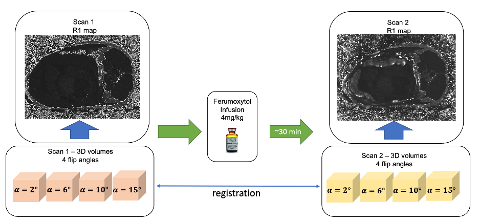
Subjects: Six rhesus macaques went through three longitudinal scans at around days 100, 115, and 145 of gestation. One day after the first imaging date, three of the subjects received a 0.5ml Tisseel injection into the anterior lobe of the placenta, and the other three received a saline injection as controls. Tisseel (Baxter Healthcare Corp) is an FDA-approved fibrin sealant used surgically to control bleeding. We hypothesized its injection would create blood clots within cotyledons in the placenta, thereby mimicking vascular infarcts. Histopathological analysis was performed on the placentas following cesarean section delivery at gestation day 155.Imaging: All subjects were sedated with isoflurane prior to imaging. The scans were acquired on a 3.0 T system (Discovery MR750, GE Healthcare) with a 32-channel phased array coil. The subjects were imaged in right-lateral position. A respiratory-gated center out, 3D radial spoiled gradient echo sequence (TR=6.0ms, TE=1.2ms, scan time=525.9s, BW=125kHz, spatial resolution=0.87×0.87×1.00mm3) covering the entire placenta was performed at four different flip angles (2°, 6°, 10°, 14°). The same protocol was performed 30 minutes after intravenous ferumoxytol (4mg/kg) infusion. Figure 1 demonstrates the imaging workflow. Following delivery, each placenta was evaluated by a pathologist.
Processing: Pixel-wise signal intensity of each flip angle datapoint was fitted via an inhouse toolbox in MATLAB (Mathworks, Natick, MA) to obtain the pre- and post-contrast R1 maps. Non-rigid registration (ANTS6) was used to register post- and pre- contrast scans. Both placental and maternal vascular segmentation of the uterine artery were performed to obtain R1 values in placenta and in blood. Maternal fractional blood volume (FBV) was calculated as the ratio between ΔR1 (difference between pre- and post-contrast R1 maps) in the placenta and ΔR1 in maternal blood4,5.
Results
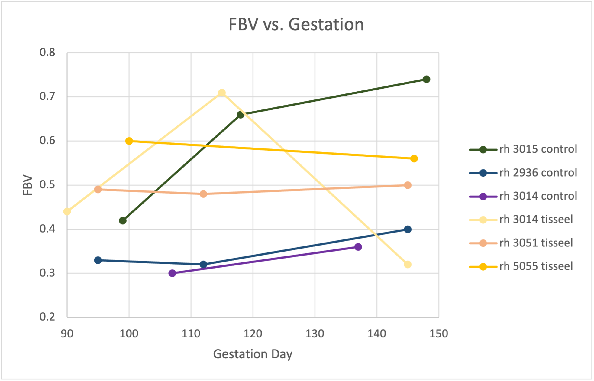
Two out of three Tisseel-treated subjects had only a single disc of the placenta; all other subjects had the rhesus typical two placental discs. Figure 2 shows longitudinal measures of mean FBV for each subject. All control subjects have a trend of increasing FBV with gestation, whereas the Tisseel-treated subjects have a relatively constant trend through gestation.A wide range of pathology was detected for each subject, including the controls; however, one Tisseel-treated placenta in particular (rh3014) was inferred from pathology seen upon histologically analysis to have significant ischemia that multifocally affects large portions of several cotyledons.
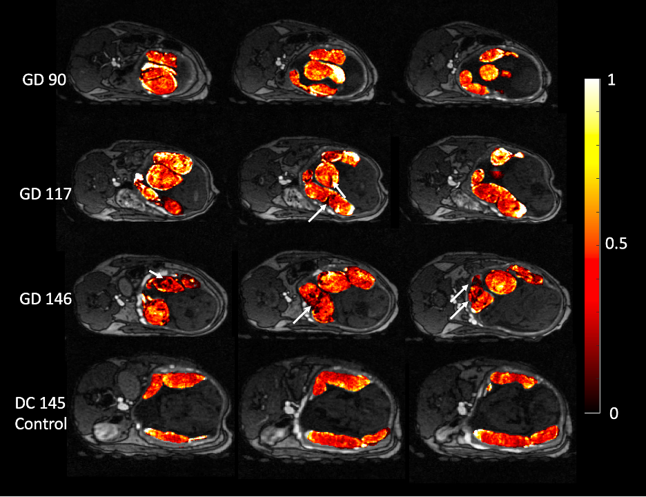
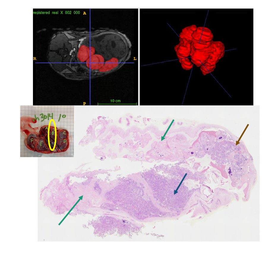
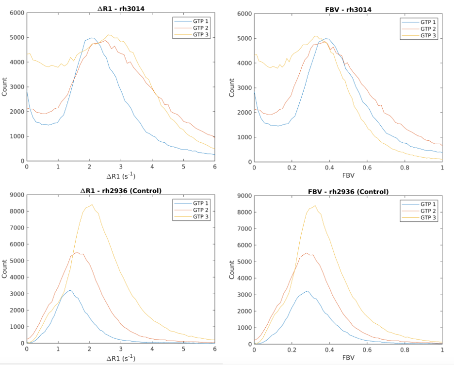
Figure 3 shows three non-consecutive slices of maternal fractional blood volume for subject rh3014, and one control subject on the bottom row. Upon visual inspection, we did not observe congregated regions of low blood volume in the MRI of the first gestation timepoint (before Tisseel injection). However, the second and third gestational timepoint show visually distinct regions of low blood volume regions, suggesting potential ischemia. Figure 4 shows post-delivery histopathological analysis of a center cut of cotyledon B in rh3014. There are significant regions of diffused ischemia and coagulative necrosis, as well as regions of villus tissue likely disrupted by fibrin injection (Tisseel). Figure 5 shows histograms of ΔR1 and FBV of subjects rh3014 and rh2936 (a representative case from the control group). In comparison, the histograms of rh3014 reflect increased regions of reduced FBV in scan 2 which further increases in scan 3.
Discussion and Conclusion
This study reports maternal fractional blood volume in a preclinical intervention model designed to create vascular infarcts in the placenta. Histopathological analysis showed diffused ischemia and coagulative necrosis in multiple cotyledons for one Tisseel-treated subject. The FBV maps reflect such conditions through visible low blood volume regions on multiple cotyledons presented throughout the slices; this condition was not reflected by the FBV on the first gestational timepoint, as Tisseel injections were not introduced then. FBV histograms also quantitatively show the low blood volume regions in the Tisseel-treated subject that are increasing in volume with gestation, likely caused by tissue ischemia and/or necrosis. These results indicate that the intervention was successful in at least one subject and that noninvasive FBV measures are sensitive to the created infarct regions. Future studies will investigate local comparisons between the in vivo FBV data and placental pathology.Acknowledgements
We gratefully acknowledge GE Healthcare for research support of UW-Madison, and funding support from NIH-NICHD (R01HD103443).References
1. Roberts JM, Escudero C. The placenta in preeclampsia. Pregnancy Hypertens. 2012;2(2):72-83. doi:10.1016/j.preghy.2012.01.0012.
2. Starekova J, Nagle SK, Schiebler ML, Reeder SB, Meduri VN. Pulmonary MRA During Pregnancy: Early Experience With Ferumoxytol. J Magn Reson Imaging. Published online October 31, 2022. doi:10.1002/jmri.285043.
3. Badachhape AA, Devkota L, Stupin I v, et al. Nanoparticle Contrast-enhanced T1-Mapping Enables Estimation of Placental Fractional Blood Volume in a Pregnant Mouse Model. Sci Rep. 2019;9(1):18707. doi:10.1038/s41598-019-55019-84.
4. Chen R, Nguyen S, Murphy M, et al. Longitudinal Placental Blood Volume Measurements on Zika-Infected Rhesus Macaques Using Variable Flip Angle T1 Mapping. In: Proc 29th Annual Meeting ISMRM. ; 2021.5.
5. Chen R, Fain S, Magness R, et al. Maternal Blood Volume Measurements of Human Placenta with Fetal Growth Restriction using Ferumoxytol-Enhanced MRI. In: Proc 30th Annual Meeting ISMRM. ; 2022.6.
6. Avants BB, Tustison NJ, Stauffer M, Song G, Wu B, Gee JC. The Insight ToolKit image registration framework. Front Neuroinform. 2014;8:44. doi:10.3389/fninf.2014.00044
Figures