2964
Evaluate the value of Curved Planar Reconstruction of 3D high-resolution T2WI for the Detection of Deep Infiltrative Endometriosis
Ye Li1, Ailian Liu1, Jiazheng Wang2, and Liangjie Lin2
1The First Affiliated Hospital of Dalian Medical University, Dalian, China, 2Clinical and Technical Support, Philips Healthcare, Beijing, China
1The First Affiliated Hospital of Dalian Medical University, Dalian, China, 2Clinical and Technical Support, Philips Healthcare, Beijing, China
Synopsis
Keywords: Pelvis, Image Reconstruction
22 DIE patients were scanned for comparison of diagnostics accuracy between ordinary reconstruction and curved planar reconstruction image of 3D-CS-HR T2WI sequence. Results indicate that Curve images improved the detection of ectopic lesions in each ligament of the pelvic floor.Synopsis
Diagnosis of deep infiltrating endometriosis (DIE) disease remains challenging, because the location of ectopic lesions can be multifocal and the pelvic anatomy may change due to the adhesion of endometriosis lesions. In this study, we investigated the value of curved planar reconstruction of compressed sensing (CS) accelerated 3D high-resolution (HR) T2WI for the detection of DIE. Results indicate that Curve images of 3D-CS-HR T2WI improved the detection of DIE lesions when compared to the ordinary slice-based image reading.Summary of Main Findings
22 DIE patients were scanned for comparison of diagnostics accuracy between ordinary reconstruction and curved planar reconstruction image of 3D-CS-HR T2WI sequence. Results indicate that Curve images of 3D-CS-HR T2WI improved the detection of ectopic lesions in each ligament of the pelvic floor.Introduction
Common clinical manifestations of deep infiltrating endometriosis (DIE) include pelvic pain and infertility. Deep endometriosis is considered the main cause of chronic pelvic pain in women of reproductive age1. Deep infiltrating endometriosis (DIE), defined as endometrial glands and stroma infiltrating the peritoneum by at least 5 mm, is the most severe form of endometriosis. In DIE patients, endometrial glands and stroma are most common in the ovaries but may also involve broad ligament, sacral ligament, round ligament, etc. Surgery to DIE is challenging as deep endometriosis plaques may be hidden under extensive adhesions or may not be visible. Laparoscopy is the gold standard for the diagnosis of endometriosis. However, it increases the risk of implantation in DIE. The 3D high resolution T2 weighted MR protocol may give a more comprehensive evaluation of pelvis lesions2. In this study, we investigated the value of curved planar reconstruction based on compressed sensing (CS)-accelerated 3D high-resolution (HR) MRI for the detection of DIE.Methods
22 DIE patient (mean age:39.96 ± 7.28 years, range: 28-52 years) were scanned using a 3.0 T MR scanner (Ingenia CX, Philips Healthcare, the Netherlands) with the body coil to transmit and a 32-channel abdominal receive coil. The MR protocol included3D-CS-HR-T2WI (FOV=370mm, Slice think=2mm, during time=3 min 10 s, CS acceleration factor 4), for which the original result imageswere further reconstructed along the direction of the broad ligament and the central direction of the bottom of uterus. For both the original images and the curved-reconstructed images, two imaging physicians reviewed the images independently to evaluate the endometriosis on the following tissues: sacral ligament, broad ligament, round ligament, rectovaginal pouch adhesion. The sensitivity, specificity, positive predictive value, negative predictive value and optimal criterion were calculated for the diagnosis of DIE by both observers, and the sensitivity and specificity for the diagnosis of DIE were compared using the McNemar test.Results
Measurement consistency between the two observers was good (kappa value > 0.75). In this set of data (Table 1), the sensitivity and specificity of original image for the detection of lesions at sacral ligament, broad ligament, round ligament, rectovaginal pouch were:70% and 74%, 25% and 89%,100% and100%, 85% and 33%, respectively. The sensitivity and specificity of reconstructed image for the detection of lesions in these tissues were:.60% and 91%, 25% and 89%,100% and 100%, 100% and 0%, respectively. The specificity of reconstructed image was significantly higher than that of original image in sacral ligament (P=0.01). However, there was no difference between the sensitivity and specificity of other sites(P>0.05)Discussion
Curved reconstructed image seemed to be more efficient than the original image in the diagnosis of DIE. Since images could be reconstructed in various directions based on 3D-CS-HR-T2WI small lesions located at different positions become more visible, potentially help the surgery plan in the future.Conclusion
Curved Planar Reconstruction of 3D-CS-HR-T2WI is potentially a promising and valuable non-invasive method in detection of DIE.Acknowledgements
No acknowledgement found.References
[1] Lanieri MM, Raimondo D, Rosati A, et al. Impact of nerve-sparing posterolateral parametrial excision for deep infiltrating endometriosis on postoperative bowel, urinary, and sexual function. Int J Gynaecol Obstet. 2022 Oct;159(1):152-159.
[2] Xu Y, Yao Y, Pylypenko D, et al. Diagnosis of pelvic endometriosis: a preliminary study on the added value of R2*MFGRE sequence in magnetic resonance imaging. Acta Radiol. 2022 Aug 8:2841851221117260.
Figures
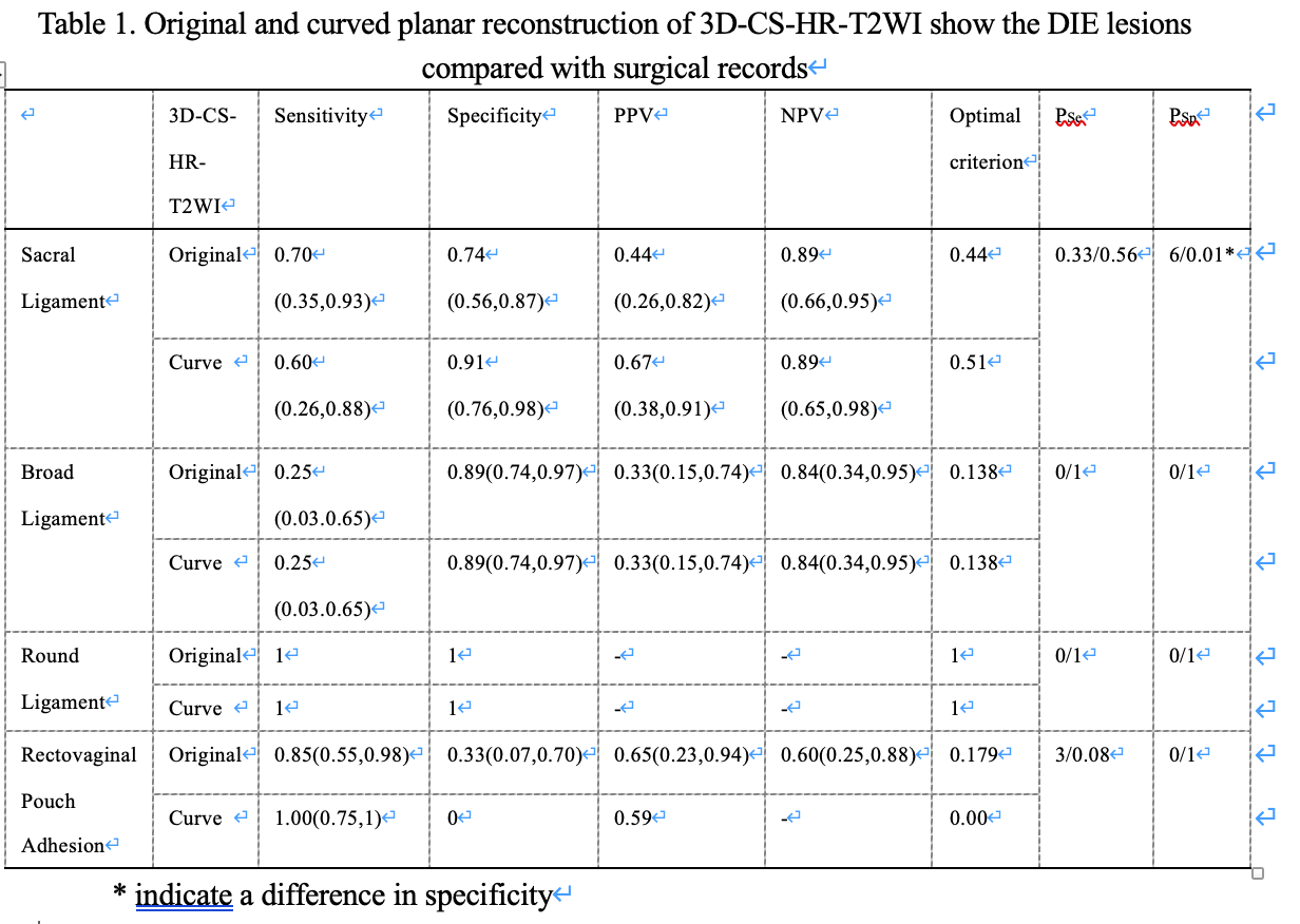
Table 1. Original and curved planar reconstruction of 3D-CS-HR-T2WI show the DIE lesions compared with surgical records
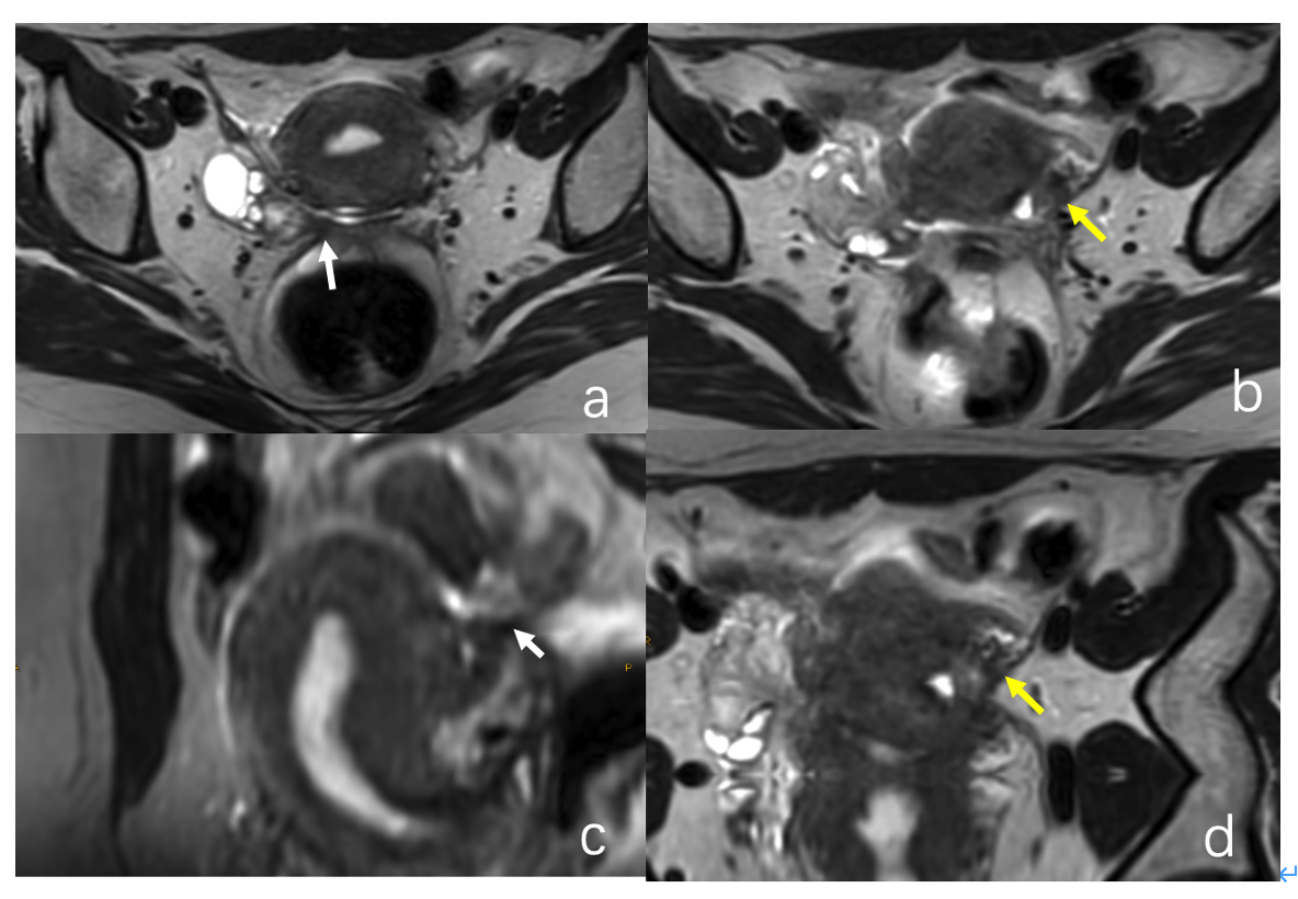
Figure 1. A 39-year-old woman with right side ovary endometriosis and DIE. Fig a and b: Original image of 3D-CS-HR-T2WI shows that there is a thickened strip of low signal on the left side of right sacral ligament (white arrow) and left broad ligament (yellow arrow). Fig c and d. Corresponding Curve reformation images of 3D-CS-HR-T2WI shows a clearly sign of localized thickening of ligaments.
DOI: https://doi.org/10.58530/2023/2964