2959
To evaluate the utility of magnetization transfer (MT) imaging in the study of normal uterus and common uterine lesions.
Qiu Bi1, Kunhua Wu1, and Yunzhu Wu2
1the First People’s Hospital of Yunnan Province, Kunming, China, 2Siemens Healthcare, Shanghai, China
1the First People’s Hospital of Yunnan Province, Kunming, China, 2Siemens Healthcare, Shanghai, China
Synopsis
Keywords: Uterus, Tumor
In this study, we explored the value of MT imaging to characterize normal uterine structures and common uterine lesions by measuring MTR values. The results showed the MTR values were significantly different among normal uterine structures, among uterine lesions of different origin, or between some uterine lesions and corresponding normal structures. MTR values were found to be effective in the diagnosis and differential diagnosis of certain uterine diseases. It might provide a preoperative basis for neoplastic histologic origin in the uterus.Objective
To evaluate the utility of magnetization transfer (MT) imaging in the study of normal uterus and common uterine lesions.Methods
This prospective study enrolled 160 consecutive patients with suspected uterine lesions. Magnetization transfer ratio (MTR) map was obtained by pelvic MT imaging on a 3.0T MRI scanner. Patients confirmed by pathology were divided into microscopic lesion group and lesion group, according to whether the maximum diameter of the lesion was less than 5mm. After evaluating and eliminating patients with poor image quality by a 3-point Likert scale, MTR values of lesions and normal endometrium, myometrium and cervix were independently measured on the MTR map by two radiologists. Inter-reader agreement was evaluated. MTR values were compared among different uterine lesions and normal uterine structures using the Mann-Whitney U test with Bonferroni correction. Receiver operating characteristic curve was performed. The correlations between age and MTR values were explored by Pearson correlation analyses.Results
A total of 96 patients with 121 uterine lesions in lesion group and 41 patients in microscopic lesion group were measured. The MTR values among normal endometrium, myometrium and cervix were statistical significant differences (P < 0.05). There were significant differences between endometrial cancer and normal endometrium and between cervical cancer and normal cervix (both P ≤ 0.001). Area under the curve (AUC) for diagnosing endometrial and cervical cancer were 0.73 and 0.86. Myometrial lesions had significantly higher MTR values than endometrial lesions and cervical cancer (both P < 0.001), and the AUC for differentiating myometrial lesions from them was 0.89 and 0.94. MTR values of endometrial cancer were significantly higher than those of cervical cancer (P = 0.02). There was a critical correlation between age and MTR values in endometrial cancer (r = 0.81, P = 0.04).Conclusions
MTR values showed significant differences among normal uterine structures. It was valuable for diagnosing and differentiating uterine cancer. MTR values could differentiate myometrial lesions from endometrial or cervical lesions.Acknowledgements
NoneReferences
1. Bray F, Ferlay J, Soerjomataram I, Siegel RL, Torre LA, Jemal A. Global Cancer Statistics 2018: GLOBOCAN Estimates of Incidence and Mortality Worldwide for 36 Cancers in 185 Countries. CA Cancer J Clin (2018) 68(6):394-424.
2. Su C, Zhao L, Li S, Jiang J, Cai K, Shi J, et al. Amid Proton Transfer (APT) and Magnetization Transfer (MT) MRI Contrasts Provide Complimentary Assessment of Brain Tumors Similarly to Proton Magnetic Resonance Spectroscopy Imaging (MRSI). Eur Radiol (2019) 29(3):1203-10.
3. Li XH, Mao R, Huang SY, Sun CH, Cao QH, Fang ZN, et al. Characterization of Degree of Intestinal Fibrosis in Patients with Crohn Disease by Using Magnetization Transfer MR Imaging. Radiology (2018) 287(2):494-503.
Figures
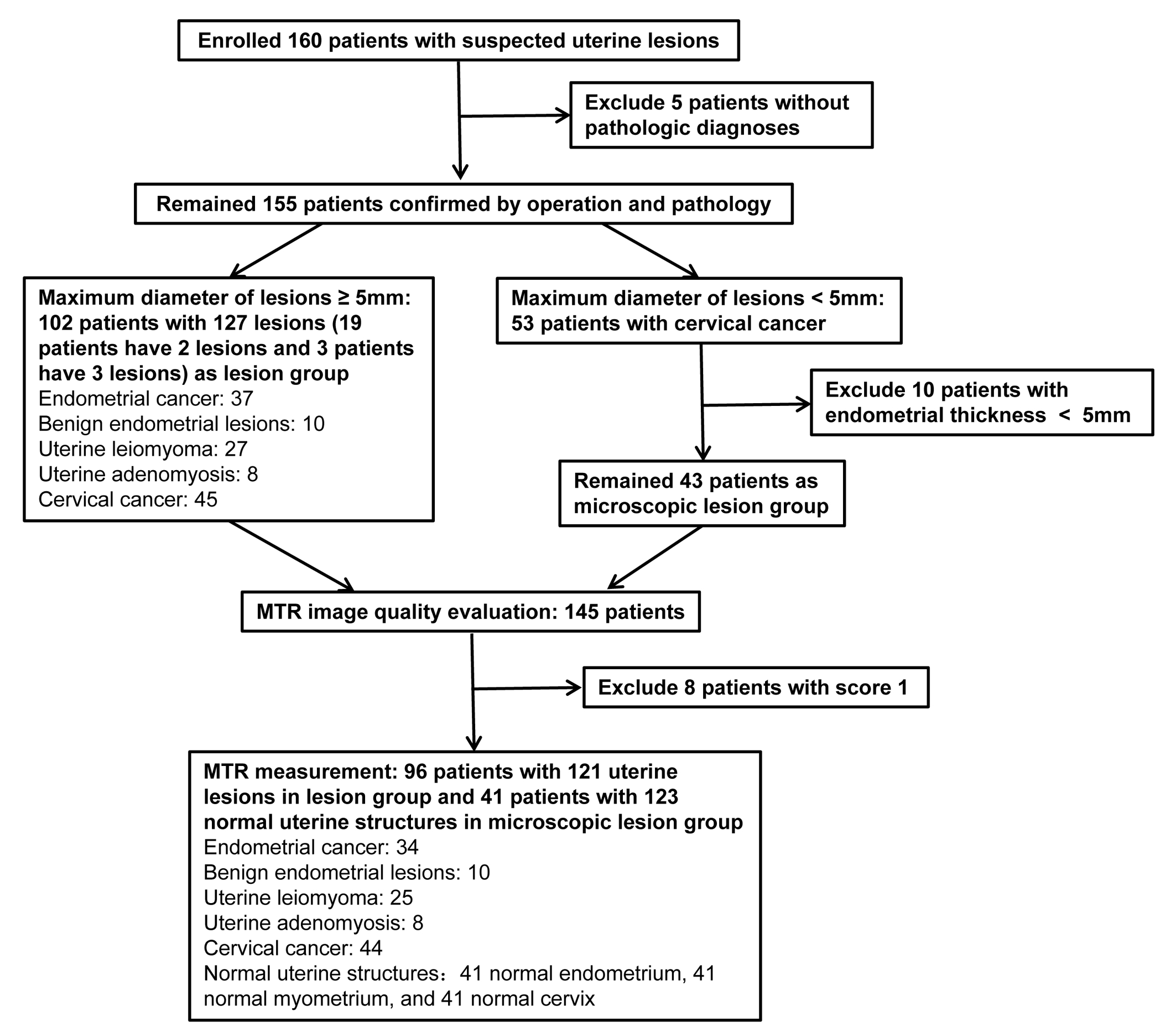
FIGURE 1 Flowchart of the study
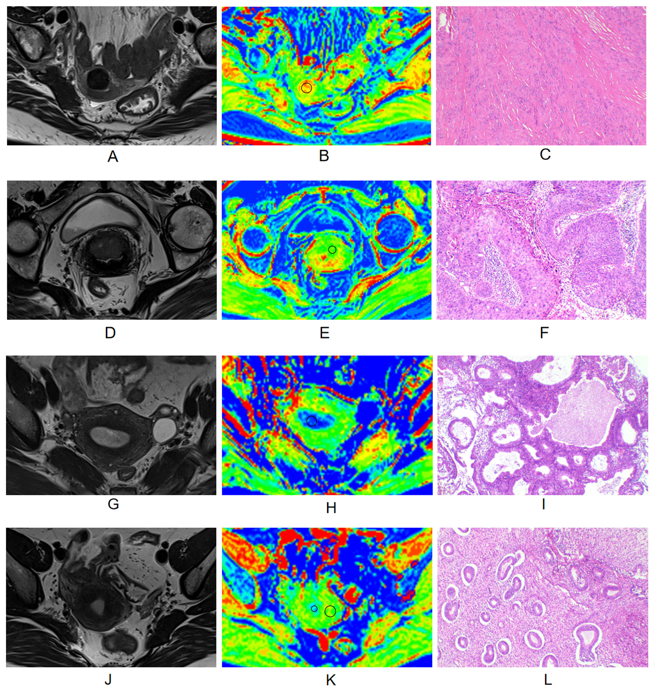
FIGURE 2 (A-C) A 64-year-old patient with Uterine leiomyoma, MTR of leiomyoma is 13.01. (D-F) A 65-year-old patient with cervical cancer, MTR of cervical cancer is 7.46. (G-I) A 49-year-old patient with endometrial hyperplasia, MTR of endometrial hyperplasia is 6.93. (J-L) A 40-year-old patient with carcinoma in situ of cervix, MTR of normal endometrium and myometrium are 7.17 and 12.03. Figure A, D, G, and J represent T2WIs. Figure B, E, H, and K represent pseudo-color MTR maps. Figure C, F, I, and L represent hematoxylin&eosin staining map (40×) of the lesions.
DOI: https://doi.org/10.58530/2023/2959