2922
High Resolution MR Reconstruction with Functionally Separate Neural Networks1MRI Systems Development Department, Canon Medical Systems Corporation, Kanagawa, Japan, 2MRI Systems Development Department, Canon Medical Systems Corporation, Tochigi, Japan
Synopsis
Keywords: Image Reconstruction, Machine Learning/Artificial Intelligence, Super resolution
The authors propose a new reconstruction method to obtain higher resolution images from an MR acquisition. The method incorporates MR physics and two neural networks, which are functionally separate, for denoising and upsampling. The proposed method was evaluated by applying it to both retrospectively and prospectively undersampled data. The result showed that the proposed technique is capable of reconstructing higher resolution images over a conventional method, by multiplying the matrix size while keeping more detail structure in the originally sampled data.INTRODUCTION
Acquiring higher resolution image faster, is the ultimate and endless demand for MR imaging. However, often the acquisition matrix cannot be large enough because a higher matrix inevitably leads to longer scan time. To compensate this limitation, a technique called zero-padding interpolation (ZIP)1 has been widely used to increase the matrix size to be displayed. Because the ZIP process, consistent with MR physics and applied in k-space, suffers from characteristic Gibbs ringing artifacts, it has been usually used with low-pass-filters (LPFs) sacrificing the resulting sharpness.On the other hand, recently there are variety of proposals so-called Super-Resolution techniques, in other applications, employing neural networks2-5. But most of them are targeted to low resolution images operating in image-space, and not always optimal to MR acquisition.
Therefore, the purpose of this study is to provide a fine way to increase the resolution of MR acquired images, by an assist of neural network technology. Furthermore, a combination of functionally separate networks is proposed to increase its applicability to realistic MR images.
METHODS
The reconstruction pipeline of the proposed method is illustrated in Figure 1. The first neural network for denoising is applied to coil combined image, which keeps complex values in each pixel. For the denoising network, we chose Soft-shrinkage denoising network in DCT domain6 as it is known for its applicability to varieties of contrasts and wide range of noise levels. The output of the denoising network is then Fourier-transformed back to k-space data to be input to the upsampling process. In the upsampling process, the input k-space data is zero-padded around its edges, Inverse-Fourier-transformed to image-space, then input to the second neural network. The network processes input image to recognize and remove characteristic artifacts introduced by the zero-padding, while keeping the detail structure which has been sampled originally.The training of the two neural networks was made with the same dataset as the ground-truth images, though they were developed separately. A dataset was collected with higher SNR and resolution compared to typical clinical images, by applying averaging technique to repeated scans of healthy volunteers. The acquisitions were made under an approval of our internal review board, and included varieties of contrasts such as T1w, T2w, FLAIR, PD, PD+FS, TOF, and variation of field strength such as 3T or 1.5T. The neural network for denoising was trained to recover that dataset as the ground truth, from simulated degraded inputs with gaussian noise added. The neural network for upsampling was trained to recover the same ground truth, from images degraded by truncation artifact (i.e., Gibbs ringing). The degraded inputs were created by replacing peripheral regions of its k-space with zeros, to simulate degradation along a ZIP process. The amount of truncated k-space was randomly selected from 0% to 97% to make single network applicable to wide range of upsampling factor.
The evaluation of the proposed method was made with both retrospectively and prospectively undersampled data. For the study with retrospective undersampling, high-SNR knee image was acquired from a healthy volunteer. The k-space data was then retrospectively undersampled at its peripheral region, gaussian noise of 5% of its peak signal was added in the image-space to simulate degraded input. As the comparison to a conventional method, ZIP with linear LPF method was also evaluated. Furthermore, we also tested proposed method without the ones of the two neural networks. The output images were quantitively evaluated in terms of PSNR7 and SSIM8 to the reference image. The undersampling factor of 2 and 3 were tested to evaluate the method’s availability to wide range of upsampling factor. For the study with prospective undersampling, T2w brain images were acquired from a healthy volunteer with two sets of scan parameters. For this study, only the visual evaluations were made because the reference images were not available and there were no chance to calculate PSNR nor SSIM.
RESULTS
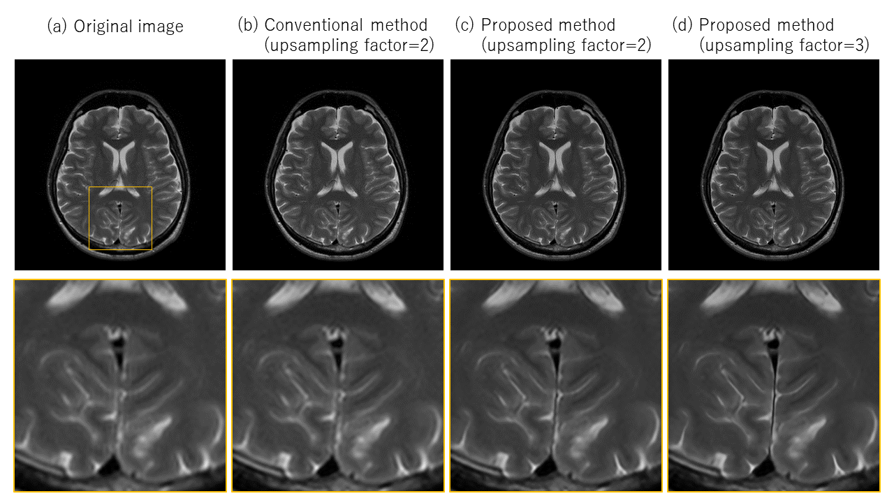
The resulted images from the retrospective study are shown in Figure 2 and 3. The PSNR and SSIM from the proposed method were higher than those from the conventional method with both upsampling factors. The images from the prospective study are shown in Figure 4 and 5.DISCUSSION
In the retrospective study, the advantage of upsampling network was more prominent at the higher upsampling factor. Interestingly, the upsampling network did not work well without the denoising network. We interpret this result that the upsampling network is very sensitive to noises. Here, the advantage of the proposed method is seen that the combination of two networks is enabling the method applicable to realistic images that usually have certain amount of noises.In the prospective study, it is certified that the proposed method works well on realistic scans. While the upsampling factor of 2 increases the sharpness of the image compared to the conventional method, upsampling factor of 3 increases the sharpness further. The method would also be helpful, when applied to more aggressive scan conditions, to increase the resolution to a practical level.
CONCLUSION
A new reconstruction method to obtain high resolution images from an MR acquisition is proposed. It is expected that the method extends the image quality and the flexibility of MR imaging.Acknowledgements
No acknowledgement found.References
- Bernstein MA, Fain SB, Riederer SJ. Effect of windowing and zero-filled reconstruction of MRI data on spatial resolution and acquisition strategy. J Magn Reason Imaging. 2001 Sep;14(3):270–280.
- Dong C, Loy CC, He K, et al. Image Super-Resolution Using Deep Convolutional Networks. IEEE Trans Pattern Anal Mach Intell. 2016;38(2):295-307
- Kim J, Lee JK, Lee KM. Accurate Image Super-Resolution Using Very Deep Convolutional Networks. Proc. The IEEE Conf. On Computer Vision and Pattern Recognition (CVPR). 2016:1646-1654
- Ledig C, Theis L, Huszar F, et al. Photo-Realistic Single Image Super-Resolution Using a Generative Adversarial Network. Proc. The IEEE Conf. On Computer Vision and Pattern Recognition (CVPR). 2017:4681-4690
- Lim B, Son S, Kim H, et al. Enhanced Deep Residual Networks for Single Image Super-Resolution. Proc. The IEEE Conf. On Computer Vision and Pattern Recognition Workshops (CVPRW). 2017:1132-1140
- Isogawa K, Ida T, Shiodera T, et al. Deep Shrinkage Convolutional Neural Network for Adaptive Noise Reduction. IEEE Signal Process Lett. 2018;25(2):224-228
- Horé A and Ziou D. Image Quality Metrics: PSNR vs. SSIM. Proc. 20th International Conf. on Pattern Recognition. 2010:2366-2369
- Wang Z, Bovik AC, Sheikh HR, et al. Image quality assessment: from error visibility to structural similarity. IEEE Trans Image Process. 2004;13(4):600-612
Figures
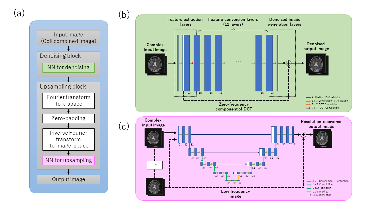
Illustration of the proposed reconstruction method
(a) Overview of the reconstruction pipeline
(b) Architecture of the neural network for denoising
(c) Architecture of the neural network for upsampling.
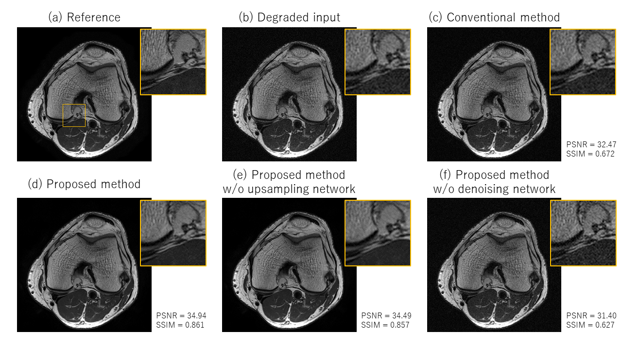
Resulted images from the retrospective study with the upsampling factor of 2
(a) Reference image
(b) Degraded input image
(c) Image by the conventional method (ZIP and LPF)
(d) Image by the proposed method
(e) Image by the proposed method, without the upsampling network
(f) Image by the proposed method, without the denoising network.
Scan parameters (Reference): FSE2D, TE=44ms, TR=3000ms, FOV=11 x 11cm, Matrix=640 x 640, Sampling-pitch=5$$$\mu$$$s, Slices=15 x 2mm, Scan-time=96s, Averaging=9.
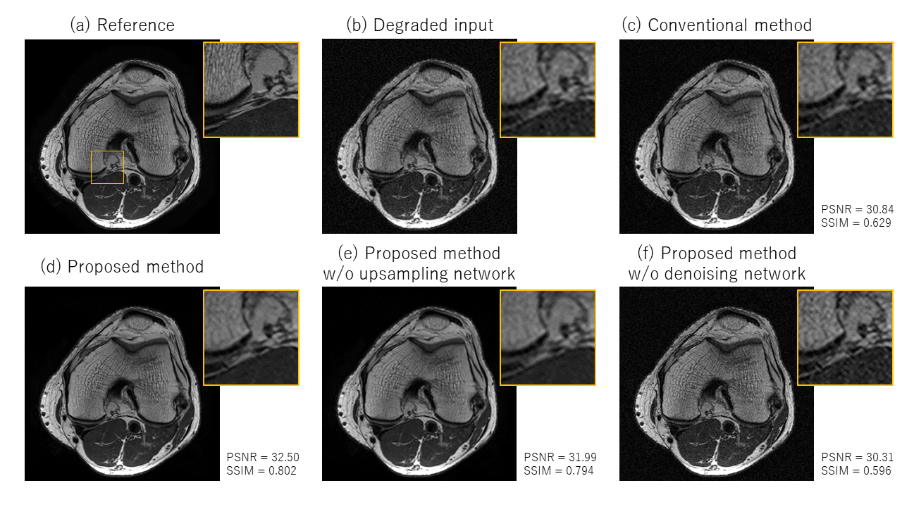
Resulted images from the retrospective study with the upsampling factor of 3
(a) Reference image (identical to Figure 2(a))
(b) Degraded input image
(c) Image by the conventional method (ZIP and LPF)
(d) Image by the proposed method
(e) Image by the proposed method, without the upsampling network
(f) Image by the proposed method, without the denoising network.
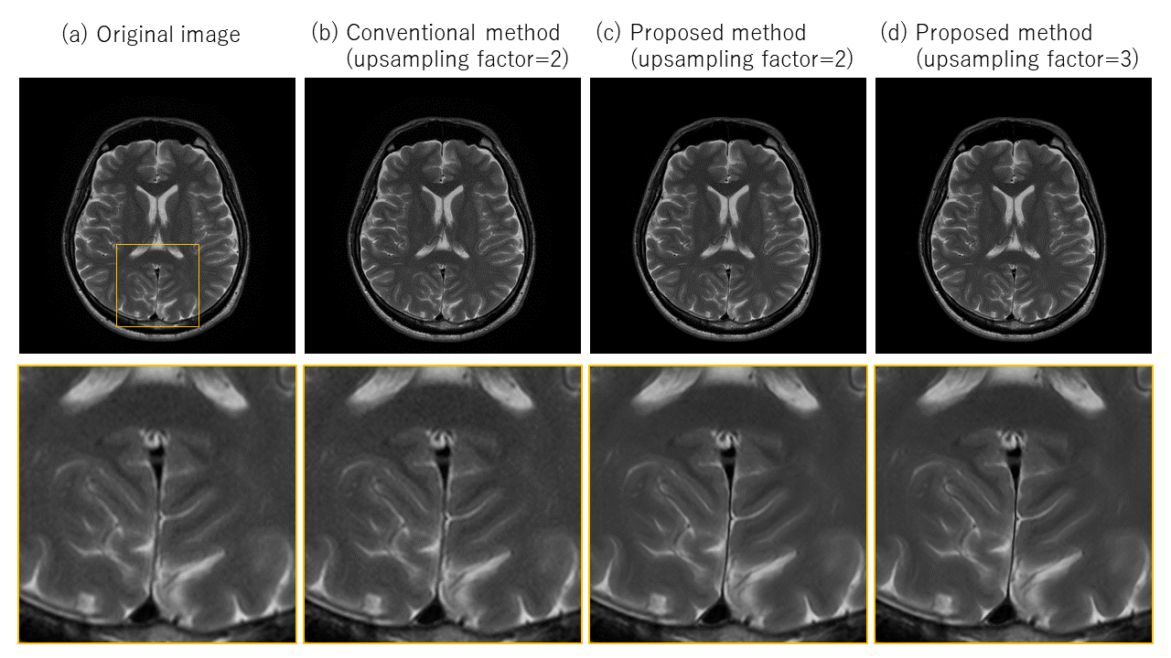
Resulted images from the prospective study, with a set of moderate scan parameters
(a) Original acquired image
(b) Image by the conventional method (ZIP and LPF), with the upsampling factor of 2
(c) Image by the proposed method, with the upsampling factor of 2
(d) Image by the proposed method, with the upsampling factor of 3.
Scan parameters: FSE2D, TE=90ms, TR=4000ms, FOV=23 x 23cm, Matrix=384 x 384, Sampling-pitch=22$$$\mu$$$s, Slices=30 x 4mm, Scan-time=208 s.
Resulted images from the prospective study, with a set of aggressive scan parameters
(a) Original acquired image
(b) Image by the conventional method (ZIP and LPF), with the upsampling factor of 2
(c) Image by the proposed method, with the upsampling factor of 2
(d) Image by the proposed method, with the upsampling factor of 3.
Scan parameters: FSE2D, TE=90ms, TR=4000ms, FOV=23 x 23cm, Matrix=256 x 256, Sampling-pitch=22$$$\mu$$$s, Slices=30 x 4mm, Scan-time=176 s.