2894
Different multiparametric MRI-based radiomics models for differentiating stage IA endometrial cancer from benign endometrial lesions: A multicenter study
Qiu Bi1, Kunhua Wu1, and Yunzhu Wu2
1the First People’s Hospital of Yunnan Province, Kunming, China, 2Siemens Healthcare, Shanghai, China
1the First People’s Hospital of Yunnan Province, Kunming, China, 2Siemens Healthcare, Shanghai, China
Synopsis
Keywords: Artifacts, Uterus
In the study, age and irregular vaginal bleeding were the valid predictive parameters in clinical model. On the basis of several common machine learning algorithms, the diverse multiparametric MRI-based radiomics models were developed to differentiate stage IA EC from benign endometrial lesions, and LR algorithm model were selected as the optimal radiomics model with the highest AUC and accuracy. Compared with clinical model and radiologist, the optimal radiomics model and the compositive models combining clinical parameters with radiomics features, like the nomogram, stacking model, and ensemble model showed better diagnostic performance and achieved good clinical net benefits. The nomogram had a higher AUC than that of the optimal radiomics model, and revealed more stable discrimination efficiency and better generalization ability than stacking and ensemble modals.Objective
To evaluate the value of different multiparametric MRI-based radiomics models in differentiating stage IA endometrial cancer (EC) from benign endometrial lesions.Methods
Data of patients with endometrial lesions from two centers were collected.The radiomics features were extracted from T2-weighted imaging (T2WI), diffusion-weighted imaging (DWI), apparent diffusion coefficient (ADC) map, and late contrast-enhanced T1-weighted imaging (LCE-T1WI). After data dimension reduction and feature selection, 9 machine learning algorithms were conducted to determine which was the optimal radiomics model for differential diagnosis. The univariate analyses and logistic regression (LR) were performed to reduce valueless clinical parameters and to develop the clinical model. A nomogram using the radscores combined with clinical parameters was developed. Two integrated models were obtained respectively by the ensemble strategy and stacking algorithm based on the clinical model and optimal radiomics model. The area under the curve (AUC), clinical decisive curve (CDC), net reclassification index (NRI), and integrated discrimination index (IDI) were used to evaluate the performance and clinical benefits of the models.Results
A total of 371 patients were incorporated. LR model was the optimal radiomics model with the highest average AUC (0.854) and accuracy (0.802) in the internal and external validation groups (AUC=0.910 and 0.798, respectively), and outperformed the clinical model (AUC=0.739 and 0.592, respectively) or radiologist (AUC=0.768 and 0.628, respectively). The nomogram (AUC=0.917 and 0.802, respectively) achieved better discrimination performance than that of the optimal radiomics model in two validation groups. The stacking model (AUC=0.915) and ensemble model (AUC=0.918) had similar performance compared with the nomogram in the internal validation group. Whereas, the AUCs of the stacking model (AUC=0.792) and ensemble model (AUC=0.794) were lower than those of the nomogram and radiomics model in the external validation group. According to the CDC, NRI and IDI, the optimal radiomics model, nomogram, stacking model, and ensemble model achieved good net benefits.Conclusions
Multiparametric MRI-based radiomics models can non-invasively differentiate stage IA EC from benign endometrial lesions, and LR is the best machine learning algorithm. The nomogram present excellent and stable diagnostic efficiency.Acknowledgements
NoneReferences
1. Rodriguez-Ortega A, Alegre A, Lago V, Carot-Sierra JM, Ten-Esteve A, Montoliu G, et al. Machine Learning-Based Integration of Prognostic Magnetic Resonance Imaging Biomarkers for Myometrial Invasion Stratification in Endometrial Cancer. J Magn Reson Imaging (2021) 54(3):987-95.
2. Hoivik EA, Hodneland E, Dybvik JA, Wagner-Larsen KS, Fasmer KE, Berg HF, et al. A Radiogenomics Application for Prognostic Profiling of Endometrial Cancer. Commun Biol (2021) 4(1):1363.
3. Chen X, Wang X, Gan M, Li L, Chen F, Pan J, et al. MRI-based Radiomics Model for Distinguishing Endometrial Carcinoma From Benign Mimics: A Multicenter Study. Eur J Radiol (2022) 146:110072.
Figures
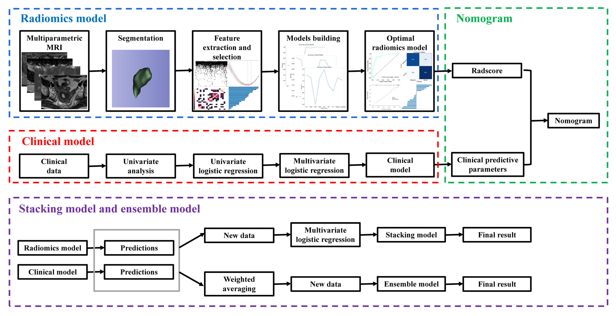
FIGURE 1 The overall workflow of this study.
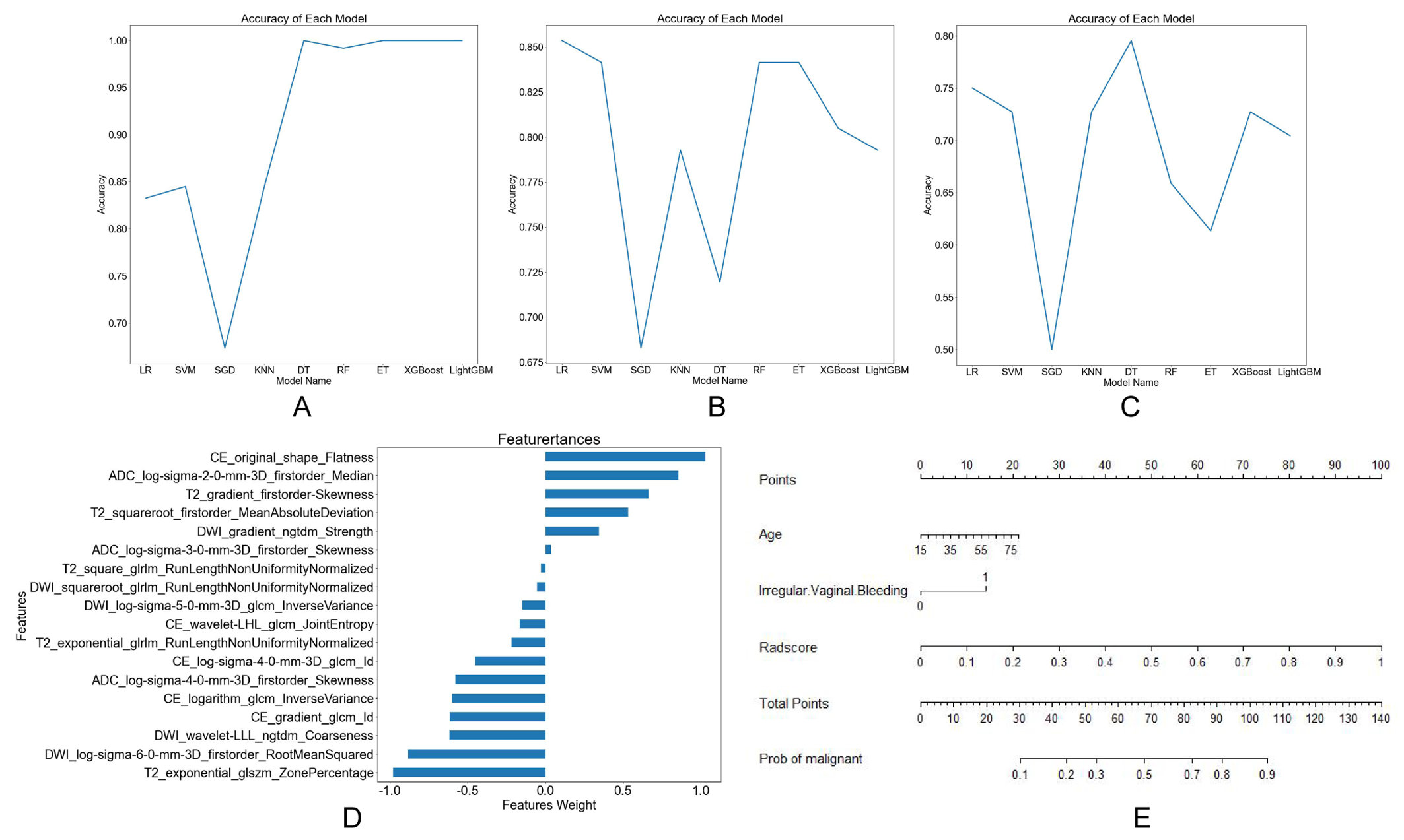
FIGURE 2 Different models buiding. Broken line graphs of accuracy for different machine learning algorithms in training group (A), internal validation group (B), and external validation group (C). Bar chart of features weight for logistic regression model (D). Nomogram of the training group (E).
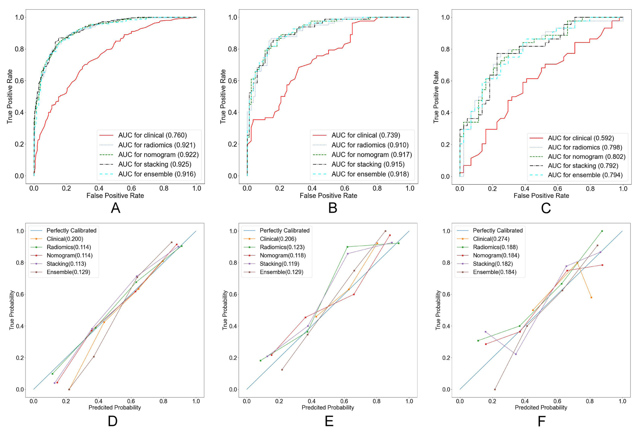
FIGURE 3 Receiver operator characteristic (ROC) curves (A-C) and calibration curves (D-F) of different models in training group (A, D), internal validation group (B, E), and external validation group (C, F).
DOI: https://doi.org/10.58530/2023/2894