2879
Influence of arm rotations on peripheral nerve stimulation thresholds for the interventionalist in MR guided procedures1Division of Medical Physics, Department of Radiology, University Medical Center Freiburg, Freiburg, Germany, Freiburg, Germany, 2Department of Radiology, University Medical Center Freiburg, Freiburg, Germany, Freiburg, Germany
Synopsis
Keywords: Safety, Safety, PNS Simulation
Simulation of peripheral nerve stimulation of MRI gradient coils for rotated arms of an interventional radiologist. The arm rotation has a direct influence on the site of stimulation in the arm.Introduction
One fundamental limitation for advancing fast MRI arises from physiology, namely by peripheral nerve stimulation (PNS). The limits for the safe operation of gradient coils are usually determined by experimental studies on several human subjects1. Predicting thresholds for peripheral nerve stimulation from simulated body models attracted broad interest recently2-5. Regulatory limits are only given for the patient, omitting further persons that may be present in the MRI bore, such as the interventionalist during MRI-guided procedures. In a preliminarily study, we evaluated the PNS safety of the interventionalist5 using one fixed position of a realistic human body model. However, the radiologist's arm is frequently moved during an interventional procedure, and translational movements and rotations are common. Impact of translational movements on PNS thresholds has been analyzed before, using a human leg model3. Up to now, PNS safety evaluation for a rotated arm has not been attempted. In this work, the thresholds for arms with different rotated angles for two human body models were simulated to reveal new insights concerning PNS risks for a radiologist.Methods
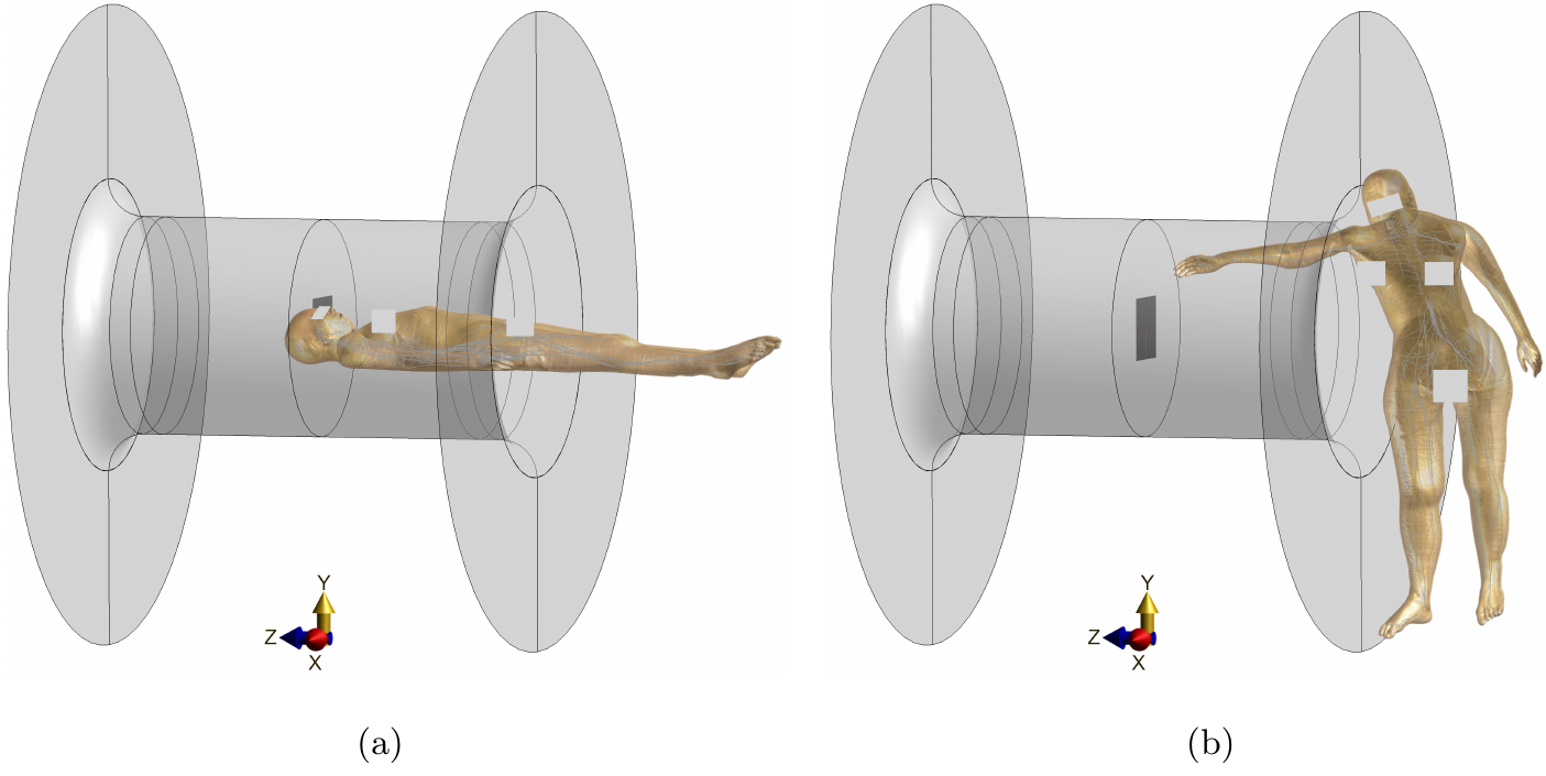
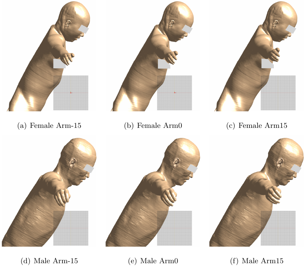
Quasi-static electromagnetic calculations combined with the neurodynamic simulator NEURON6 were used to find PNS thresholds. All calculations were performed using Sim4Life (ZMT Zurich MedTech AG, Zurich, Switzerland). The two virtual human models: female Yoon-Sun V 4.0 [DOI: 10.13099/ViP11016-04-0] and male Jeduk V 4.0 [DOI: 10.13099/ViP11017-04-0] with the embedded trajectories of the main peripheral nerves (IT'IS foundation, Zurich, Switzerland) were used for gender balance. PNS thresholds of a typical patient position (Fig. 1.a) were simulated as a reference. A board-certified interventional radiologist was consulted for replicating realistic positions as shown in Fig. 1.b. Three different rotational arm positions of both models are depicted in Fig. 2. The wire tracks of the Gx, Gy and Gz gradient coil axis of a 70 cm 1.5T Aera scanner (Siemens Healthcare, Erlangen, Germany) were used for the calculation of all components of the magnetic vector potential. To cover all scenarios, four combined-axes operation modes of these potentials (X+Y+Z, X-Y+Z, X-Y-Z and X+Y-Z) were applied as sources for the electromagnetic field. In the neurodynamic simulation, trapezoidal waveforms with pulse durations of 0.1 ms to 1.2 ms were employed.Results and discussions
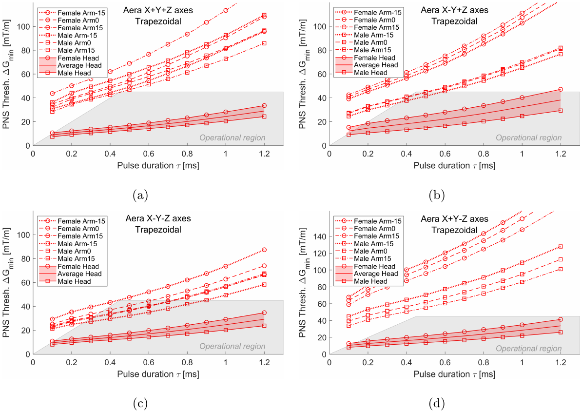
Figure 3 displays PNS threshold curves for the interventionalist position with different arm rotations and patient position for reference. All the thresholds increase with the pulse duration. Moreover, compared to imaged subject positions, the average thresholds for all positions of the interventionalist increased by a factor of at least 3.63, 2.69, 2.4 and 4.48 times in X+Y+Z, X-Y+Z, X-Y-Z and X+Y-Z combined-axes modes, respectively. The thresholds for the male model in X-Y-Z mode, which were the lowest of all modes studied in the interventionalist (Fig 2), were at least two times higher, compared to the average threshold of the patient position. Therefore, in the operation mode that is safe for the imaged subject, it is unlikely to achieve PNS in interventionalists for any of the studied arm rotations.Figure 3 also shows that the female interventionalist had at least 1.07, 1.48, 1.14 and 1.61 times higher average PNS thresholds than the male interventinalist in X+Y+Z, X-Y+Z, X-Y-Z and X+Y-Z combined-axes modes, respectively. However, the threshold for the female interventionalist with 15 degrees of arm rotation (Arm15 in Fig. 2) was a little lower than the male one with 15 degrees of arm rotation in X-Y-Z mode. Moreover, compared to the male interventionalist with -15 degrees of arm rotation (Arm-15 in Fig. 2) in X+Y+Z mode, the female interventionalist with the same arm rotation decreased the threshold by a factor of at least 1.45 times.
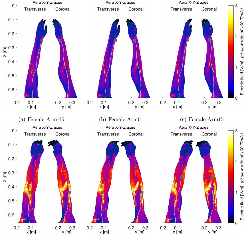
Also seen in Figure 3, male and female interventionalist thresholds might show different trends as the angles of arm rotation changed. This trend is related to combined-axes modes. For example, in the X-Y+Z mode, both male and female threshold levels became larger as the angle changed from -15 degrees, to 0 degrees and further to 15 degrees, while in the X+Y-Z mode, for both models, the thresholds became smaller as the angle changed. However, in the X-Y-Z mode, female interventionalist thresholds decreased with the change of the angle while male thresholds increased. These opposite tendencies might be due to the differences of the primary PNS sites. Figure 4 depicts the locations of such stimulation sites. The female interventionalist stimulation site was near the wrist, while the male interventionalist stimulation site was close to the elbow.
The electrical field distributions and stimulated nerve tracks in the arm regions are also presented in Figure 4. The stimulation sites might occur at a hotspot of the electric field (Fig. 4d and 4e) or at a non-hotspot location (Fig. 4a-c and 4f). Moreover, the rotations of the arm could lead to a change in the locations of the stimulation sites (Fig. 4e and 4f).
Conclusions
Two posable human body models were used to predict the effects of arm rotations on PNS thresholds for interventionalists. The preliminary results suggest that PNS is unlikely to appear in the interventionalist before stimulating the patient, regardless the arm rotation. However, the arm rotation has a direct influence on the site of stimulation in the arm.Acknowledgements
The authors are deeply grateful to Dr. Axel Vom Endt, Dr. Heiko Rohdjess and Dr. Martino Leghissa at Siemens Healthcare (Erlangen/Forchheim) for their generous support. Financial support by the Federal Ministry of Education and Research (BMBF) (project number 13GW0356B) is also gratefully acknowledged.References
1. W. Irnich, F. Schmitt, Magnetostimulation in MRI, MRM, 1995;33: 619-623;
2. Davids et al. Predicting Magnetostimulation Thresholds in the Peripheral Nervous System using Realistic Body Models. Sci. Rep. 2017;7:5316;
3. Klein et al., Sensitivity analysis of neurodynamic and electromagnetic simulation parameters for robust prediction of peripheral nerve stimulation. Phys. Med. Biol. 2019;64:015005;
4. Davids et al. Prediction of peripheral nerve stimulation thresholds of MRI gradient coils using coupled electromagnetic and neurodynamic simulations. MRM 2019;81:686;
5. Jia et al. Are interventionalists prone to nerve stimulation during MR interventions? PNS simulation study with posable human body models. ISMRM 2021;2478;
6. Hines and Carnevale, The NEURON simulation environment. Neural Comput. 1997;9:1179;
Figures