2871
RF exposure of patient assistants at a low-field open MRI system1Korea Basic Science Institute, Cheongju, Korea, Republic of, 2Electronics and Telecommunications Research Institute, Daejeon, Korea, Republic of, 3Chungbuk National University, Cheongju, Korea, Republic of, 4BioBrain, Inc., Daejeon, Korea, Republic of, 5Daewon University College, Jechoen, Korea, Republic of
Synopsis
Keywords: Safety, Safety
Numerical investigations on the safety of patient assistants exposed to a 1.2T open MRI are performed at different poses of patient assistants (including wearing latex gloves), because current regulations are concerned with only patient RF safety. Compared with the patient, up to 29.8% of the patient 10-gram SAR was observed in the patient assistant. To prevent possible RF hazards of a patient assistant during MRI scans, certain clauses regarding the patient assistant’s poses or wearing gloves must be added to the existing MRI screening forms.Introduction
Patient assistants’ supports during MR scans can afford considerable psychological comfort to patients, enabling timely completion of all the required scans at an open MRI system. It is one of the important advantages of an open MRI system comparing with a closed magnet. Most regulations or RF safety guidelines for MRI concern only the RF exposure of a patient1. However, there have been few studies and guidelines on RF exposure of patient assistants during MRI scan2. Access of patient assistants to patients is enabled by the shape of the open MRI system, even during MRI scans. Therefore, it is necessary to evaluate RF energy exposure of patient assistants. In this study, we numerically investigated RF energy exposures of patient assistants using planar–shaped RF coil, which is widely used transmit RF coil type, particularly at 1.2 T in an open MRI system.Methods
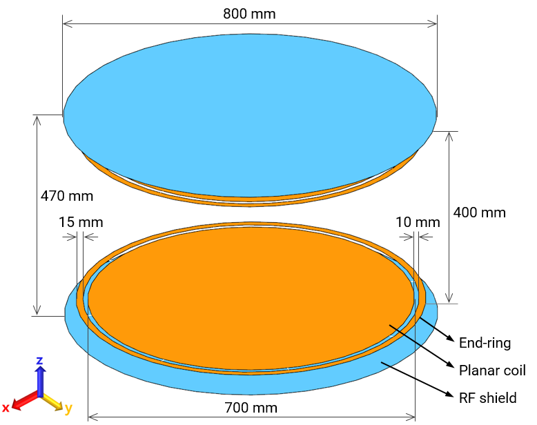
The planar Tx coil3,4 is top–down symmetrically (in the z-direction) composed of RF shielding plates (800 mm in diameter), end-ring (15 mm in width), and planar RF coils (with 10 mm gap between end-ring and coil, with 400 mm space between the coils), as shown in Fig. 1. Eight constant current sources were circularly placed at the gap in equispacing order to create a B1+ field around the z-axis in a CP mode at 51 MHz. The amount of applied RF power of the Tx RF coil was scaled to create the same magnitude of B1+, for example, 2 μT5, at the isocenter of the magnet for each pose of the patient assistant. Four different poses of patient assistants were defined by changing the poses and patient contact conditions using the posable Duke model (age: 34, sex: male, BMI: 23.1 kg/m3, tissues: 77)6,7 as shown in Fig. 2. Including sitting pose (Fig. 2(a)), the patient assistant also touched the patient (age: 11, sex: female, BMI: 16.7 kg/m3, tissues: 75) with one hand (Fig. 2(b)), two hands stacked up (Fig. 2(c)), and finally two hands on the patient’s hand and thigh (Fig. 2(d)). EM field simulations of RF exposure of patient assistants were performed using Sim4Life (Zurich Med Tech AG, Switzerland) with GPUs (two NVDIA Quadro RTX A6000s), CPU (Intel Xeon 6126), and 704 GB of system memory. The 10-g averaged SAR was analyzed from the patient assistant for each pose.Results and Discusstion
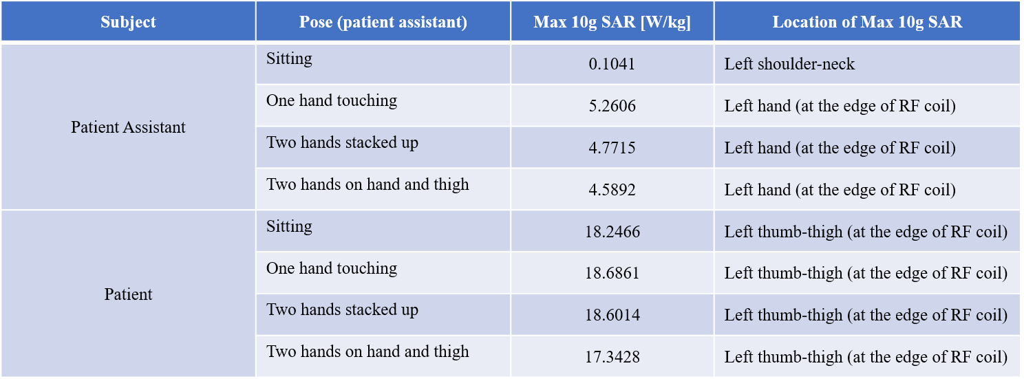
The lowest max10-gram SAR was found for sitting pose (Fig. 2(a), 0.1041 W/kg). The highest max 10-gram SAR was measured for the pose of one hand of the patient’s assistant touched the patient (Fig. 2(b), 5.2606 W/kg). It is approximately 50.5 times higher than the lowest max 10-gram SAR of the patient assistant. The other two poses (Fig. 2(c) & Fig. 2(d)) of the patient assistant showed an approximately 11% lower max 10-gram SAR level than one-hand touching pose. This is because of the total mass difference within the exposed electric field of the planar Tx RF coil, particularly around the edge of the coil. A smaller mass tends to result in greater RF exposure. In addition, the induced SAR level of the patient assistant would worsen if a patient assistant was electrically connected to the patient (i.e., touching each other). No significant differences in the max 10-gram SAR levels in patients were found, regardless of the pose of the patient assistant. The max 10-gram SARs were found at the finger tip of the left hand, which is electrical connected to the patient. This signifies level of RF exposure in the patient assistant. For this reason, we suggest wearing electrically insulated gloves, such as latex, to prevent direct contact of the patient body. While assisting patients by holding their body parts during MRI scans, the patient assistants should be aware of the possible risk of RF exposure. In addition, insulating gloves must be worn to minimize the possibility of unwanted adverse events.Conclusion
We investigated the amount of RF exposure in four different poses of a patient assistant during MRI scans at 1.2 T in an open MRI system. The planar Tx RF coil induces a higher max 10-gram SAR level in the patient assistant, up to 50 times higher when the patient assistant holds the patient’s body comparing with sitting pose. Based on our study, we suggest adding certain clauses to the existing MRI screening forms to prevent or minimize any possible RF hazards.Acknowledgements
This work was supported by the IT R&D program of MSIP/IITP [2019-0-00102, A Study on Public Health and Safety in a Complex EMF Environment].References
1. International Electrotechnical Commission (IEC). IEC 60601-2-33: Medical electrical equipment-Part 2–33: Particular requirements for the basic safety and essential performance of magnetic resonance equipment for medical diagnosis, 2015.
2. Hong SE, Oh S, Choi HD. RF exposure assessment for various poses of patient assistant in open MRI environment. Appl. Sci. 2021;11:4947.
3. Wei S, Yang W, Wang H, Chen L. Analysis of a planar transmit coil used for vertical field MRI. In Proceedings of the 4th International Conference on Biomedical Engineering and Informatics. Shanghai, China, 2011;118–121.
4. Boskamp, EB. Flat RF body coil design for open MRI. In Proceedings of the 22nd Annual EMBS International Conference. Chicago, USA, 2000;2387–2389.
5. Collins CM, Smith MB. Signal–to–noise ratio and absorbed power as functions of main magnetic field strength, and definition of “90°” RF pulse for the head in the birdcage coil. Magn. Reson. Med. 2001;45:684–691.
6. Christ A, Kainz W, Hahn EG, Honegger K, Zefferer M, Neufeld E, Rascher W, Janka R, Bautz W, Chen J. The virtual family-development of surface-based anatomical models of two adults and two children for dosimetric simulations. Phys. Med. Biol. 2010;55:N23–N38.
7. Gosselin MC, Neufeld E, Moser H, Huber E, Farcito S, Gerber L, Jedensjö M, Hilber I, Di Gennaro FD, Lloyd B. Development of a new generation of high-resolution anatomical models for medical device evaluation: The virtual population 3.0. Phys. Med. Biol. 2014;59:5287–5303.
Figures