2852
Increased regional homogeneity and correlation with clinical indicators and scales in type 2 diabetes1Department of Radiology, the First Affiliated Hospital of Dalian Medical University, Dalian, China
Synopsis
Keywords: Brain Connectivity, Diabetes, regional homogeneity
To further investigate the changes in brain function of type 2 diabetes we prospectively enrolled 38 patients clinically diagnosed with T2DM and 24 age-matched healthy controls and underwent blood oxygen level-dependent fMRI scans. The image data was processed to obtain regional homogeneity values and compared by two sample t test. Results found that the local synchronization of spontaneous neural activity of the supramarginal gyrus in patients with T2DM was abnormal and correlated with cognitive function, which may be involved in the pathophysiological mechanism of T2DM.Introduction
Diabetes and its complications have a major impact on individuals and families, health systems and national economies. Therefore, it is of great significance to comprehensively understand the health status of T2DM patients and explore the pathophysiological mechanism of T2DM for the prevention and control of the disease. At present, there is a lack of clear imaging indicators as markers for the diagnosis and assessment of diabetic encephalopathy. Therefore, we conducted this prospective study to analyze the functional changes and further analyze its correlation with clinical and cognitive scores to explore the neural mechanism of cognitive decline in T2DM.Methods
We recruited 38 patients (21 males, 17 females, T2DM group) clinically diagnosed with T2DM and 24 age-matched healthy controls (10 males, 14 females, HC group). Informed consent was obtained from each participant. The clinical examination data and laboratory examination data of all subjects were recorded separately, and a series of neuropsychological tests were carried out by a well-trained neurologist. The Mini-Mental State Examination (MMSE) and the Montreal Cognitive Assessment (MoCA) were used to assess the cognitive status of all subjects. The two cognitive domains of memory and execution, which are more vulnerable to early impairment in T2DM, were evaluated using the California verbal learning test (CVLT) and the Symbol-digital mode test (SDMT).All patients were scanned using a Philips Ingenia CX 3.0T MR scanner (Philips Healthcare, Best, the Netherlands) with 32-channel head coil, and instructed to keep their eyes closed but to remain awake during the scanning. Functional images were obtained using a gradient-echo planar (EPI) sequence. A total of 200 time points were scanned. Structural images were acquired using multishot turbo Field Echo sequence.
Functional data were analyzed using DPARSF [1] and statistical parametric mapping (SPM12, http:// www.fil.ion.ucl.ac.uk/spm) on MATLAB software. The first 10 points were discarded to eliminate nonequilibrium effects of magnetization and allow subjects to acclimate to the scanning environment. The remaining functional images were processed using the following steps: data were slice time corrected, realigned, coregistered, and regressed to remove nuisance covariates including head motion as well as the cerebrospinal fluid signal and white matter signals, then the functional images were normalized to the Montreal Neurological Institute (MNI), resampled to a voxel size of 3 × 3 × 3 mm3 and temporal bandpass filtered (0.01–0.1 Hz) to reduce the effects of low-frequency drift and high-frequency noise. Data from individuals with an estimated maximum displacement in any direction larger than 2 mm or head rotation larger than 2◦ were discarded from the study.
Regional homogeneity (ReHo) values were quantified by calculating the Kendall’s coefficient of concordance value between a given voxel and its neighbors in a voxel-wise way and then zReHo values were calculated through fisher r-z transformation. Finally, spatial smoothing was conducted with an 6mm FWHM Gaussian kernel.
A two samples t-test were implemented to test the differences of zReHo between HC and T2DM group at the whole brain grey matter level, respectively. A Family-wise error (FWE) correction was applied for multiple comparison corrections at cluster level with age, gender, education level as covariates; p < 0.05 was considered statistically significant.
Results
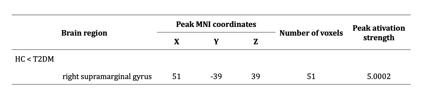
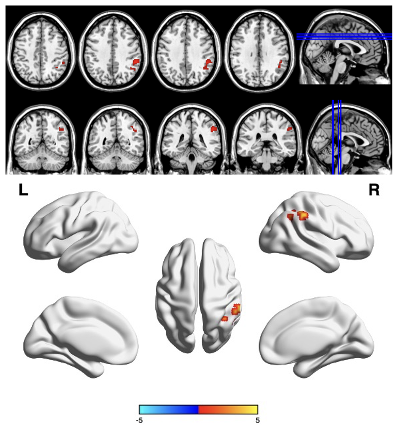
According to the results of two samples t-test, compared with HC group, the ReHo value of the right supramarginal gyrus of T2DM group was significantly increased. Pearson correlation analysis showed MoCA score was positively associated with ReHo value(r = 0.32,p = 0.047).Discussion
The supramarginal gyrus belongs to the parietal-inferior lobule, and is an important part of the default mode network (DMN). The DMN exhibits strong spontaneous activity in the resting state and participates in many functions related to memory or abstract thinking [2]. Zhang et al. [3] found that functional connectivity between the bilateral ventral anterior insula and the right supramarginal gyrus was reduced in the T2DM group compared with the control group, and conclude that distinct functional connectivity patterns may affect cognitive, emotional, and sensorimotor functions in patients with T2DM. Li et al. [4] conducted a multimodal meta-analysis of voxel-based morphometry and regional resting-state functional MRI studies in T2DM, and found that in patients with T2DM compared to controls, conjoint decreased regional gray matter volume and altered intrinsic activity including the supramarginal gyrus and other brain regions mainly in the default mode network. Combined with the finding of abnormal ReHo in supramarginal gyrus of T2DM patients in this study, these all indicate the mechanism by which this brain region may be involved in the deep changes of the T2DM brain. In addition, this study found that the ReHo of the supramarginal gyrus was positively correlated with the MoCA score, and MoCA mainly assessed the cognitive function of the subjects, suggesting that the local synchronization of spontaneous neural activity of the supramental gyrus may be involved in the change of the cognitive function of the patients' overall cognitive state. But it is worth noting that this correlation is weak. And this may help provide insights into the neuropathology of T2DM.Conclusion
This study found that the local synchronization of spontaneous neural activity of the supramarginal gyrus in patients with T2DM was abnormal and correlated with cognitive function, which may be involved in the pathophysiological mechanism of T2DM.Acknowledgements
Thanks to all colleagues involved in this study.
References
[1] Yan, C.-G., Wang, X.-D., Zuo, X.-N. & Zang, Y.-F. DPABI: Data Processing & Analysis for (Resting-State) Brain Imaging. Neuroinformatics 14, 339-351, doi:10.1007/s12021-016-9299-4 (2016).
[2] Smallwood, J. et al. The default mode network in cognition: a topographical perspective. Nat Rev Neurosci 22, 503-513, doi:10.1038/s41583-021-00474-4 (2021).
[3] Zhang, D. et al. Altered Functional Connectivity of Insular Subregions in Type 2 Diabetes Mellitus. Front Neurosci 15, 676624, doi:10.3389/fnins.2021.676624 (2021).
[4] Yao, L. et al. A multimodal meta-analysis of regional structural and functional brain alterations in type 2 diabetes. Front Neuroendocrinol 62, 100915, doi:10.1016/j.yfrne.2021.100915 (2021).
Figures