2848
Time-Varying Functional Connectivity of the Hippocampus Is Associated With Cognitive Performance in Multiple Sclerosis Patients
Paola Valsasina1, Olga Marchesi1, Carmen Vizzino1, Damiano Mistri1, Maria Assunta Rocca1,2,3, and Massimo Filippi1,2,3,4,5
1Neuroimaging Research Unit, Division of Neuroscience, IRCCS San Raffaele Scientific Institute, Milan, Italy, 2Neurology Unit, IRCCS San Raffaele Scientific Institute, Milan, Italy, 3Vita-Salute San Raffaele University, Milan, Italy, 4Neurorehabilitation Unit, IRCCS San Raffaele Scientific Institute, Milan, Italy, 5Neurophysiology Service, IRCCS San Raffaele Scientific Institute, Milan, Italy
1Neuroimaging Research Unit, Division of Neuroscience, IRCCS San Raffaele Scientific Institute, Milan, Italy, 2Neurology Unit, IRCCS San Raffaele Scientific Institute, Milan, Italy, 3Vita-Salute San Raffaele University, Milan, Italy, 4Neurorehabilitation Unit, IRCCS San Raffaele Scientific Institute, Milan, Italy, 5Neurophysiology Service, IRCCS San Raffaele Scientific Institute, Milan, Italy
Synopsis
Keywords: Brain Connectivity, Multiple Sclerosis
In this study, we explored hippocampal static and time-varying functional connectivity and its association with cognition in patients with multiple sclerosis. We found decreased static and time-varying connectivity of the hippocampus with temporo-parietal regions, and increased connectivity of the hippocampus with pre- and postcentral gyri, inferior temporal gyrus, precuneus and frontal regions. Better visuospatial and verbal memory were associated with higher hippocampal time-varying connectivity with precentral and inferior temporal gyri, while better attention scores were associated with higher hippocampal time-varying connectivity with the superior frontal cortex.Introduction
The hippocampus is a key structure of the central nervous system, which has an important role in cognition and mood regulation. Several clinical manifestations of multiple sclerosis (MS), including cognitive impairment and depression, can be explained by focal hippocampal damage, but also by disconnection of the hippocampus from several brain circuits [1]. Abnormalities of hippocampal activation and resting state (RS) functional connectivity (FC) are known to be present in MS. However, most of previous studies [2-5] investigating hippocampal RS FC in MS used a static FC (sFC) approach. Dynamic FC, also known as time-varying connectivity (TVFC), of the hippocampus in MS has been explored only by one preliminary study so far [6], which found that a lower hippocampal TVFC could explain some variance of memory perfomance in these patients. Against this background, aim of this study was to explore hippocampal sFC and TVFC abnormalities in patients with MS and assess their association with performances in several cognitive domains.Methods
Structural and RS functional MRI scans were acquired from 108 right-handed MS patients and 63 right-handed healthy controls (HC). Subjects underwent a neuropsychological evaluation, comprising the Brief Repeatable Battery of Neuropsychological Tests (BRB-N). Sliding-window correlation analysis using the left (L) and right (R) hippocampus as seed regions (extracted from FSL FIRST) was used to assess TVFC, which was quantified by the standard deviation of connectivity across windows. Conversely, mean connectivity across windows indicated sFC.Results
MS patients had decreased sFC vs HC between L hippocampus and temporo-parietal regions, and increased sFC between L and R hippocampus and thalamus, precuneus and superior frontal regions. TVFC was decreased in MS patients between L hippocampus and temporo-parietal regions. Conversely, TVFC was increased in MS patients between L and R hippocampus and L pre- and postcentral gyri, cuneus, orbitofrontal cortex and inferior temporal gyrus (ITG) (Figure 1). Correlations between abnormalities of TVFC and neuropsychological performances are reported in Figure 2. In details, in MS patients, better global cognition correlated with higher TVFC between L hippocampus and L pre- and postcentral gyri (r=0.21-0.28; p=0.04-0.006). Better verbal memory correlated with higher TVFC between L hippocampus and L precentral gyrus (r=0.21, p=0.03), and better visuospatial memory correlated with higher TVFC between L and R hippocampus and L cuneus, pre- and postcentral gyri and ITG (r=0.19-0.23, p=0.02-0.04). Better information processing speed correlated with higher TVFC between L hippocampus and L postcentral gyrus (r=0.21, p=0.03) and with higher sFC between R hippocampus and L superior frontal cortex (SFC) (r=0.21, p=0.03). Finally, better attention scores correlated with higher TVFC between L hippocampus and L temporal cortex (r=0.25, p=0.02), with higher TVFC between L hippocampus and L SFC (r=0.22, p=0.03) and with higher sFC between R hippocampus and L SFC (r=0.20, p=0.05).Discussion
In MS patients, a complex pattern of increased and decreased hippocampal sFC/TVFC was detected. Increased TVFC between the bilateral hippocampus and parietal, temporal and frontal regions was correlated with better performances in several neuropsychological tests, with some correspondence between involved brain regions and cognitive domains (i.e., regions of the dorsal stream for visuospatial memory, and fronto-temporal regions for attention).Conclusions
Increased hippocampal connectivity contributed to explain better cognitive performances in MS, with a peculiar association between higher hippocampal TVFC and memory scores.Acknowledgements
No acknowledgement found.References
[1] Rocca et al., Lancet Neurol 2018; [2] Roosendaal et al., Radiology 2010; [3] Hulst et al., MSJ 2015; [4] Gonzalez-Torre et al., MSJ 2017; [5] Rocca et al., Hum Brain Mapp 2015; [6] Van Geest et al., Brain Behav 2018.Figures
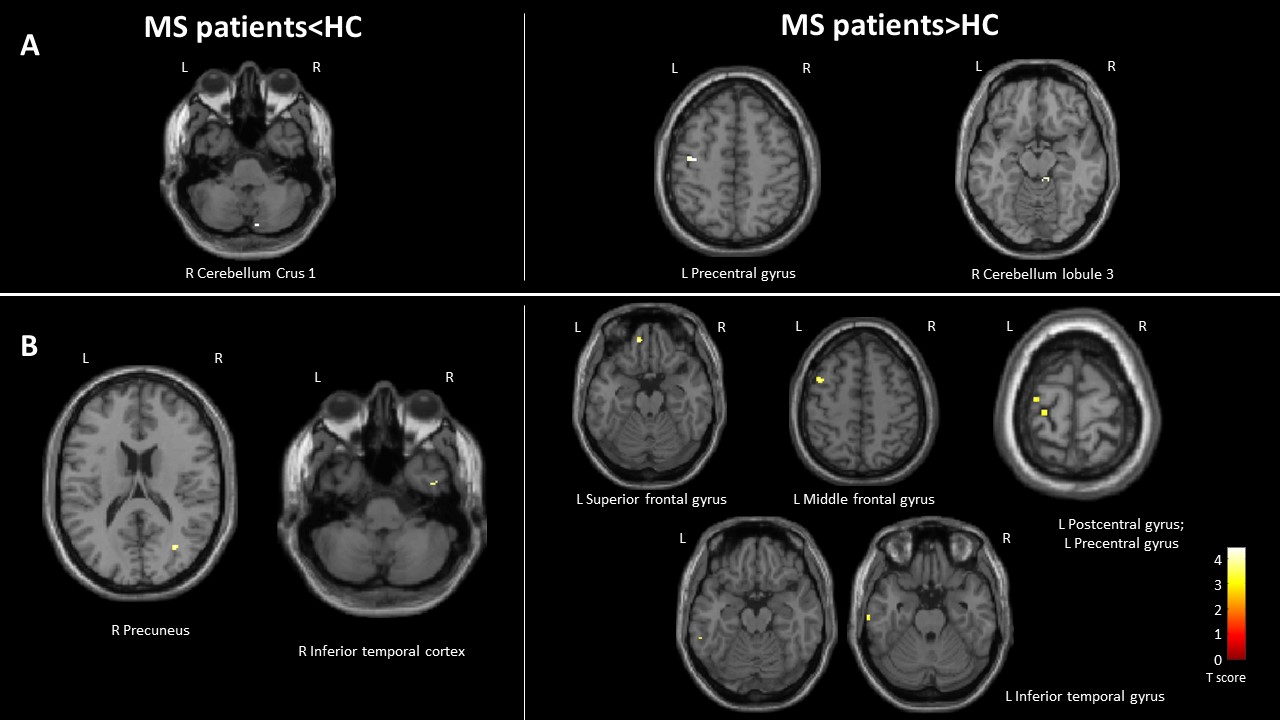
Figure 1. Abnormalities
of time-varying functional connectivity (TVFC) in multiple sclerosis (MS)
patients compared to healthy controls (HC) for the right (A) and the left (B)
hippocampus. Abbreviations: L=left, R=right.
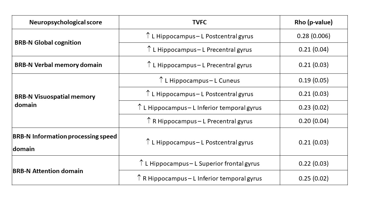
Figure 2. Correlations between time-varying
functional connectivity (TVFC) abnormalities and neuropsychological scores in
multiple sclerosis patients. Abbreviations: L=left; R=right; BRB-N=Brief
repeatable battery of neuropsychological tests.
DOI: https://doi.org/10.58530/2023/2848