2835
Lesional and non-lesional cortical integrity in cognitive networks are linked to cognitive impairment in multiple sclerosis.1MS Center Amsterdam, Anatomy and Neurosciences, Amsterdam Neuroscience, Amsterdam UMC location VUmc, Amsterdam, Netherlands, 2Department of Neurology, Massachusetts General Hospital, Harvard Medical School, Boston, MA, United States, 3Multiple Sclerosis Specialist Center, Neurology B, Department of Neurosciences, Biomedicine and Movement Sciences, University of Verona, Verona, Italy, 4MS Center Amsterdam, Radiology and Nuclear Medicine, Amsterdam Neuroscience, Amsterdam UMC location VUmc, Amsterdam, Netherlands, 5Institutes of Neurology and Healthcare Engineering, University College London, London, United Kingdom, 6MS Center Amsterdam, Neurology, Amsterdam Neuroscience, Amsterdam UMC location VUmc, Amsterdam, Netherlands
Synopsis
Keywords: Multiple Sclerosis, Gray Matter, Diffusion/Other Diffusion Imaging Techniques
Cognitive functioning in people with multiple sclerosis (MS) is strongly related to cortical lesions (CLs), although the severity of demyelination remains difficult to assess in-vivo. In addition, how the pathological microstructural changes in normal-appearing gray matter relate to cognition also remains unclear. This study assessed how microstructural integrity (based on diffusion MRI) in CLs and normal-appearing cortex relates to cognition in MS. Microstructural integrity changes were most evident in cognitively-impaired MS, especially in the normal-appearing cortex. Regionally, damage was especially related to cognition within cognitive functional networks such as the ventral attention network.Introduction
Cognitive impairment is recognized as a prevalent and debilitating symptom in people with multiple sclerosis (MS), especially related to gray matter damage within lesions and normal-appearing tissues.1 Using diffusion tensor MRI, the severity of cortical damage can be assessed in MS using measures like mean diffusivity (MD) and fractional anisotropy (FA).2-5 Despite these technological advances, the differential impact of focal lesional cortical demyelination and normal-appearing microarchitectural changes on cognition remains unclear in MS. Functional MRI studies have shown the importance of certain functional networks in the development of cognitive decline.6,7 Microstructural integrity might reflect this spatial predominance for specific networks as well. Our aim was to evaluate integrity changes within cortical lesions and normal-appearing tissue on a regional basis, and to assess their relationship to cognitive impairment. The latter was explored after grouping regions according to literature-based networks of gray matter areas.Methods
One-hundred seventy-six participants with MS of the Amsterdam MS cohort and 48 healthy controls (HC) underwent MRI (3D-FLAIR, 3D-T1, double inversion recovery [DIR] and diffusion-weighted [DW] sequences at 3T) and neuropsychological assessment (expanded Brief Repeatable Battery of Neuropsychological tests). A single-shell diffusion MRI protocol was acquired using a b-value of 1000 s/mm2 in 30 directions. The cerebral cortex was parcellated into 212 regions, which were classified as either a region with cortical lesions or normal-appearing cortex based on visual inspection of DIR images. Cortical gray matter regions were also spatially divided into seven networks of functionally-related regions using the Yeo atlas. Diffusion tensor fitting was applied to the pre-processed DW images using DSI studio, yielding FA and MD maps. To minimize the potential effect of partial volumes in our analyses, we calculated partial volume-weighted mean FA and MD values per region. Integrity changes within cortical lesions and normal-appearing cortex were compared within-subjects and between cognitive groups: cognitively preserved (CP; N=83), mildly cognitively impaired (MCI; ≥two domains at Z<-1.5 below controls; N=34), or cognitively impaired (CI; ≥two domains at Z<-2; N=59). Comparisons were adjusted for the effects of age, sex and level of education. All p-values are Bonferroni-corrected for multiple comparisons.Results
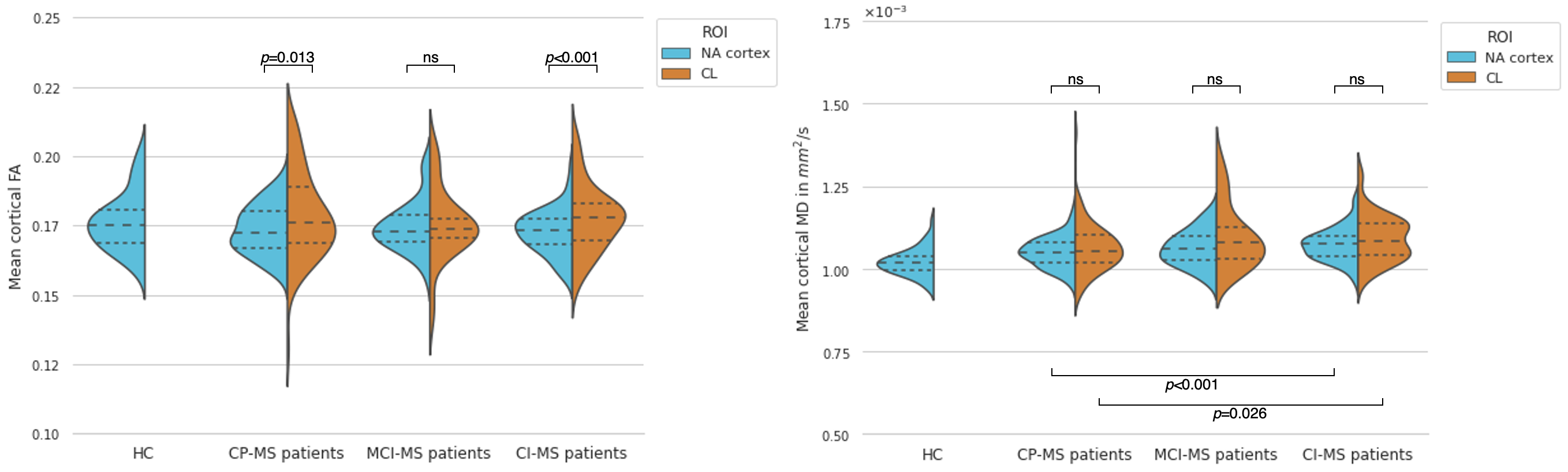
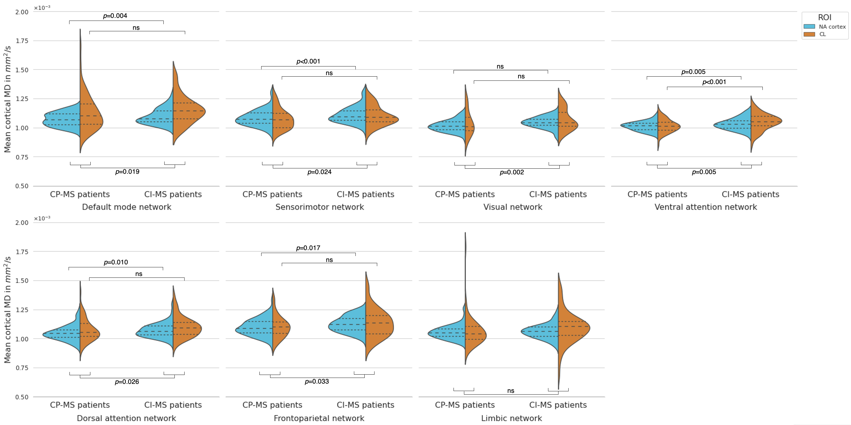
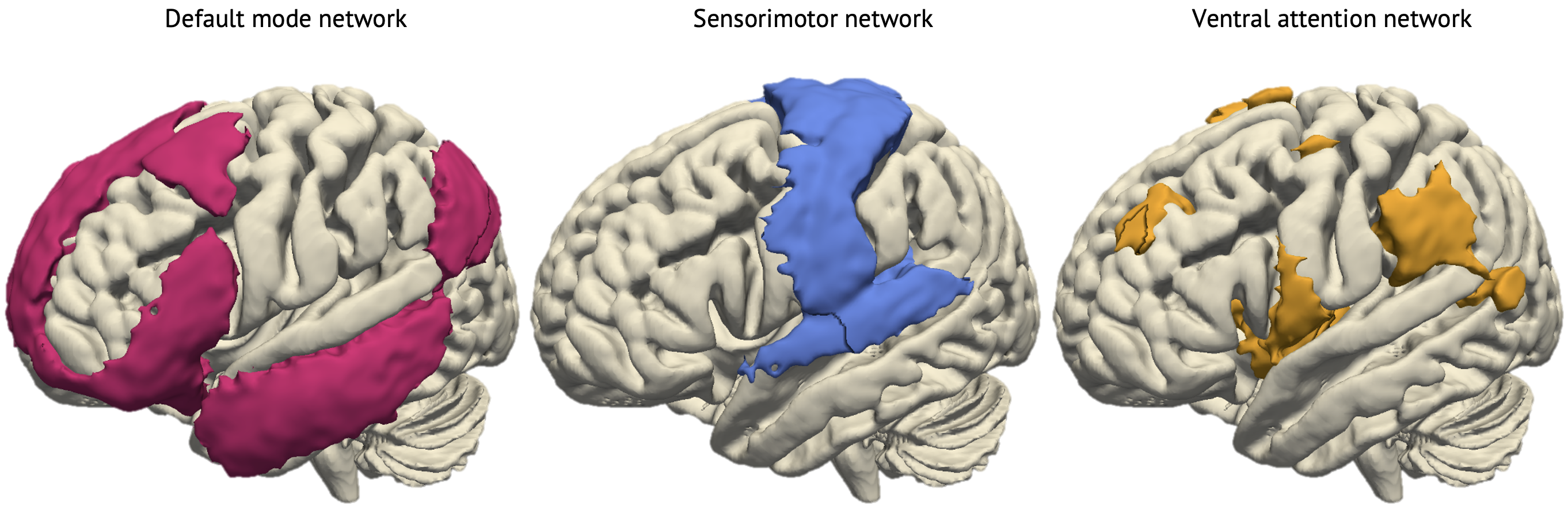
Cortical lesions were present in 87.5% of people with MS, with highest relative lesion load in the ventral attention network, followed by the limbic and default mode networks. CI-MS had significantly higher number of areas with cortical lesions as well as total cortical lesion volume compared to CP-MS (p<0.001). Mean MD was significantly increased in normal appearing cortex in MS compared to HC (p<0.001), but mean FA did not differ (p=0.17). Within-subject analysis showed an increase in both mean FA and MD in regions with cortical lesions compared to normal-appearing cortex in MS (p<0.001 and p=0.007, respectively). Cognitive subgroup analysis showed a stronger increase in mean FA in regions with cortical lesions compared to normal-appearing cortex in CI-MS than in CP-MS (Cohen’s d=0.53, p<0.001 and d=0.38, p=0.013, respectively) (Figure 1). Between cognitive subgroups, CI-MS showed higher cortical MD compared to CP-MS in both lesional and normal-appearing cortex (p<0.001 and p=0.026, respectively), with highest effect size in normal-appearing cortex (Hedges’ g=0.64 vs. g=0.38 in lesional cortex). FA showed no significant differences between subgroups and was not explored further (Figure 1). At network-level, cortical MD was increased in CI-MS compared to CP-MS in all networks, except limbic (p-range=0.002–0.033) (Figure 2). Region-specific analysis showed increases in MD of normal-appearing cortex in CI-MS compared to CP-MS in ventral attention, sensorimotor and default mode networks (p-range=0.006–0.037), whereas mean MD in areas with cortical lesions showed significant differences only in the ventral attention network (p=0.002) (Figure 2 and 3).Discussion
This study showed the relevance of cortical microstructural integrity changes in MS, reflected by lesional FA and extra-lesional MD. In line with previous literature,2,4,8 areas with cortical lesions had increased FA compared to normal-appearing cortex. This could be at least partly explained by an additional loss of parallel axons in the lesional cortex along with the loss of perpendicular axons present in normal-appearing cortex, influencing cortical microstructural coherence and being responsible for the FA changes.8 Whereas an FA increase seemed specific to focal cortical damage, an increase in MD was mainly observed in normal-appearing tissue, thus might reflect more diffuse damage to microstructural barriers.8,9 Interestingly, while cortical lesions are known to relate to cognition as well,10,11 microstructural integrity of normal-appearing cortex was even more strongly distinct between cognitive phenotypes, which is supported by previous literature.2,4 We observed that regions in the ventral attention, sensorimotor and default mode network were particularly vulnerable to integrity loss. Previous functional MRI studies highlighted the importance of alterations to particularly these cognitive networks in disease progression and cognitive decline, which might be driven by these structural effects.6,7Conclusion
Cortical diffusion changes were more prominent in CI-MS compared to CP-MS patients. FA was clearly increased in cortical lesions compared to normal-appearing cortex, but MD within normal-appearing cortex seemed more relevant for cognitive impairment in MS. CI-related changes to gray matter integrity of normal-appearing cortex were most severe in ventral attention, sensorimotor and default mode networks, probably indicating a preferential spatial susceptibility for cortical pathology relevant for cognitive decline.Acknowledgements
No acknowledgement found.References
1. Sumowski JF, Benedict R, Enzinger C, et al. Cognition in multiple sclerosis: State of the field and priorities for the future. Neurology. 02 06 2018;90(6):278-288. doi:10.1212/WNL.0000000000004977
2. Preziosa P, Pagani E, Morelli ME, et al. DT MRI microstructural cortical lesion damage does not explain cognitive impairment in MS. Mult Scler. Dec 2017;23(14):1918-1928. doi:10.1177/1352458516689147
3. Filippi M, Preziosa P, Pagani E, et al. Microstructural magnetic resonance imaging of cortical lesions in multiple sclerosis. Mult Scler. Apr 2013;19(4):418-26. doi:10.1177/1352458512457842
4. Yaldizli Ö, Pardini M, Sethi V, et al. Characteristics of lesional and extra-lesional cortical grey matter in relapsing-remitting and secondary progressive multiple sclerosis: A magnetisation transfer and diffusion tensor imaging study. Mult Scler. Feb 2016;22(2):150-9. doi:10.1177/1352458515586085
5. Calabrese M, Rinaldi F, Seppi D, et al. Cortical diffusion-tensor imaging abnormalities in multiple sclerosis: a 3-year longitudinal study. Radiology. Dec 2011;261(3):891-8. doi:10.1148/radiol.11110195
6. Huiskamp M, Eijlers AJC, Broeders TAA, et al. Longitudinal Network Changes and Conversion to Cognitive Impairment in Multiple Sclerosis. Neurology. 08 24 2021;97(8):e794-e802. doi:10.1212/WNL.0000000000012341
7. Koubiyr I, Besson P, Deloire M, et al. Dynamic modular-level alterations of structural-functional coupling in clinically isolated syndrome. Brain. 11 01 2019;142(11):3428-3439. doi:10.1093/brain/awz270
8. Preziosa P, Kiljan S, Steenwijk MD, et al. Axonal degeneration as substrate of fractional anisotropy abnormalities in multiple sclerosis cortex. Brain. 07 01 2019;142(7):1921-1937. doi:10.1093/brain/awz143
9. Stock B, Shrestha M, Seiler A, et al. Distribution of Cortical Diffusion Tensor Imaging Changes in Multiple Sclerosis. Front Physiol. 2020;11:116. doi:10.3389/fphys.2020.00116
10. Curti E, Graziuso S, Tsantes E, Crisi G, Granella F. Correlation between cortical lesions and cognitive impairment in multiple sclerosis. Brain Behav. 06 2018;8(6):e00955. doi:10.1002/brb3.955
11. Roosendaal SD, Moraal B, Pouwels PJ, et al. Accumulation of cortical lesions in MS: relation with cognitive impairment. Mult Scler. Jun 2009;15(6):708-14. doi:10.1177/1352458509102907
Figures