2816
Investigation of iron content changes in Alzheimer's disease and mild cognitive impairment using quantitative susceptibility mapping
Chuan-Bin Huang1, Yong Zhang2, Ke-Xue Deng1, and Chang Liu1
1Radiology, The First Affiliated Hospital of University of Science and Technology of China, Hefei, China, 2GE Healthcare, Shanghai, China
1Radiology, The First Affiliated Hospital of University of Science and Technology of China, Hefei, China, 2GE Healthcare, Shanghai, China
Synopsis
Keywords: Alzheimer's Disease, Quantitative Susceptibility mapping
This study investigated iron content changes using Quantitative Susceptibility Mapping (QSM) technique in AD and MCI patients, as compared with normal controls, and correlated iron deposit levels with cognitive scores to assess the clinical values of QSM in the diagnosis of AD and MCI. Bilateral caudate nucleus and right putamen showed significantly increased QSM values in AD and MCI patients as compared to healthy controls. The QSM values of right caudate nucleus correlated with the MMSE scores of AD patients. These results might indicate QSM as the potential biomarker for clinical diagnosis of AD and MCI.Introduction
Alzheimer's disease is a neurodegenerative disease that impairs cognition, memory, and language functions. Recent studies have shown that elevated levels of cortical iron concentration may cause brain damage, further leading to Alzheimer’s disease (AD)1-2. Exploring the interaction between aberration iron homeostasis of gray matter may provide new insights into AD and mild cognitive impairment (MCI) pathogenesis. This study investigated iron content changes using Quantitative Susceptibility Mapping (QSM) technique in AD and MCI patients, as compared with normal controls (NC), and correlated iron deposit levels with cognitive scores to assess the clinical values of QSM in the diagnosis of AD and MCI.Methods
All subjects agreed to participate in writing and the study was approved by the local ethics committee. Ten AD patients (aged 64.1±6.3 years, 3 males), 11 MCI patients (aged 62.7±4.8 years, 4 males) and 12 age-matched normal controls (aged 62.9±8.7 years, 6 males) with no prior history of neurological damage were recruited. AD patients were diagnosed based on the National Institute on Aging-Alzheimer’s Association (NIA-AA) and MCI patients were diagnosed based on the Petersen criteria. Both AD and MCI patients took the Mini-Mental State Examination (MMSE) questionnaire survey for quick assessment of cognitive ability. The QSM scans were conduct with a 3.0-T MR750w scanner (GE Healthcare, Milwaukee, WI) using a 24-channel phase array head and neck coil. Imaging parameters were as follows: repetition time=31.4 ms, flip angle=12°, field of view=25.6 cm, matrix size=256×256, acceleration factor=2, number of echoes=12, first echo time=2.0 ms, echo time spacing=2.35 ms, slice thickness=1 mm, slice number=136, the total acquisition time=7 minutes. QSM calculation was performed using vendor provided Functool software on Advantage Workstation. Bilateral caudate nucleus (CN), putamen (PUT), globus pallidus (GP), substantia nigra (SN) and red nucleus (RN) were manually outlined on all the continuous levels to measure QSM values (Figure 1). The statistical analysis was conduct using one-way analysis of variance (ANOVA) among three groups. Multiple Comparisons was carried out using Bonferroni correction. QSM values within individual brain regions were correlated with MMSE scores while adjusting for age.Results
Right caudate nucleus showed significantly increased QSM values in both AD and MCI patients as compared to healthy controls (corrected P<0.05). Left caudate nucleus and right putamen showed significantly increased QSM values in AD patients only as compared to healthy controls (corrected P<0.05). No significant differences were observed in other brain regions (Figure 2). The QSM values were negatively correlated with the MMSE scores in the right caudate nucleus for the AD patients (r=-0.805, P=0.016) (Figure 3).Discussion and Conclusion
Bilateral caudate nucleus and right putamen showed significantly increased QSM values in AD and MCI patients as compared to healthy controls. The QSM values of right caudate nucleus correlated with the MMSE scores of AD patients. These results might indicate QSM as the potential biomarker for clinical diagnosis of AD and MCI.Acknowledgements
No acknowledgement found.References
1. Krebs N, Langkammer C, Goessler W, et al. Assessment of trace elements in human brain using inductively coupled plasma mass spectrometry. J Trace Elem Med Biol. 2014;28(1):1-7.
2. Ayton S, Wang Y, Diouf I, et al. Brain iron is associated with accelerated cognitive decline in people with Alzheimer pathology. Mol Psychiatry. 2020;25(11):2932-2941.
Figures
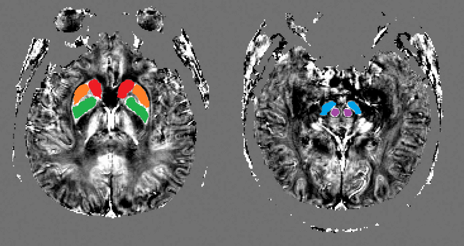
Fig 1. Regions of interest for QSM measurement (caudate nucleus, red; putamen,
orange; globus pallidus, green; substantia nigra, blue and red nucleus, purple)
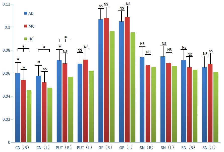
Fig 2. Comparison of QSM values of different brain regions among AD, MCI and healthy controls
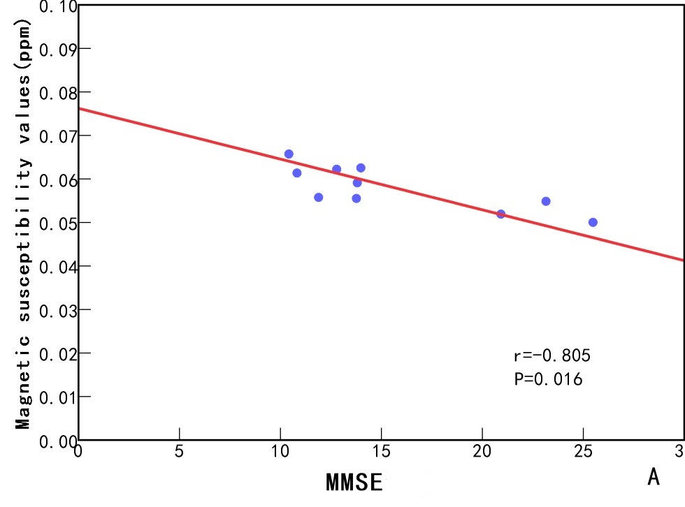
Fig 3. Correlation of QSM values of right caudate nucleus with MMSE scores in
AD patients
DOI: https://doi.org/10.58530/2023/2816