2806
Glymphatic Dysfunction Mediates the Influence of White Matter Hyperintensities on Episodic Memory in Cerebral Small Vessel Disease1Department of Neurology, Nanjing Drum Tower Hospital Clinical College of Nanjing Medical University, Nanjing 210008, China, nanjing, China
Synopsis
Keywords: Dementia, Dementia
In this study, we used the diffusion tensor image analysis along the perivascular space (ALPS)-index to evaluate glymphatic function and highlighted the reliability of using the ALPS-index in the early recognition of cognitive impairment (CI) in cerebral small vessel disease (CSVD) patients. We reported that 1) ALPS-index was a sensitive indicator to distinguish mild CI (MCI); 2) ALPS-index was an independent influencing factor of episodic memory in CSVD patients with MCI; 3) ALPS-index mediated the relationship between white matter hyperintensities and episodic memory in CSVD patients with MCI.Abstract
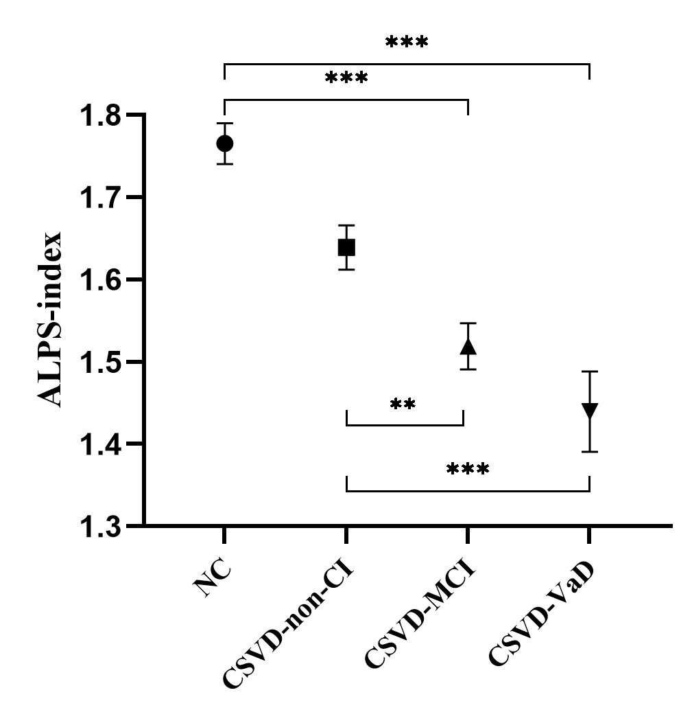
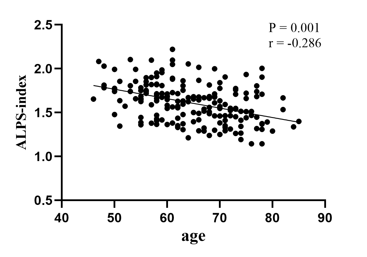
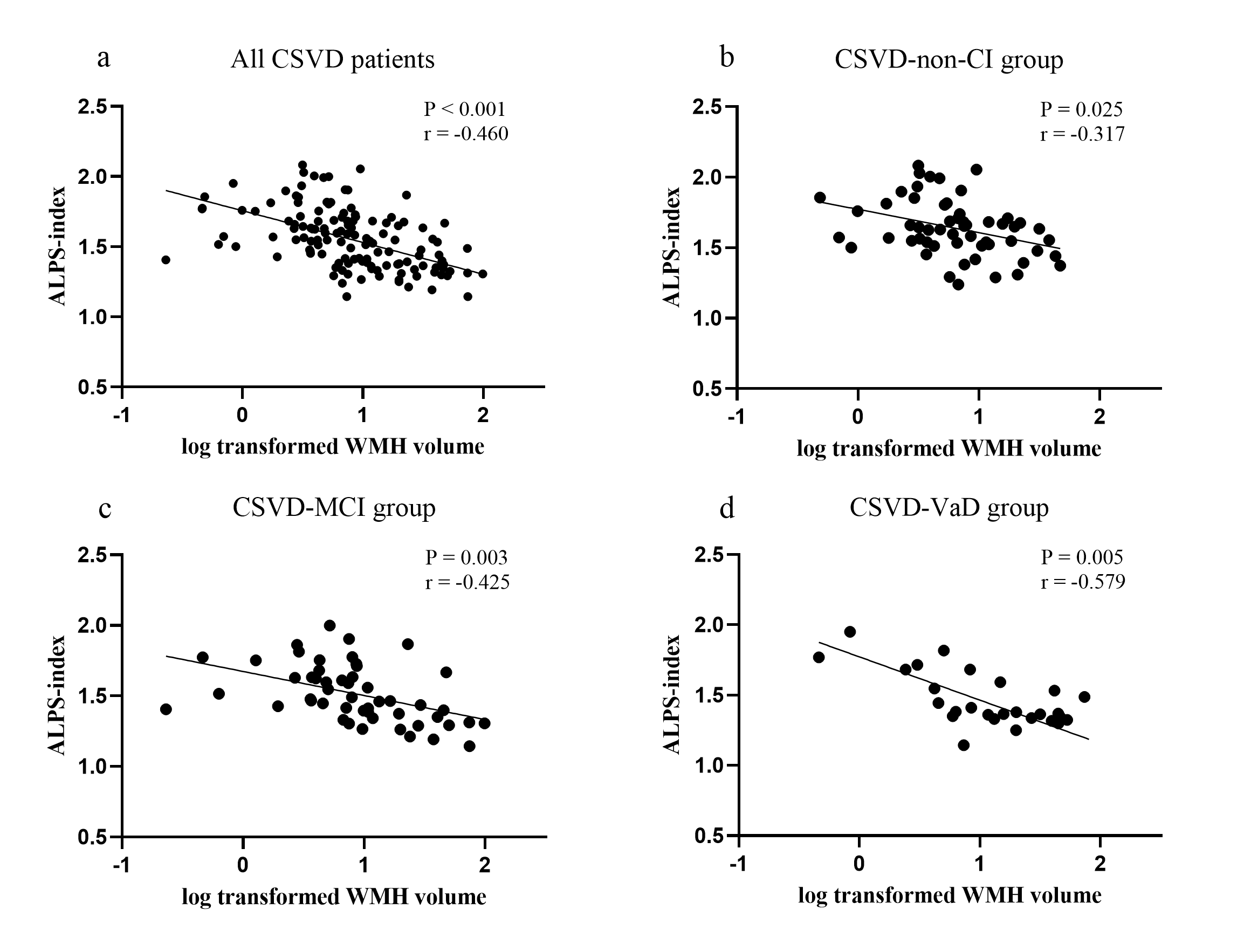
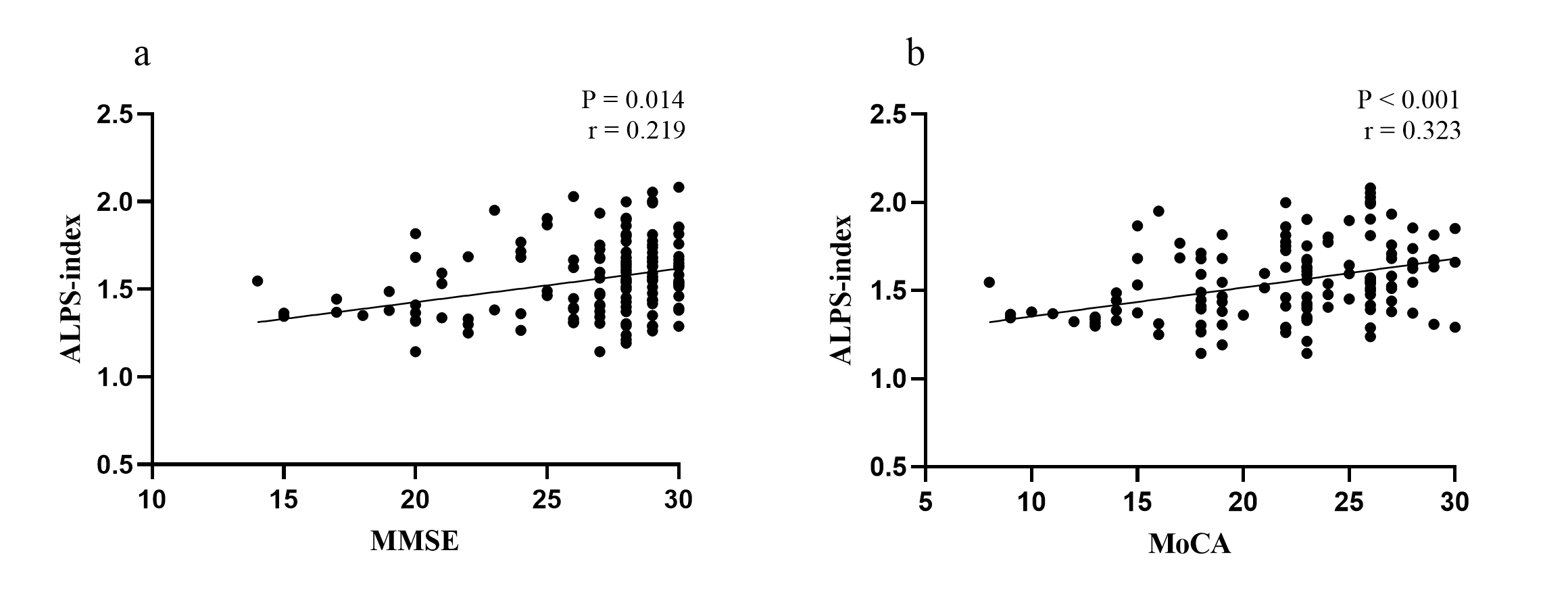
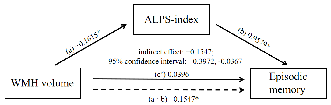
Glymphatic dysfunction has been linked to cognitive decline in several neurodegenerative diseases. In cerebral small vessel disease (CSVD), the mechanism of white matter hyperintensities (WMH)-related cognitive impairment (CI) is still under investigation. The diffusion tensor image (DTI) analysis along the perivascular space (ALPS) method has been used as a reliable parameter to assess glymphatic function. Therefore, we applied the ALPS-index to determine the influence of glymphatic function on CI in CSVD. Totally 137 CSVD patients (normal cognitive group, mild CI group, and dementia group) and 52 normal controls were recruited in this study. The ALPS-index was calculated based on the DTI. Correlation analyses and mediation analysis were performed to examine the relationship between glymphatic function and cognition. Significant differences in the ALPS-index were observed between subjects with and without CI. The ALPS-index was negatively correlated with age, WMH volume, and general cognitive function in all CSVD patients. In the mild CI group, the ALPS-index was independently positively correlated with episodic memory, and mediated the relationship between WMH volume and episodic memory. In conclusion, the ALPS-index is a potential marker for early recognition of CI in CSVD. Glymphatic dysfunction mediates the relationship between WMH and CI in CSVD.Methods
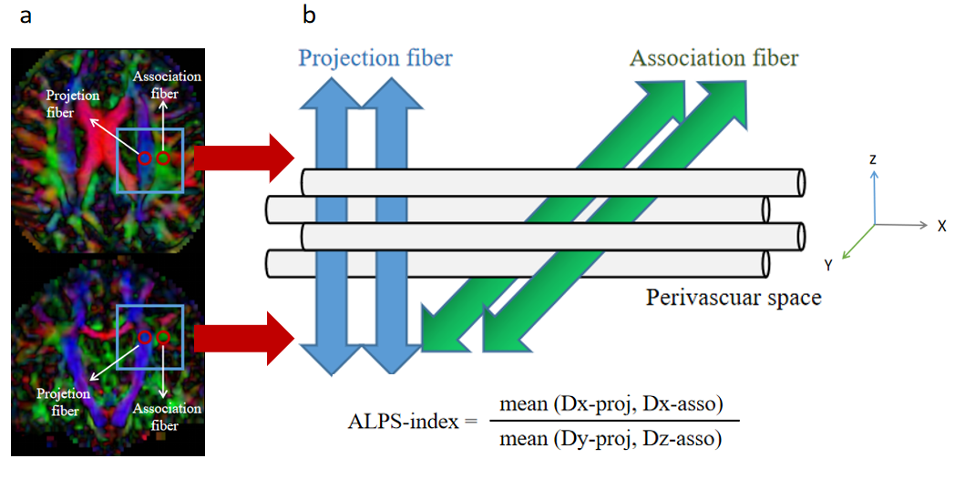
The ALPS-index was calculated as described previously [12]. In the paraventricular horizontal position, the perivascular space was horizontal from left to right, and perpendicular to the lateral ventricular wall. In this position, the projection fibers of the cortex were in the craniocaudal direction, and the associated fibers (superior longitudinal fasciculus) were anteroposterior. The three structures near the lateral ventricle were perpendicular to each other. Since the fiber bundles in this region were not parallel to the perivascular space, the diffusivity along the perivascular space could be independently analyzed in this region. Based on this structure, a spherical ROI containing 12 voxels was placed in the blue area (projection fiber) of the color FA diagram (Figure 2). The average diffusivities along the x-axis and y-axis were calculated and recorded as Dx-proj and Dy-proj, respectively. A spherical ROI containing 12 voxels was also placed in the green area (associated fiber) parallel to the ROI on the projection fiber (Figure 2), and the average diffusivities along the x-axis and z-axis in the ROI were calculated and recorded as Dx-asso and Dz-asso, respectively. In the projection fiber area, the fibers were shaped along the z-axis, while the x-axis and y-axis were perpendicular to the fiber direction. In the association fiber area, the fibers were oriented along the y-axis, while both the x-axis and z-axis were perpendicular to the fiber direction. The perivascular space of these two regions was shaped along the x-axis, so that Dx-proj and Dx-asso could represent the diffusivity along the perivascular space without the interference of nerve fibers. Since paraventricular WMHs can increase the diffusivity level in all directions as a whole, this effect was eliminated by using the following formula to calculate the ALPS-index: ALPS-index = (mean (Dx-proj, Dx-asso)) / (mean (Dy-proj, Dz-asso)).Result
Significant differences in the ALPS-index were observed between subjects with and without CI. The ALPS-index was negatively correlated with age, WMH volume, and general cognitive function in all CSVD patients. In the mild CI group, the ALPS-index was independently positively correlated with episodic memory, and mediated the relationship between WMH volume and episodic memory.Acknowledgements
No acknowledgements found.References
1. Wardlaw, J. M.; Smith, C.; Dichgans, M., Small vessel disease: mechanisms and clinical implications. Lancet Neurol. 2019, 18 (7), 684-696.
2. Cannistraro, R. J.; Badi, M.; Eidelman, B. H.; Dickson, D. W.; Middlebrooks, E. H.; Meschia, J. F., CNS small vessel disease A clinical review. Neurology 2019, 92 (24), 1146-1156.
3. Kasper, S.; Bancher, C.; Eckert, A.; Forstl, H.; Frolich, L.; Hort, J.; Korczyn, A. D.; Kressig, R. W.; Levin, O.; Palomo, M. S. M., Management of mild cognitive impairment (MCI): The need for national and international guidelines. World J. Biol. Psychiatry 2020, 21 (8), 579-594.
4. Iliff, J. J.; Wang, M. H.; Liao, Y. H.; Plogg, B. A.; Peng, W. G.; Gundersen, G. A.; Benveniste, H.; Vates, G. E.; Deane, R.; Goldman, S. A.; Nagelhus, E. A.; Nedergaard, M., A Paravascular Pathway Facilitates CSF Flow Through the Brain Parenchyma and the Clearance of Interstitial Solutes, Including Amyloid beta. Sci. Transl. Med. 2012, 4 (147), 11.
5. Plog, B. A.; Nedergaard, M., The Glymphatic System in Central Nervous System Health and Disease: Past, Present, and Future. In Annual Review of Pathology: Mechanisms of Disease, Vol 13, Abbas, A. K.; Aster, J. C., Eds. Annual Reviews: Palo Alto, 2018; Vol. 13, pp 379-394.
6. Nedergaard, M.; Goldman, S. A., Glymphatic failure as a final common pathway to dementia. Science 2020, 370 (6512), 50-+.
7. Mestre, H.; Kostrikov, S.; Mehta, R. I.; Nedergaard, M., Perivascular spaces, glymphatic dysfunction, and small vessel disease. Clin. Sci. 2017, 131 (17), 2257-2274.
8. Zhang, Y.; Zhang, R. T.; Ye, Y. Q.; Wang, S. Y.; Jiaerken, Y.; Hong, H.; Li, K. C.; Zeng, Q. Z.; Luo, X.; Xu, X. P.; Yu, X. F.; Wu, X.; Yu, W. K.; Zhang, M. M.; Huang, P. Y., The Influence of Demographics and Vascular Risk Factors on Glymphatic Function Measured by Diffusion Along Perivascular Space. Front. Aging Neurosci. 2021, 13, 8.
Figures