2783
Rapid high-resolution musculoskeletal MR imaging with a super resolution deep learning constrained Compressed SENSE reconstruction1Shanghai Sixth People's Hospital, Shanghai, China, 2Philips Healthcare, Shanghai, China, 3Philips Healthcare, Hong Kong, China, 4Philips Health Technology, Suzhou, China, 5Philips Healthcare, Best, Netherlands
Synopsis
Keywords: Skeletal, Bone
The integration of compressed-SENSE and artificial intelligence allows to acquire high-resolution images within relatively short scan times The purpose of this study is to compare the image quality of the musculoskeletal images reconstructed with Compressed -SENSE (CS), CS with integrated artificial intelligence (CS-AI) and CS-AI combined with superresolution(CS-AI-HR) . We found that the image quality was significantly improved by using CS-AI-HR compared to CS. This study provides supportive information for the application of CS-AI-HR in routine clinical practice.Introduction
Equipment efficiency is a constant challenge for imaging devices, which mainly balances two aspects: speed and image quality. With traditional acceleration techniques, speed is increased, but at too high acceleration factors image quality can be severely jeopardized. High-resolution MRI images can provide excellent anatomical information for precise diagnosis and for treatment guidance. However, high-resolution image scanning requires considerable time, which may cause patient discomfort and, potentially, motion.In order to reduce the scan time without compromising the image quality, a series of techniques represented by Compressed-SENSE (CS) have been implemented in the imaging field. Recently, the technique of applying artificial intelligence (AI) to CS for acceleration, named as CS-AI, has been introduced1. CS-AI can increase the acceleration multiplier more than conventional CS while obtaining the same image quality. Its advantage has been demonstrated in the imaging of the ankle2 and brain3,4.
Also, superresolution AI models have been introduced to improve the sharpness of images. The combination of superresolution with acceleration based on CS-AI promises to acquire high resolution images from highly undersampled k-sapce data. This combination is denoted here as CS-AI-HR. The purpose of this study is to compare the image quality of the musculoskeletal images reconstructed with CS, CS-AI and CS-AI-HR.
Methods
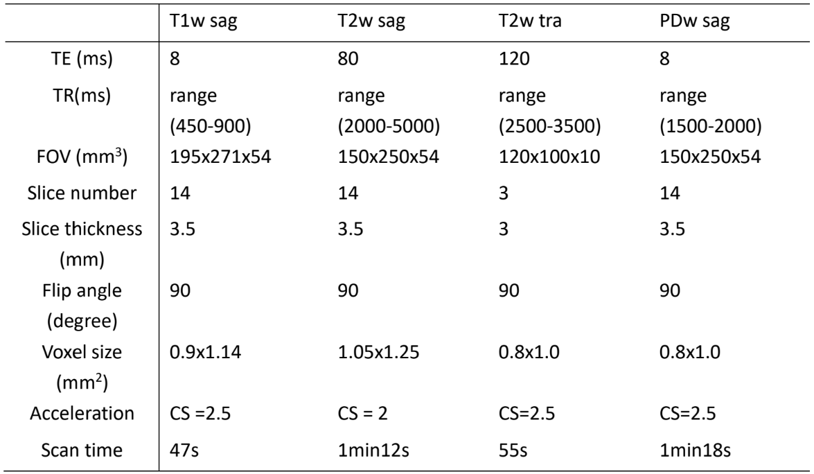
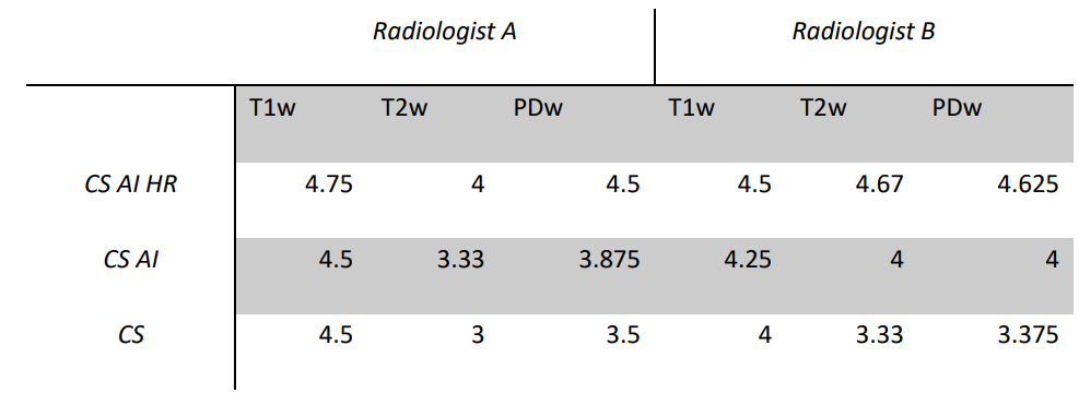
5 subjects were included in this study. Routine MR sequences were acquired in all subjects on a 3.0T MRI system (Elition, Philips Healthcare, Best, the Netherlands). T1-weighted (T1w), T2-weighted (T2w) and Proton density-weighted imaging (PD) sequences were acquired with the musculoskeletal system as the main targets. The parameters used are shown in Table 1.These sequences were reconstructed by CS, CS-AI, and CS-AI-HR.Image quality was evaluated by two radiologists with more than 5 years of experience. All images were rated according to an ordinal 5-point Likert scale (1 = poor, 2 = below average, 3 = fair, 4 = good, 5 = excellent), evaluating the following criteria: partial volume effect, blurring, discrimination from adjacent structures, and signal homogeneity. Friedman test with Dunn’s correction for multiple comparisons was used to compare the semi-quantitative image quality rates between CS, CS-AI and CS-AI-HR sequences. p-values <0.05 were considered significant.
Results
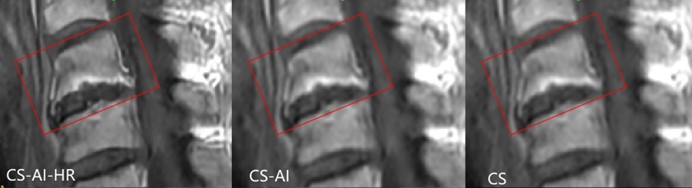
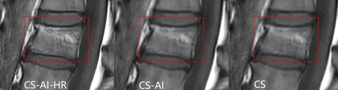
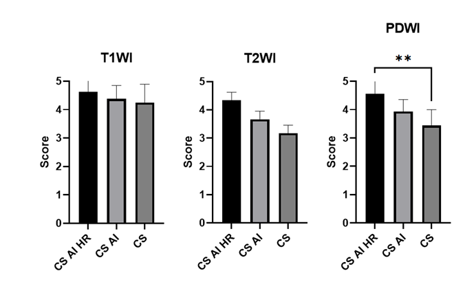
Examples of CS-AI-HR, CS-AI and CS images at lumbar region are shown in Figure 1 and Figure 2. In Figure 1, CS-AI-HR has a much clearer depiction of the hyperplasia and grossness of the vertebral body. In Figure 2, the presentation of an old compression fracture was better visible on the CS-AI-HR image compared to CS-AI or CS.. The image quality assessment of the two radiologists is provided in Table 2. The image quality of the routine scanned PDw images using CS-AI-HR was significantly higher than using CS (Figure 3, P = 0.0035).Discussion
In our results, CS, CS-AI and CS-AI-HR images are all adequate for diagnosis without missed lesions. However, in terms of image clarity, CS-AI-HR enhanced the sharpness of the images, offers better image quality and is more descriptive for details.While CS and CS-AI improve the SNR through the sparse representation of MR images, CS-AI-HR adds sharpening of image by its low- to high-resolution feature. This sharpening is present in all image features including artifacts that might exist e.g. due to motion. Even with the above limitation, CS-AI-HR was still scored better thanthan CS-AI or CS, which is in line with the finding from previous study2.
The above results suggest that scan acceleration by CS-AI-HR reconstruction may help to obtain high quality images that can beimplemented in clinical routine.
Conclusion
CS-AI-HR reconstruction produce MR images with higher quality than that of CS images. It has potential to be integrated in clinical routine.Acknowledgements
No acknowledgement found.References
1.Quan TM, Nguyen-Duc T, Jeong WK. Compressed Sensing MRI Reconstruction Using a Generative Adversarial Network With a Cyclic Loss. IEEE Trans Med Imaging. 2018 Jun;37(6):1488-1497. doi: 10.1109/TMI.2018.2820120. PMID: 29870376.
2.Foreman SC, Neumann J, Han J, Harrasser N, Weiss K, Peeters JM, Karampinos DC, Makowski MR, Gersing AS, Woertler K. Deep learning-based acceleration of Compressed Sense MR imaging of the ankle. Eur Radiol. 2022 Jun 25. doi: 10.1007/s00330-022-08919-9. Epub ahead of print. PMID: 35751695.
3.Mönch S, Sollmann N, Hock A, Zimmer C, Kirschke JS, Hedderich DM. Magnetic Resonance Imaging of the Brain Using Compressed Sensing - Quality Assessment in Daily Clinical Routine. Clin Neuroradiol. 2020 Jun;30(2):279-286. doi: 10.1007/s00062-019-00789-x. Epub 2019 May 16. PMID: 31098666.
4.Kayvanrad M, Lin A, Joshi R, Chiu J, Peters T. Diagnostic quality assessment of compressed sensing accelerated magnetic resonance neuroimaging. J Magn Reson Imaging. 2016 Aug;44(2):433-44. doi: 10.1002/jmri.25149. Epub 2016 Jan 18. PMID: 26777856.
Figures