2778
Quantification of soft tissue of the buttocks in a seated position
Jill M Slade1, Nkhensani Mogale2, Daniel Schanz1, Viktor Ilyasov1, Justin Scott3, Albert van Schoor2, and Tamara R Bush3
1Radiology, Michigan State University, East Lansing, MI, United States, 2University of Pretoria, Pretoria, South Africa, 3Mechanical Engineering, Michigan State Univerisity, East Lansing, MI, United States
1Radiology, Michigan State University, East Lansing, MI, United States, 2University of Pretoria, Pretoria, South Africa, 3Mechanical Engineering, Michigan State Univerisity, East Lansing, MI, United States
Synopsis
Keywords: MSK, Muscle
MRI is well suited to quantify soft tissue but challenges remain in using MRI to address soft tissue in a seated position and for studying how tissue responds to loading. IDEAL and T1 weighted images were used to quantify apparent fat fraction and soft tissue thicknesses of the buttocks in a side lying position with the tissue unloaded and loaded. Tissue compression during loading predominantly reflected changes in the muscle and tendon. Intramuscular fat was not related to the change in muscle/tendon. These methods may be useful to study soft tissue in populations at risk for pressure ulcer development.INTRODUCTION
Numerous factors contribute to the development of pressure ulcers that occur with prolonged sitting including soft tissue composition and thickness of tissues below the ischial tuberosity (IT) of the pelvis. Soft tissue contributions may include muscle atrophy and increased fat infiltration of skeletal muscles. MRI is well suited to study soft tissue but presents challenges for addressing a seated position and posture and additionally for studying how soft tissue responds to loading.PURPOSE
The purpose of the study was three-fold. 1) to develop a protocol to examine seated soft tissue in a horizontal bore MRI 2) to develop an MRI compatible loader to mimic loading during seating and 3) to evaluate the impact of unloaded tissue thickness and intramuscular fat on loaded tissue.METHODS
Soft tissue of the buttocks was measured in able-bodied male (n=11, 26±5yrs old, BMI=24±4) and female participants (n=11, 20±2yrs old, BMI=24±4). Subjects were positioned in a lateral decubitus position with hip flexion at 100°-120° and knee flexion at 90° and imaged at 3T (GE Excite). IDEAL images (512x224 matrix, 40 FOV, 4mm slice, FA=5˚) were used to evaluate the apparent fat fraction of the gluteus maximus in unloaded tissue. A custom designed loading seat was applied to the buttocks with loads reflecting 30% of body weight with a load cell mounted in the center of a 30 x 12.7cm seat to quantify load; the loader consisted of a brass rod on a crank to apply the load. T1 weighted images (4mm slices, TE=7.9ms, TR=1216ms, 320x224 matrix, 40cm FOV) were used to measure tissue thickness during loading. The tissue thickness was measured over 3 slices from the peak of the ischial tuberosity (IT). The apparent fat fraction was quantified from the portion of the gluteus maximus below the IT over 3 slices from the IDEAL fat and water images. Sample images are shown in Figure 1. Paired t-tests were used to examine changes in tissue compression with load. Pearson’s correlation was used to explore relationships between loaded and unloaded tissue.RESULTS
Total soft tissue thickness was reduced by 48±14% with the application of 30% body weight load (5.39±0.93 cm unloaded, 2.89 ±0.61 cm loaded) including a reduction in the muscle+tendon of 60±16% (3.76±0.71cm unloaded, 1.58 ± 0.59 loaded) and 16±23% reduction in skin+fat thickness (Figure 2). The muscle+tendon thickness reduction with load was greater than the skin+fat reduction (p<0.001). The total unloaded tissue was correlated with loaded tissue thicknesses (r= 0.47-0.79, p≤0.027) with the total thickness changes influenced largely by the muscle+tendon thickness (r=0.76). Fat fraction of the unloaded gluteus maximus ranged from 9-25%. There was no significant correlation between loaded tissue thickness and the apparent fat fraction of the gluteus maximus (r=0.05).DISCUSSION
A novel approach was successfully used to examine soft tissue of the buttocks using joint angles that reflect a seated position. The MRI compatible loader was used to successfully apply a 30% body weight load to all participants. Loading the buttocks resulted in a large compression of soft tissue that impacted the thickness of the muscle and tendon more than the subcutaneous tissue. These results may partly be explained by the differences in viscoelastic properties between muscle and tendon compared to fat. The loading resulted in both compression and sliding of the gluteus maximus away from the IT. The composition of the gluteus maximus muscle (fat fraction) did not contribute to the change in tissue thickness.CONCLUSION:
In summary, a side lying position and custom MR compatible seat loader can be used to study soft tissue changes in a seated body position in a horizontal bore MRI. In this population of healthy participants, fatty infiltration did not predict soft tissue compression. These methods can be applied to evaluate optimal seating design and the soft tissue compression across populations prone to pressure injuries. Further, these methods may be combined with MR elastography to quantify additional tissue properties of the buttocks soft tissue.Acknowledgements
This work was supported by the Alliance for African Partnership, the Department of Radiology and the College of Osteopathic Medicine at Michigan State University.References
No reference found.Figures
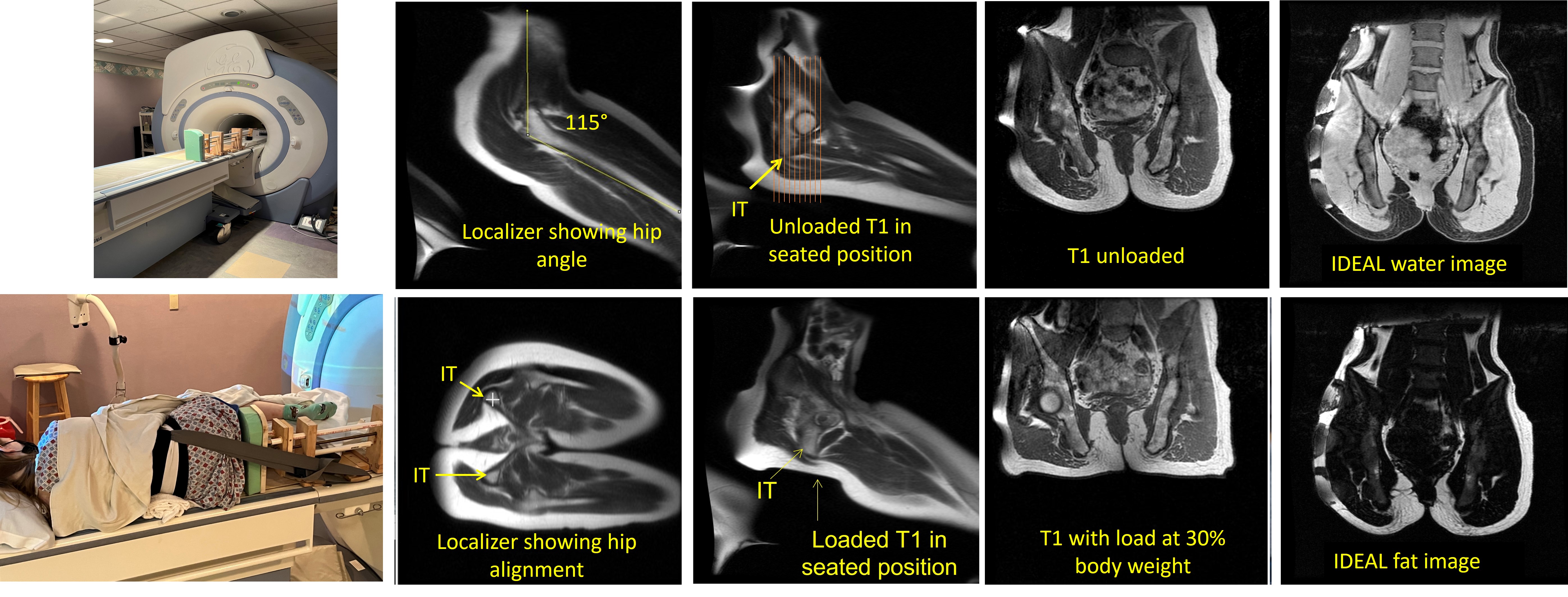
The figure shows the MRI loader (left panel) and sample images of the pelvis during a seated unloaded and loaded position. IT indicates the ischial tuberosity.
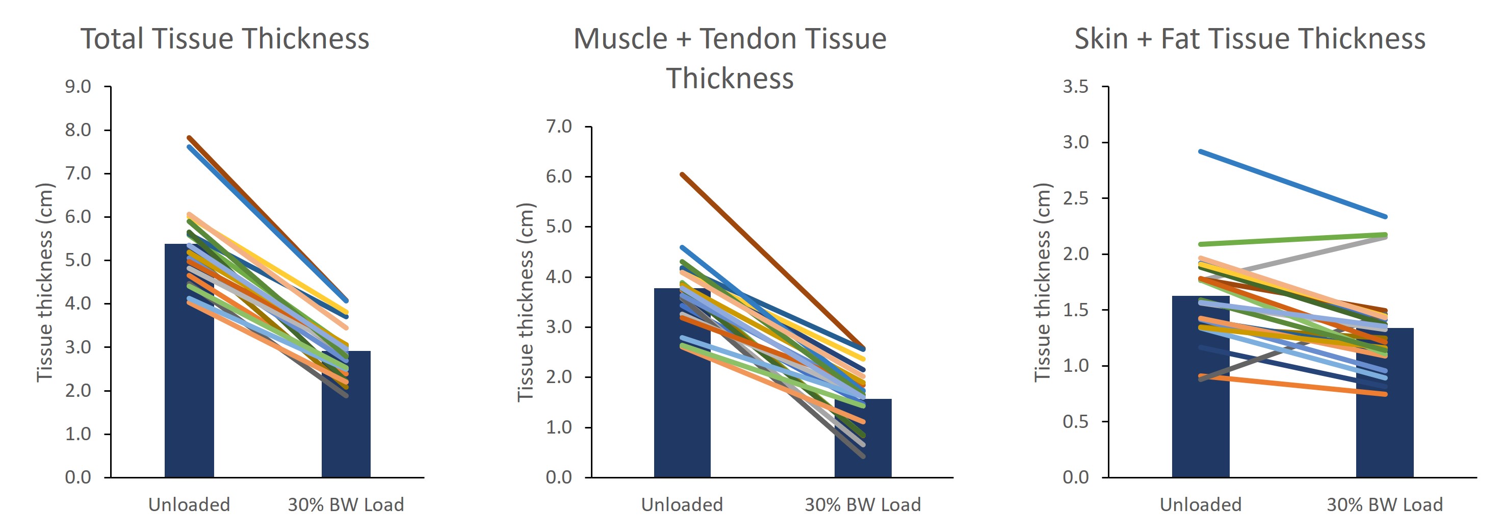
Soft tissue thicknesses of the buttocks shown for the group (bar) and individual participants (lines). Tissue measures reflect soft tissue below the ischial tuberosity in a seated unloaded position and a seated loaded position with a 30% body weight load applied.
DOI: https://doi.org/10.58530/2023/2778