2775
A Comparison of High-Resolution Simultaneous Multi-Slice Accelerated Turbo Spin-Echo Knee Imaging with Routine Turbo Spin-Echo Imaging
Yanping Xue1, Li Wang1, Hua Gu1, Jiyang Zhang1, Chen Zhang2, Xiuqin Jia1, and Qi Yang1
1Radiology, Beijing Chao-Yang Hospital, Capital Medical University, Beijing, China, 2MR Scientific Marketing, Siemens Healthineers, Beijing, China
1Radiology, Beijing Chao-Yang Hospital, Capital Medical University, Beijing, China, 2MR Scientific Marketing, Siemens Healthineers, Beijing, China
Synopsis
Keywords: MSK, Joints
MRI has been widely used in the early detection of osteoarthritis (OA), especially in knee, as an important technique. However, the long acquisition time of MRI is a barrier in improving the quality of imaging and the comfort of patients, especially for pediatric and disabled people. Many imaging acceleration factors have been implied in order to improve the condition. This study focuses on the clinical application of simultaneous multi-slice (SMS) imaging, a newly developed acceleration technique using multi-band RF pulses, which can simultaneously excite, acquire, and reconstruct multiple slices and readout with two-dimensional images.Introduction
Osteoarthritis (OA) of the knee joint is one of the most common forms of arthritis, which affects millions of people and has a substantial impact on the economy and the health care system worldwide [1]. Therefore, it is crucial for clinicians to detect structural and functional changes of the knee OA at the early stages using a non-invasive and sensitive method. In musculoskeletal (MSK) imaging, turbo spin echo (TSE) is widely used as it offers excellent depiction of cartilage, ligaments, menisci, and periarticular soft tissues. However, high spatial resolution with a large number of slices is rarely used clinically because of the prolonged acquisition time for complete coverage when using a conventional 2D-TSE sequence. Integrated Parallel Imaging Technology (PAT) generalized autocalibrating partially parallel acquisitions (GRAPPA) improves the speed of TSE sequences by undersampling k-space [2-4], although this is usually related to a loss of signal-to-noise ratio (SNR). Simultaneous multi-slice (SMS) with integration of the controlled aliasing in parallel imaging result in higher acceleration (CAIPIRINHA) is a promising parallel imaging method to increase the acquisition speed without a significant decrease to the SNR [5]. Although SMS had been used in several knee joint imaging studies [6], the clinical evaluation of SMS in accelerating and providing high resolution, expectable SNR and contrast-to-noise ratio (CNR) in knee joint, compared to GRAPPA-TSE have still not been investigated. Therefore, this study aimed to quantify and compare the image quality and diagnostic value in lesion of SMS 2D TSE sequences with gradient-based CAIPIRINHA and GRAPPA-TSE in knee joint imaging.Methods
A total of 11 human subjects (aged 16-66 years; 44±12 years; 4 males, 7 females) was were prospectively recruited for this study. Informed consent was obtained from all subjects in accordance with the guidelines of the local Institutional Review Board. The whole knee joint (6 left knees, 5 right knees) was scanned using 2D sagittal SMS-TSE PDWI with fat-suppressed (FS) sequence and GRAPPA- TSE PDWI-FS sequence on a 3.0 T Vida MR scanner (MAGNETOM Vida , SIEMENS Healthcare Technologies, Germany). The detailed parameters of the two MR sequences are listed in Table 1. Quantitative signal-to-noise ratio (SNR ) for distal femur, cartilage, joint fluid, ligaments, menisci, tendons and muscle, and the contrast-to-noise ratio (CNR ) for joint fluid/cartilage, distal femur /cartilage, ligament/ cartilage, tendon/cartilage, muscle/cartilage, and meniscus/cartilage of the optimized SMS-TSE-PDWI sequence were calculated, and compared with GRAPPA- PDWI sequence using a paired t-test. As another objective index, Tthe detection and visualization of cartilage and meniscus lesions in a patient cohorts was were evaluated in the two protocols as another objective index. The imaging quality was graded by two musculoskeletal radiologists (with 30 and 21 years of experience, respectively) according the four-point scale: clarity of cruciate ligaments, bone marrow, cartilage, and tendon (4=excellent: optimal diagnostic value and clearly shows the structure, 3=good: good for the majority of diagnoses, 2=acceptable: for the majority of diagnoses and the evaluation of the structure was somewhat limited, 1=poor: poor for the majority of diagnoses and the evaluation of the structure was substantially limited) [7]. The inter-reader agreement of the SNR measurements and qualitive evaluation was also assessed using intraclass correlation efficient (ICC) and Cohen’s kappa analysis. All statistical analyses were performed using SPSS (IBM, Armonk, NY, USA) version 26.0 and P<0.05 was considered statistically significant.Results and Discussion
As shown in Table 2, the SNRs of distal femur, tendons, ligaments gotten from the SMS-2D TSE PDWI-FS were higher than the SNRs of the same position obtained from the GRAPPA-TSE PDWI-FS sequence, and the SNRs of cartilage, menisci, and muscle didn’t show statistical difference between the two sequences. The CNRs between the two sequences did not have significant difference except the CNR for joint fluid to cartilage, which may explain the clear boundary between the joint fluid and cartilage, and the capability to show subtle defect in cartilage with the SMS-TSE PDWI-FS sequence as shown in figure 1-2. The subjective evaluation of image quality with respect to diagnostic value and clearly displayed structure showed no significant differences between the two protocols.Conclusion
The SMS technique can accelerate the conventional TSE sequence with high SNR and CNR. Compared with GRAPPA, it can be used to improve spatial resolution in the same scan time, which is helpful to find minor lesions, or further reduce acquisition time, which is beneficial for patients, especially for pediatric and disabled people.Acknowledgements
No acknowledgement found.References
1. Cross M, Smith E, Hoy D, et al. The global burden of hip and knee osteoarthritis: estimates from the Global Burden of Disease 2010 study. Ann Rheum Dis. 2014, 73:1323-1330. 2. Griswold MA, Jakob PM, Heidemann RM, et al. Generalized autocalibrating partially parallel acquisitions (GRAPPA). Magn Reson Med. 2002, 47:1202–1210. 3. Fritz J, Fritz B, Zhang J, et al. Simultaneous Multislice Accelerated Turbo Spin Echo Magnetic Resonance Imaging: Comparison and Combination With In-Plane Parallel Imaging Acceleration for High-Resolution Magnetic Resonance Imaging of the Knee. Invest Radiol. 2017, 52:529-537. 4. Fritz J, Guggenberger R, Del Grande F. Rapid Musculoskeletal MRI in 2021: Clinical Application of Advanced Accelerated Techniques. AJR Am J Roentgenol. 2021, 216:718-733. 5.Gao F, Wen Z, Dou S, et al. High-Resolution Simultaneous Multi-Slice Accelerated Turbo Spin-Echo Musculoskeletal Imaging: A Head-to-Head Comparison With Routine Turbo Spin-Echo Imaging. Front Physiol. 2021, 12:759888. 6. Benali S, Johnston PR, Gholipour A, et al. Simultaneous multi-slice accelerated turbo spin echo of the knee in pediatric patients. Skeletal Radiol. 2018, 47:821-831. 7. Li G, Wu D, Xu Z, et al. Evaluation of an accelerated 3D modulated flip-angle technique in refocused imaging with an extended echo-train sequence with compressed sensing for imaging of the knee: comparison with routine 2D MRI sequences. Clin Radiol. 2021, 76:158.e13-158.e18.Figures
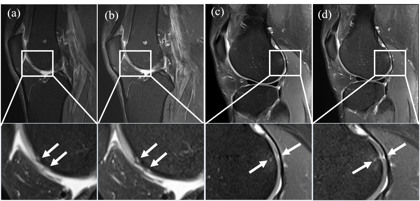
Figure
1. (a, c) Sagittal 2D GRAPPA-TSE
PDWI and (b, d) SMS-TSE PDWI with fat-suppressed
image show the focal subtle defect of the femoral trochlear cartilage (a and b,
right
knee of a 46-year-old male patient ) and
the dorsal lateral femoral condyle cartilage with a subchondral cyst (c and d,
left knee of a 46-year-old male patient
), (arrow).
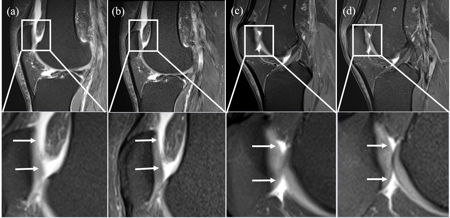
Figure
2. (a, c) Sagittal 2D GRAPPA-TSE
PDWI and (b, d) SMS-TSE PDWI with fat-suppressed
image demonstrate the difference in showing the margin between the cartilage
and the joint fluid (arrow). (a) and (b) come from a right knee of a 49-year-old female
patient, (c) and (d) come from a right knee of a 46-year-old male patient.

TR=repetition
time, TE= echo time, FOV= field of view, SMS= simultaneous multi-slice, GRAPPA=
generalized autocalibrating
partially parallel acquisitions, PDWI= proton density weighted imaging, TSE=
turbo spin-echo.

CNR=contrast
to noise ratio, SNR=signal to noise ratio, SMS= simultaneous multi-slice,
GRAPPA= generalized autocalibrating partially parallel acquisitions, PDWI=
proton density weighted imaging, TSE= turbo spin-echo.
DOI: https://doi.org/10.58530/2023/2775