2771
Clinical feasibility of synthetic T1-weighted images in evaluation of rotator cuff injury using ZTE-MRI as reference1Biomedical Engineering College, Hubei University of Medicine, Shiyan Hubei, China, 2GE Healthcare,, Beijing, China, 3Taihe Hospital, Hubei University of Medicine,, Shiyan Hubei, China
Synopsis
Keywords: Tendon/Ligament, Aging, synthetic
Magnetic resonance imaging is the gold standard for evaluation of rotator cuff injury in non-invasive examination. And 90% occur in supraspinatus tendons. In this study, synthetic MRI (SyMRI) generated both structural and functional images to qualitatively and quantitatively evaluate supraspinatus tendon injury using zero-echo magnetic resonance imaging (ZTE-MRI) as structural reference images. Despite superior quality and inter-reader agreement on critical shoulder angle measurements of ZTE-MRI to SyMRI-generated T1WI, SyMRI showed excellent intra-modality agreement of acromion index and CSA to ZTE-MRI and also provided quantitative information for better identifying injured sites at the early stage of rotator cuff injury.Introduction
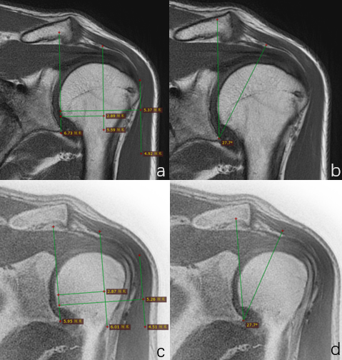
Rotator cuff (RC) injury is characterized by fiber disarrangement and cartilage degeneration due to decreased type Ⅰ collagen fibers, increased type Ⅲ collagen fibers, and accumulated glycosaminoglycans in thinning fibers and inflammatory cells1.Early detection and treatment slow down tendon injury to some extent. Structural MRI is clinically used to subjectively assess the degree of injury and shoulder joint function according to the Zlatkin scale of RC injury2. Acromion index (AI) and critical shoulder angle (CSA) are measured on conventional radiographs and can predict RC pathology3. In contrast to radiograph, conventional T1-weighted spin echo sequence is reckoned as the preferred alternative approach in diagnosis of musculoskeletal joint diseases while ZTE-MRI4, a kind of proton density weighted image, has recently been found to better display better displays periosteum and cortical bone for its nominal echo time of zero. However, objective assessment of RC injury lacks despite many quantitative studies attempted to apply T1, T2, T1rho, QSM techniques in diagnosis of joint degeneration or degree of injury5. A synthetic magnetic resonance technology can not only provide multi-contrast images but also five different quantitative maps via a newly-developed multi-delay and multi-echo fast spin echo sequence6. In this study, we aimed to explore the feasibility of synthetic magnetic resonance technology in qualitative evaluation of rotator cuff injuries using ZTE as standard reference and in quantitative assessment of injury degrees.Materials and methods
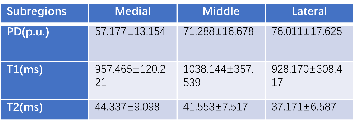
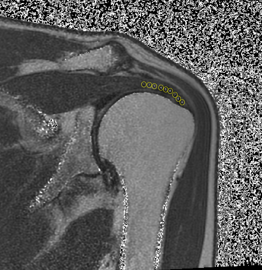
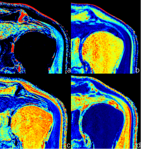
18 patients underwent MRI examination, including with fat-saturation T2-weighted images (T2WI FS), synthetic MRI (MAGiC) and ZTE-MRI on 3.0T MRI scanner (Signa Architect, GE Healthcare). The supraspinatus tendon injury was evaluated on T2WI FS sequence according to Zlatkin magnetic resonance classification criteria, and the injury showed high signal on T2WI FS sequence. The supraspinatus tendon was divided into three parts, medial, intermediate and lateral subregions, on T2WI oblique coronal images generated by Synthetic MRI. Three regions of interest (ROIs) were delineated in each subregion along the course of the supraspinatus tendon (Figure[MOU1] 1). Both AI and CSA were measured on ZTE-MRIs and synthetic T1-weighted images by two radiologists with 3- and 10-year experience in musculoskeletal diagnosis, and shoulder joint morphology and its correlation with rotator cuff injury were also assessed. Intra-group correlation coefficient (ICC) was used to test inter-rater and intra-rater consistency. Spearman rank correlation test was used to calculate the correlation between different T1, T2 and PD values, AI and CSA measurements and different grades. Paired t or Wilcoxon signed test were tested for AI and CSA measurements depending on data normality and equality of variance. P < 0.05 was considered statistically significant.Results
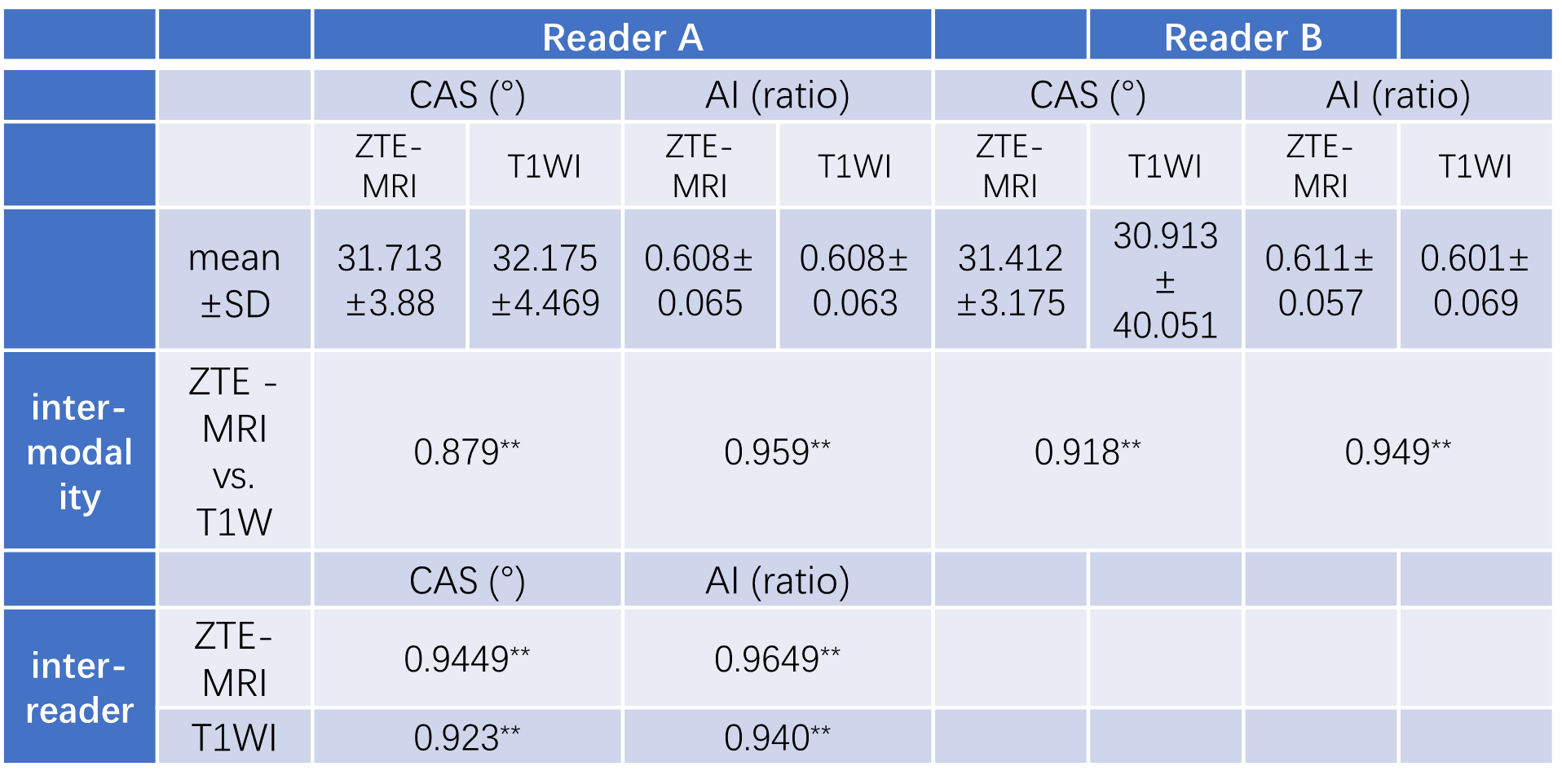
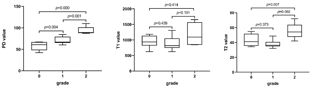
Inter-reader and inter-modality agreement of AI and CAS measurements between T1W and ZTE-MRI were good to excellent (ICC: 0.879-0.959, all P < 0.05, Table 1). The PD and T2 values of patients with supraspinatus tendon injury were different among different grades of sub regions (Figure 4), In the injury classification, PD values were significantly different between grade 0 and grade 1, grade 1 and grade 2, and the PD values of grade 0 were slightly lower than grade 1, while the PD values of grade 1 were significantly lower than grade 2. There was no statistical difference in T1 at all grade of injury. There are differences in T2 values between grade 0 and grade2, grade 1 and grade 2, and grade 2 injury is significantly higher than grade 0 and grade 1 injury. And the outer and middle sub regions of the injury site were mainly.Discussion and conclusions
In our study, synthetic MRI can generate multiple contrast images that meet the diagnostic requirements and also quantitative parameters for classifying function or injury grade6. In terms of structural images, synthetic T1WI display cortical membrane and almost the same distance and angle measurements comparative to ZTE-MRI. Good agreement of critical shoulder angle and acromion index may attribute to less shoulder movement during shorter scan time4. However, the accurate edge of the cortical bone displayed more clearly, leading to ZTE-MRI possessing better inter-reader agreement on CSA measurements. In terms of quantitative images, relaxometry maps showed tiny difference of PD values between subregions, indicating quantitative parameters elevate diagnosis precision on early rotator cuff injury7 . PD and T2 values show differences in different levels of injury, and PD and T2 values were significantly different between the lateral subzone and the medial subzone of supraspinatus tendon, so the injury in the lateral subzone was significantly higher than that in the medial subzone. It may be that the traction force on the outer sub region is higher than that on the inner sub region, and the outer sub region is more vulnerable to external impact. Therefore, synthetic magnetic resonance imaging has certain advantages in quantitative evaluation of supraspinatus tendon injury,Short-scan-time synthetic MRI could be a useful imaging technique for diagnosing rotator cuff (RC) injury.Acknowledgements
No acknowledgement found.References
1 Millar, N. L. et al. Tendinopathy. Nat Rev Dis Primers 7, 1, doi:10.1038/s41572-020-00234-1 (2021).
2 Zlatkin MB, Iannotti JP, Roberts MC, et al. Rotator cuff tears: diagnostic performance of MR imaging. Radiology. 1989;172(1):223-229. doi:10.1148/radiology.172.1.2740508
3 Spiegl, U. J., Horan, M. P., Smith, S. W., Ho, C. P. & Millett, P. J. The critical shoulder angle is associated with rotator cuff tears and shoulder osteoarthritis and is better assessed with radiographs over MRI. Knee Surg Sports Traumatol Arthrosc 24, 2244-2251, doi:10.1007/s00167-015-3587-7 (2016).
4 Breighner, R. E. et al. Technical Developments: Zero Echo Time Imaging of the Shoulder: Enhanced Osseous Detail by Using MR Imaging. Radiology 286, 960-966, doi:10.1148/radiol.2017170906 (2018).
5 Guo, T. et al. Assessment of an in vitro model of rotator cuff degeneration using quantitative magnetic resonance and ultrasound imaging with biochemical and histological correlation. Eur J Radiol 121, 108706, doi:10.1016/j.ejrad.2019.108706 (2019).
6 Arita, Y. et al. Quantitative Assessment of Bone Metastasis in Prostate Cancer Using Synthetic Magnetic Resonance Imaging. Invest Radiol 54, 638-644, doi:10.1097/RLI.0000000000000579 (2019).
7 Lockard, C. A. et al. Quantitative T2 mapping of the glenohumeral joint cartilage in asymptomatic shoulders and shoulders with increasing severity of rotator cuff pathology. Eur J Radiol Open 8, 100329, doi:10.1016/j.ejro.2021.100329 (2021).
Figures