2767
Evaluation of a deep learning-based acceleration technique for ankle MRI protocol in clinical applications1Peking University Third Hospital, Beijing, China, 2United Imaging Research Institute of Intelligent Imaging, Beijing, China, 3Central Research Institute, United Imaging Healthcare, Shanghai, China
Synopsis
Keywords: Joints, Joints, Machine Learning/Artificial Intelligence
A deep learning-based compressed sensing (ACS) technology was recently introduced for an integrative MR acceleration solution. This study assessed the effectiveness of using ACS to evaluate ankle injuries. The ACS acceleration technique allows faster imaging than conventional acceleration methods, providing adequate image quality and diagnosis performance.
Introduction
Ankle injury is one of the most frequent musculoskeletal injuries in the general public and physically active individuals across all sports participation levels. High in-plane resolution 2D Fast Spin Echo (FSE) sequences are essential in imaging the ankle joints to depict the various anatomical structures and diseases in clinical practice. However, their long acquisition time still limits their clinical applications.A deep learning-based compressed sensing (ACS) technology was recently introduced for an integrative MR acceleration solution. The ACS technique has been previously reported in the clinical applications of fast T2-weighted abdominal imaging for the liver and kidney 1,2. Nevertheless, the use of ACS has remained limited in 2D musculoskeletal imaging.
The purposes of this study are: 1) to find the optimal acceleration factor with ACS for a clinical ankle protocol; and 2) to assess the effectiveness of using ACS to evaluate ankle injuries, compared to conventional acceleration methods of compressed sensing (CS) and parallel imaging (PI).
Methods
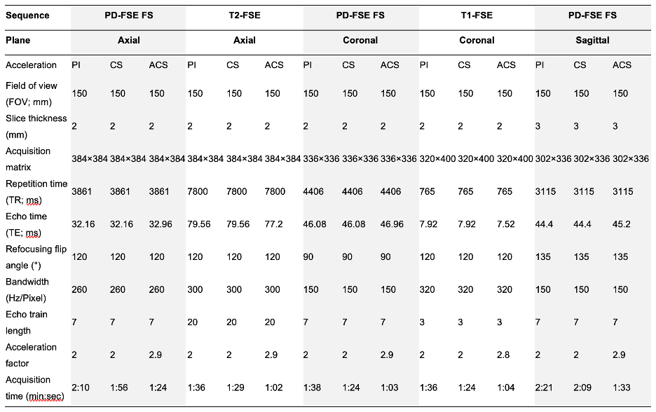
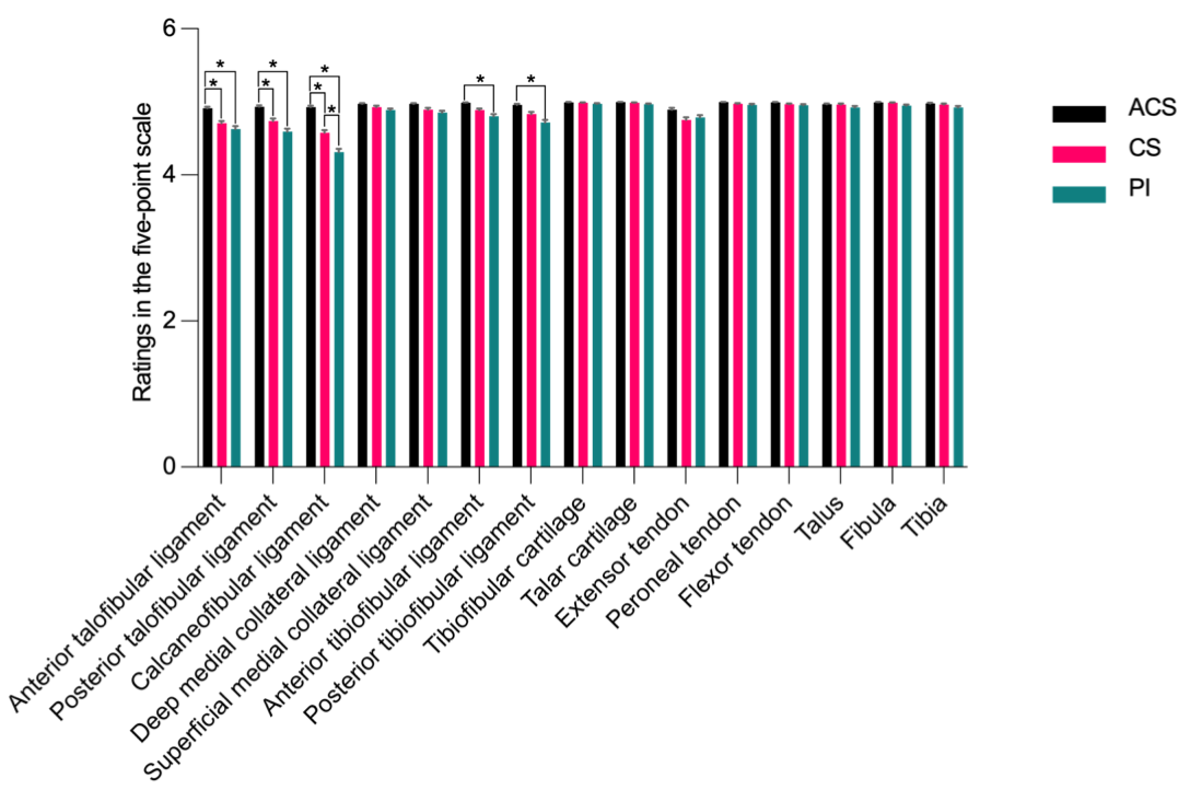
All subjects were examined with a clinical 3.0 T MR scanner (uMR 880, United Imaging Healthcare, Shanghai, China) with a dedicated 24-channel receive ankle coil. The MR protocol included a T2-weighted FSE sequence, a T1-weighted FSE sequence, and three proton density (PD) weighted FSE sequences with fat saturation (FS).This study consisted of two steps: 1) a pilot study on healthy volunteers to explore the optimal acceleration factors for ACS by rating the image quality of images acquired with ACS acceleration factors ranging from 2.3–3.8×; 2) 105 ankle-injured patients were scanned with ACS and conventional accelerated methods for clinical assessment. The images acquired with PI (acceleration factor of 2.0×) and CS (acceleration factor of 2.0×) were used as baselines. Quantitative analysis was performed by calculating the SNR and CNR of the structures within the ankle. The subjective image quality of each sequence was rated using a five-point Likert scale regarding the depiction of anatomic structures (5=best, 1=worst). The ligament injuries and osteochondral lesions were assessed for diagnostic agreement analysis.
Results
The acceleration factors of 2.8–2.9× for ACS were determined for routine ankle protocol in the pilot study. The total acquisition times of PD, T1, and T2 sequences were reduced by 24 – 36% by the ACS acceleration compared with conventional CS and PI (Fig. 1).The SNR and CNR measurements for ankle structures were shown in Fig. 2. Similar image quality was noticed in general across three acceleration methods (Fig. 3). Images acquired with ACS showed significantly higher ratings for structures of the anterior and posterior talofibular ligaments and calcaneofibular ligament than CS and PI (p < 0.05) (Fig. 4). The evaluation of pathologies of the ankle analyzed resulted in an agreement of k = 1.00 across sequences acquired with ACS, CS, and PI (Fig. 5).
Discussion
Recent developments in machine learning techniques enable faster imaging operation in image space, incorporate measured coil sensitivities in the reconstruction, and were designed to generalize the concept of compressed sensing by learning the entire reconstruction procedure for MR data 3-5. Our results further demonstrate that the deep learning-based reconstruction method has superior potential to reduce MR examination time to conventional PI and CS in routine clinical practice.The ACS sequences yielded almost the same image quality as the conventional PI and CS sequences. Moreover, slightly higher average subjective image quality ratings for ligaments were found in ACS images than in CS and PI images. The quantitative analysis also indicated that the ACS sequences could yield better SNR and CNR for most tissues than the CS and PI sequences. However, the differences in SNRs and CNRs (< 3 a.u.) and image quality ratings (< 5%) were minor, resulting in a high agreement in the assessment of pathologies of the ankles analyzed between acceleration methods.
Common lesions within the anterior talofibular ligament, calcaneofibular ligament, and cartilage were evaluated separately in this study, leading to more specific results. Our results provide reliable and comprehensive proof of the potential clinical application of this method.
Conclusions
This study presented a structured approach to reduce scan time for MR imaging protocol of the ankle. We concluded that using ACS acceleration factors of 2.8 – 2.9× to acquire 2D FSE MR imaging sequences of the ankle is feasible with a reduction in scan time of 24 - 36% without a significant decrease in diagnostic performance. The ACS acceleration is a reliable alternative for conventional PI and CS and could potentially improve the productivity of MRI systems and patient comfort in musculoskeletal radiological practices.Acknowledgements
No acknowledgement found.References
1. Sheng R-f, Zheng L-y, Jin K-p, et al. Single-breath-hold T2WI liver MRI with deep learning-based reconstruction: a clinical feasibility study in comparison to conventional multi-breath-hold T2WI liver MRI. Magnetic Resonance Imaging. 2021;81:75-81.
2. Zhao Y, Peng C, Wang S, Liang X, Meng X. The feasibility investigation of AI-assisted compressed sensing in kidney MR imaging: an ultra-fast T2WI imaging technology. BMC Medical Imaging. 2022;22(1):1-6.
3. Liu F, Samsonov A, Chen L, Kijowski R, Feng L. SANTIS: Sampling-Augmented Neural neTwork with Incoherent Structure for MR image reconstruction. Magn Reson Med. Nov 2019;82(5):1890-1904. doi:10.1002/mrm.27827
4. Aggarwal HK, Mani MP, Jacob M. MoDL: Model-Based Deep Learning Architecture for Inverse Problems. IEEE Trans Med Imaging. Feb 2019;38(2):394-405. doi:10.1109/TMI.2018.2865356
5. Hammernik K, Klatzer T, Kobler E, et al. Learning a variational network for reconstruction of accelerated MRI data. Magn Reson Med. Jun 2018;79(6):3055-3071. doi:10.1002/mrm.26977Figures
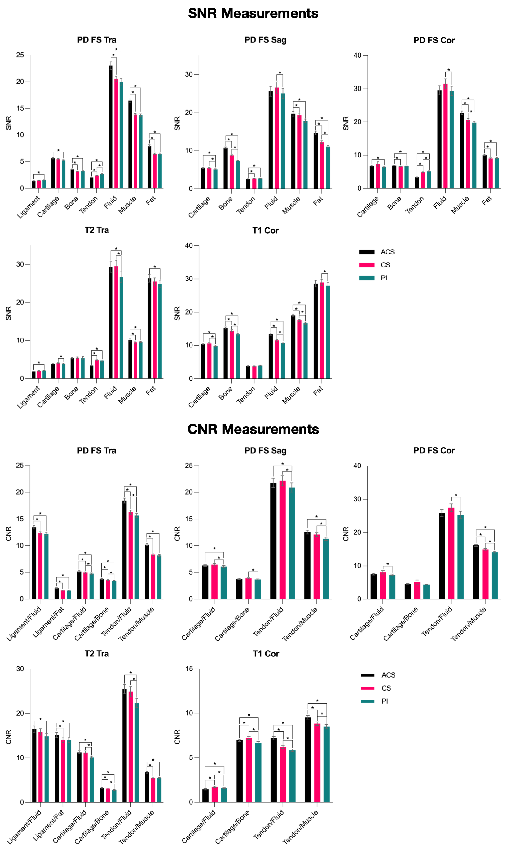
SNR and CNR measurements of different anatomical structures derived from sequences with accelerations of ACS, CS, and PI. Statistically different pairs (p < 0.05) were marked with the star signs.
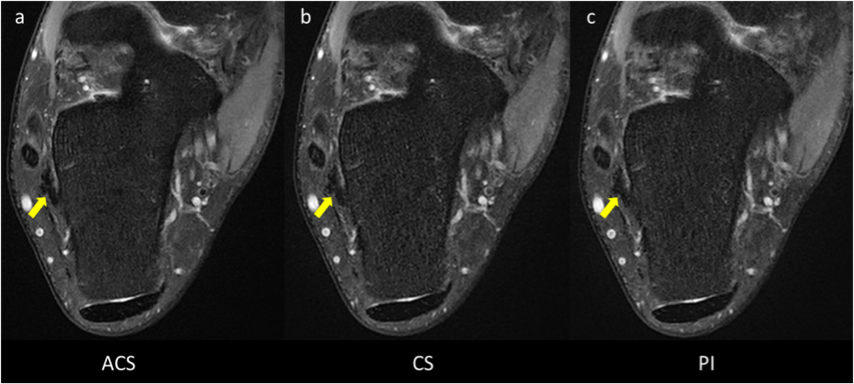
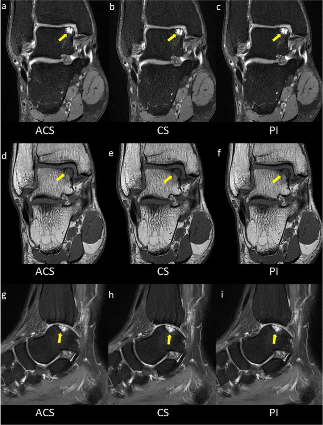
Coronal PD FS and T1-weighted images acquired using (a, d) ACS, (b, e) CS, and (c, f) PI showing the ankle of a 31-year-old male patient with an osteochondral lesion (arrows). Sagittal PD FS images acquired with (g) ACS, (h) CS, and (i) PI showing bone marrow changes adjacent to the osteochondral lesion (arrows).