2744
Efficient 3D cone trajectory design for improved combined angiographic, structural and perfusion imaging using arterial spin labelling1Wellcome Centre for Integrative Neuroimaging, FMRIB, Nuffield Department of Clinical Neurosciences, University of Oxford, Oxford, United Kingdom, 2Physical Sciences, Sunnybrook Research Institute, Toronto, ON, Canada, 3Department of Medical Biophysics, University of Toronto, Toronto, ON, Canada
Synopsis
Keywords: Arterial spin labelling, Arterial spin labelling
4D combined angiographic, structural and perfusion radial imaging using arterial spin labelling (CASPRIA) provides a tool to simultaneously acquire information about brain structure and blood flow. However, the radial trajectory used limits the resolution and SNR, especially when the data is highly undersampled. In this work, an optimised 3D cone trajectory is presented with 3D golden means rotation to improve sampling at the k-space periphery whilst maintaining flexibility in spatiotemporal resolution. A locally low rank reconstruction was used to leverage spatiotemporal correlations and improve image quality. In vivo results showed considerable improvements in resolution and SNR over the radial approach.Introduction
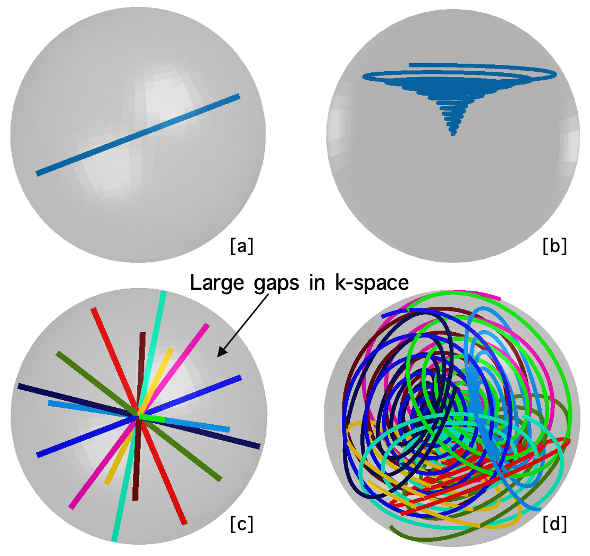
4D combined angiographic, structural and perfusion radial imaging using arterial spin labelling (CASPRIA)1 is a non-contrast scheme to simultaneously map the dynamics of blood flow through the arteries and at the level of the brain tissue, as well as T1-weighted structural signals, which could allow more information to be obtained in a short time for busy clinical settings. However, the radial trajectory used in CASPRIA leaves gaps that violate the Nyquist theorem in peripheral k-space which, in turn, limits the SNR and spatial resolution. Increasing the number of spokes to reduce these gaps without increasing the scan time requires the repetition time of the excitation pulses to be short, which leads to rapid attenuation of the arterial spin labelling (ASL) signal. Therefore, a flexible curved trajectory that could more evenly sample k-space with fewer excitations is desired. Numerous 3D curves like 3D Cones2 and Seiffert Spiral3 have been suggested, yet we would like a more flexible design to allow variable spatial and temporal resolution images to be reconstructed.In this work, we describe how we optimise a curved cone trajectory for the CASPRIA framework and compare it to the original radial trajectory in healthy volunteers.Methods
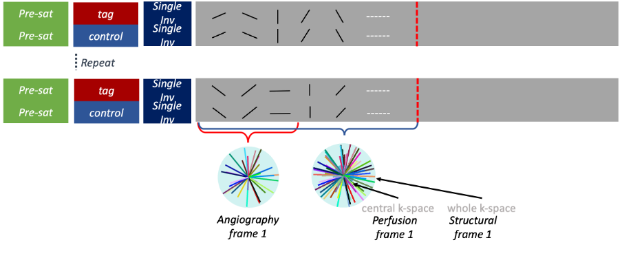
Multiple 3D cones can be stacked along a single axis 2 but this does not allow flexibility in temporal resolution during image reconstruction. Instead, we rotate the cone’s central axis around the k-space center using the 3D golden angle4 giving approximately uniform coverage of k-space for any number of cones, and rotate each cone about its central axis by a random angle to reduce overlap.The final sampling pattern is then largely decided by the shape of the base cone. Spending more time at the central region of k-space could improve robustness against motion and signal variation whereas more samples in peripheral k-space is helpful to improve spatial resolution. Incoherent sampling is also crucial for reducing structured artefacts, so radial distance from one turn of the cone to the next was varied along the trajectory. The shape of our cone trajectory was defined in a parametric form first and the exact gradient was then calculated7 to traverse the curve. Thanks to its flexibility, we could heuristically modify the characteristics of the final cone trajectory to adapt to different purposes. Based on the preliminary numerical simulations and phantom scans, our final cone trajectory is shown in Fig.1.a schematic sequence diagram is shown in Fig.2. Each preparation period (pseudo-continuous ASL labelling and background suppression pulses) is followed by the acquisition of many radial spokes or cones across a 2s readout period. We adopted the Song’s method5,6 to arrange the order of the golden means sampling across multiple ASL preparation periods, so that the temporal resolution could be arbitrarily selected retrospectively. Lower spatial resolution images (for perfusion imaging) can be reconstructed using the more densely sampled centre of k-space. Structural images can be reconstructed using the mean label/control data, rather than the difference signal.Data were acquired on a 3T Siemens Prisma scanner using 32-channel head coil in 1 healthy volunteer. Preparation pulses were repeated for 48 times (corresponding to 48 rows of tag/control in fig.2), followed by 6 frames with 24 readouts in each of them. Since the TR of each readout is 14.7 ms, the total time after each tag/control is 24×6×14.7=2016ms. Angiography images were reconstructed with 1.13mm spatial resolution, and 14.7×12=176.2ms temporal resolution. Perfusion images were reconstructed with 3.4mm spatial resolution and 14.7×24=352.8ms temporal resolution. The spatial resolution for the structural images were also 1.13mm, and the temporal resolution was 352.8ms.As the signal varies smoothly and the sampling is complementary across temporal frames, we could utilize spatiotemporal correlations during reconstruction for further improvement in image quality. Therefore, a locally low rank constraint was enforced in our iterative reconstruction.Results and discussion
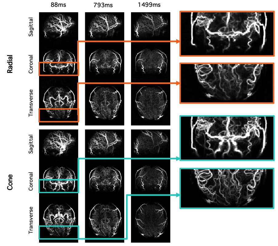
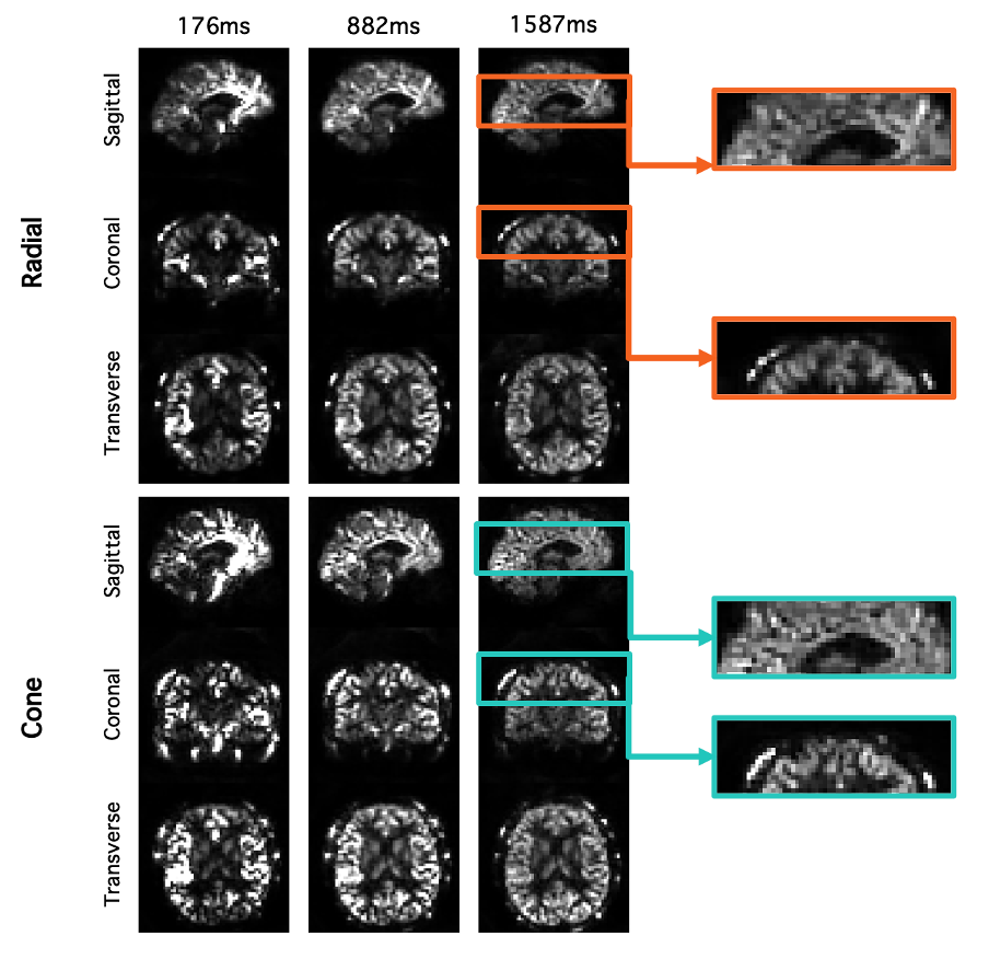
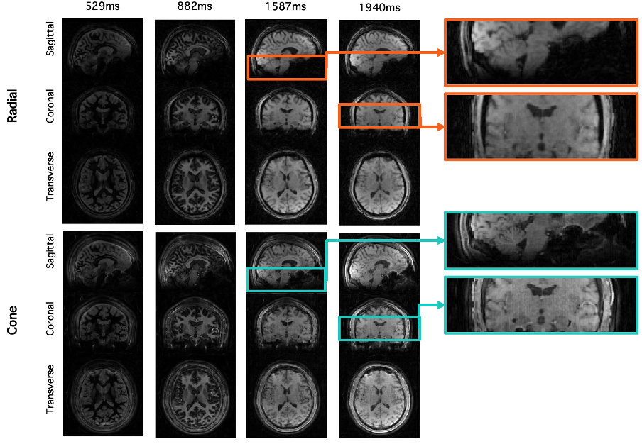
Fig. 3 compares angiographic images acquired in the same subject using cone and radial trajectories. The cone trajectory allowed depiction of the proximal arteries much better than the radial trajectory, probably due to reduced flow or B0-based dephasing at the much shorter echo time of the cone trajectory. The small vessels are also better visualized by the cone trajectory thanks to better sampling in the k-space periphery.For perfusion images reconstructed from the same raw k-space data (Fig. 4), the cone trajectory also provides better spatial resolution than the radial trajectory with an additional increase in SNR. The small structures are now clearly visible in the cone case.Using the mean rather than difference between label and control data, structural images were also reconstructed (Fig. 5). Images with varying T1-weighted contrast are produced at different times after the inversion pulse, but as above, the cone trajectory provides better resolution and SNR.In conclusion, a 3D cone trajectory can be integrated into the 4D CASPRIA framework, improving spatial resolution and SNR and allowing high quality time-resolved angiograms, perfusion images and T1-weighted structural images to be obtained from a single 6-minute scan. A quantitative comparison in a larger number of healthy controls to confirm these results will be performed in future work.Acknowledgements
No acknowledgement found.References
1. Thomas W. Okell. 4D Combined Angiography and Perfusion using Radial Imaging and Arterial Spin Labeling. In: Singapore; 2016. p. 1001.
2. Gurney PT, Hargreaves BA, Nishimura DG. Design and analysis of a practical 3D cones trajectory. Magn. Reson. Med. 2006;55:575–582 doi: 10.1002/mrm.20796.
3. Speidel T, Metze P, Rasche V. Efficient 3D Low-Discrepancy ${k}$ -Space Sampling Using Highly Adaptable Seiffert Spirals. IEEE Trans. Med. Imaging 2019;38:1833–1840 doi: 10.1109/TMI.2018.2888695.
4. Chan RW, Ramsay EA, Cunningham CH, Plewes DB. Temporal stability of adaptive 3D radial MRI using multidimensional golden means. Magn. Reson. Med. 2009;61:354–363 doi: 10.1002/mrm.21837.
5. Song HK, Yan L, Smith RX, et al. Noncontrast enhanced four-dimensional dynamic MRA with golden angle radial acquisition and k-space weighted image contrast (KWIC) reconstruction: Noncontrast Enhanced Radial 4D dMRA. Magn. Reson. Med. 2014;72:1541–1551 doi: 10.1002/mrm.25057.
6. Plas MCE, Schmid S, Versluis MJ, Okell TW, Osch MJP. Time-encoded golden angle radial arterial spin labeling: Simultaneous acquisition of angiography and perfusion data. NMR in Biomedicine 2021;34 doi: 10.1002/nbm.4519.
7. Lustig M, Kim S-J, Pauly JM. A fast method for designing time-optimal gradient waveforms for arbitrary k-space trajectories. IEEE Trans. Med. Imaging 2008;27:866–873 doi: 10.1109/TMI.2008.922699.
Figures