2740
Accelerated DWI with deep learning reconstruction in 3T breast MRI: initial clinical experiences and image quality1Departement of Diagnostic and Interventional Radiology, University Medical Center Freiburg, Faculty of Medicine, University of Freiburg, Freiburg im Breisgau, Germany, 2MR Application Predevelopment, Siemens Healthcare GmbH, Erlangen, Germany, 3EMEA Scientific Partnerships, Siemens Healthcare GmbH, Erlangen, Germany, 4MR Physics, University Hospital Freiburg, Freiburg im Breisgau, Germany
Synopsis
Keywords: Breast, Diffusion/other diffusion imaging techniques
This prospective study evaluated image quality features of a novel accelerated DWI with deep learning image reconstruction in 3T breast MRI in a clinical setting in direct comparison to conventional DWI. Deep learning DWI (DL-DWI) shows a drastically shortened acquisition time of 46% compared to standard DWI, while maintaining a high image quality. Even though some image quality features were rated superior in standard DWI, image quality remained good for DL-DWI and lesion conspicuity scores were rated superior for DL-DWI compared to conventional breast DWI. Therefore, DL-DWI seems a feasible technique for accelerated breast DWI.Introduction
Deep learning (DL) reconstruction of diffusion-weighted imaging (DWI) is a novel method that allows for a substantial faster acquisition time than conventional DWI. In this study we evaluate image quality of DL-DWI in direct comparison to standard DWI. The relevance of DWI in breast imaging is given due to the advantages to distinguish between different lesion types (1, 2) and possible advantages for early prediction of neoadjuvant therapy success (3).Material and Methods
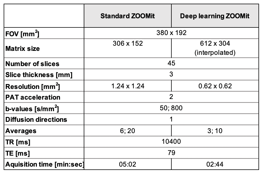
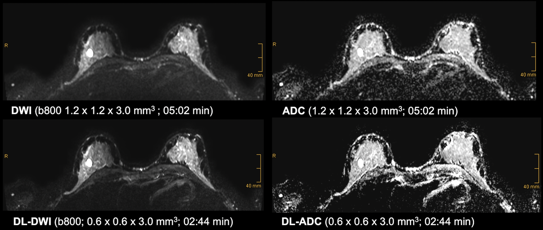
Prospective, pseudo-anonymized randomized study with inclusion of 35 patients with a mean age of 54 years (range: 29 – 85) who underwent 3T breast MRI (MAGNETOM Vida, Siemens Healthcare, Erlangen, Germany) using an 18-channel breast coil. The study was approved by the institutional review board (EK22-1185) and is listed in the German Clinical Trials Register (DRKS-ID: DRKS0002955). Informed written consent was given by all participants. A standard diagnostic contrast-enhanced breast MRI protocol with single-shot echo-planar DWI combined with reduced field-of-view excitation (ZOOMitPRO; Siemens Healthcare, Erlangen, Germany) was scanned in accordance with EUSOBI guidelines (4). Two b-values (50 and 800 s/mm2) with 6 and 20 averages, respectively, were acquired in 5:02 min using a spatial resolution of 1.2 x 1.2 x 3.0 mm3. Additionally, an accelerated DWI sequence was acquired in 2:44 min with a reduced number of averages (3 and 10, respectively) and reconstructed with a research application DL technique. For this, following the idea of a variational network (5), a k-space to image reconstruction has been trained in a supervised manner on about 500.000 DWI images which were collected in volunteer scans, covering different body regions and applications. Additionally, image interpolation based on a research application super resolution approach, again trained on MR images of volunteers, was used to obtain a spatial resolution of 0.6 x 0.6 x 3.0 mm3. Detailed acquisition parameters are shown in Table 1.Image quality was evaluated ROI-based (signal intensity) for CV (coefficient of variation) and SNR (signal-to-noise ratio). Qualitative analysis was performed by two radiologists with 5 and 11 years of experience in breast imaging, who individually rated overall image quality features, artifacts, and lesion conspicuity separately using a Likert-Scale ranging von 1 = non-diagnostic to 5 = excellent. Scoring “good” and “excellent” were considered high diagnostic image quality. Both DWI and DL-DWI were presented randomized in intraindividual comparison. To avoid bias through spatial resolution differences, DL-DWI was down sampled to 1.2 x 1.2 x 3.0 mm3 spatial resolution for reading purposes. A consensus reading was performed for all discrepant scorings few days after the first reading session. Statistical analysis was performed using SPSS 28.0 (IBM SPSS Statistics, Armonk, NY, USA). Normality was tested with Shapiro-Wilk test. Paired-samples Wilcoxon-Signed-Ranks test was applied to test for differences. In case of significance, a Wilcoxon-Signed-Ranks test for non-normally distributed data was performed. Two-sided p-values of < 0.05 were considered statistically significant. To determine interrater agreement, interrater reliability analysis was performed using a weighted Cohen’s Kappa (k) with a 95 % confidence interval.
Results
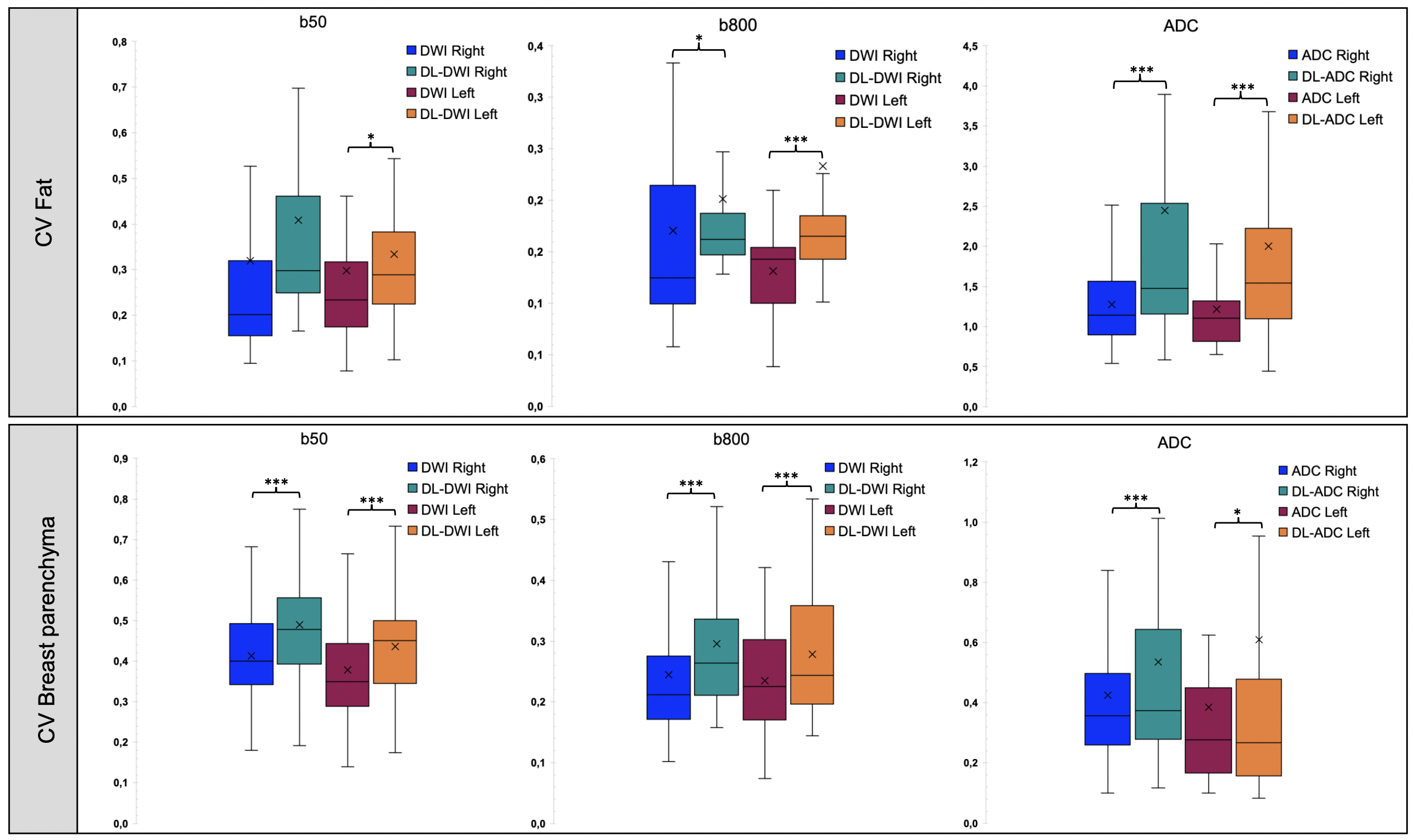
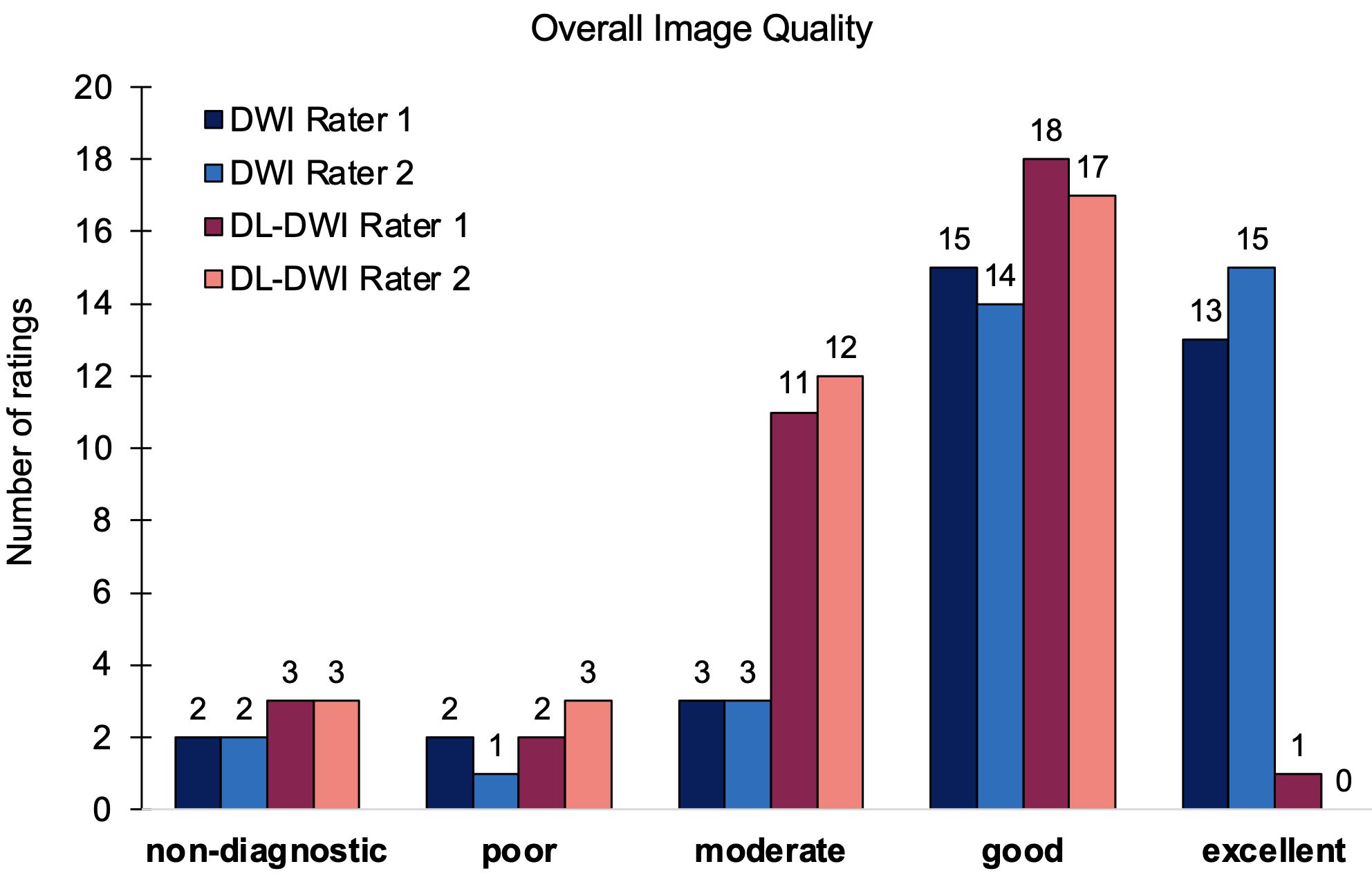
Acquisition time was 05:02 min for standard DWI and 02:44 min for DL-DWI. CV was higher for DL-DWI compared to DWI (see figure 1). SNR was higher for standard DWI. Interrater reliability was almost perfect (k =0.88 to 1.0). Both raters were able to detect DL-DWI with high certainty in 98 % of all cases. When both sequences were compared intra-individually, even though standard DWI was rated towards the higher image quality score, both sequences revealed a high image quality (see figure 2). The highest lesion conspicuity score (5 = excellent) was observed more often for DL-DWI (27,5 % of all ratings for DL-DWI vs. 20 % for DWI, figure 3). Artifacts were rated one score higher towards the stronger artifact level (p < 0.001) for DL-DWI and were on average minimal to moderate in DWI and moderate to very strong in DL-DWI (p < 0.001).Discussion
This study investigated image quality features of the novel DL-DWI in a clinical setting and compared it to standard DWI. DL-DWI shows a drastically shortened acquisition time of 46 % compared to conventional DWI in 3T breast MRI, which has a high relevance for clinical implementation in diagnostic breast MRI protocols. Even though some differences in image quality parameters were noted between standard DWI and DL-DWI and conventional DWI was rated towards the higher image quality scores, a high quality was maintained for DL-DWI. Lesion conspicuity scores of DL-DWI were superior compared to standard DWI. In summary, the preliminary results underline the potential of improving acquisition time and efficiency by application of DL based image reconstruction. Further analysis on diagnostic accuracy and interchangeability should be addressed in the future.Conclusion
DL-DWI allows for a good image quality and a high lesion conspicuity. Therefore, DL-DWI seems a feasible technique for accelerated breast DWI.Acknowledgements
Thanks to all patients, who participated in the study.References
1. Iima M, Kataoka M, Kanao S, Onishi N, Kawai M, Ohashi A, Sakaguchi R, Toi M, Togashi K. Intravoxel incoherent motion and quantitative non-Gaussian diffusion MR imaging: evaluation of the diagnostic and prognostic value of several markers of malignant and benign breast lesions. Radiology 2018;287(2):432-441.
2. Chen X, Li W-l, Zhang Y-l, Wu Q, Guo Y-m, Bai Z-l. Meta-analysis of quantitative diffusion-weighted MR imaging in the differential diagnosis of breast lesions. BMC cancer 2010;10(1):1-11.
3. Partridge SC, Zhang Z, Newitt DC, Gibbs JE, Chenevert TL, Rosen MA, Bolan PJ, Marques HS, Romanoff J, Cimino L. Diffusion-weighted MRI findings predict pathologic response in neoadjuvant treatment of breast cancer: the ACRIN 6698 multicenter trial. Radiology 2018;289(3):618-627.
4. Baltzer P, Mann RM, Iima M, Sigmund EE, Clauser P, Gilbert FJ, Martincich L, Partridge SC, Patterson A, Pinker K, Thibault F, Camps-Herrero J, Le Bihan D, group EiBD-WIw. Diffusion-weighted imaging of the breast-a consensus and mission statement from the EUSOBI International Breast Diffusion-Weighted Imaging working group. Eur Radiol 2020;30(3):1436-1450. doi: 10.1007/s00330-019-06510-3
5. Hammernik K, Klatzer T, Kobler E, Recht MP, Sodickson DK, Pock T, Knoll F. Learning a variational network for reconstruction of accelerated MRI data. Magn Reson Med 2018;79(6):3055-3071. doi: 10.1002/mrm.26977
Figures