2725
Contrasts between DWI and DCE curve in diagnosing malignancies of breast non-mass enhancement lesions based on morphology1Tongji Hospital, Wuhan, China
Synopsis
Keywords: Breast, Cancer
The diagnosis of NMEs is a challenge for most radiologists based only on morphological information. The value of DWI or DCE curves for discriminating malignant lesions from benign lesions was controversial. We estabished and compared the ADC model (ADC+morphology) and the TIC (TIC+morphology) for detecting malignancy with a relatively large sample size of NME lesions. We found that the TIC model was superior than the ADC model for differentiating between benign and malignant NME lesions. A whole DCE-MRI scan for NMEs is recommended without the need to acquire additional DWI data.Introduction
Non-mass enhancement (NME) is defined as an enhanced lesion without space-occupying effect based on contrast-enhanced magnetic resonance imaging (CE-MRI) 1. The diagnosis of NMEs is a challenge for most radiologists based only on morphological information 2-4. The value of DWI or DCE curves for discriminating malignant lesions from benign lesions was controversial 5-10. It is not clear which of these approaches is more accurate for decreasing time consumption. The aim of this study is to assess and compare the role of DWI and TIC for detecting malignancy with a relatively large sample size of NME lesions.Methods
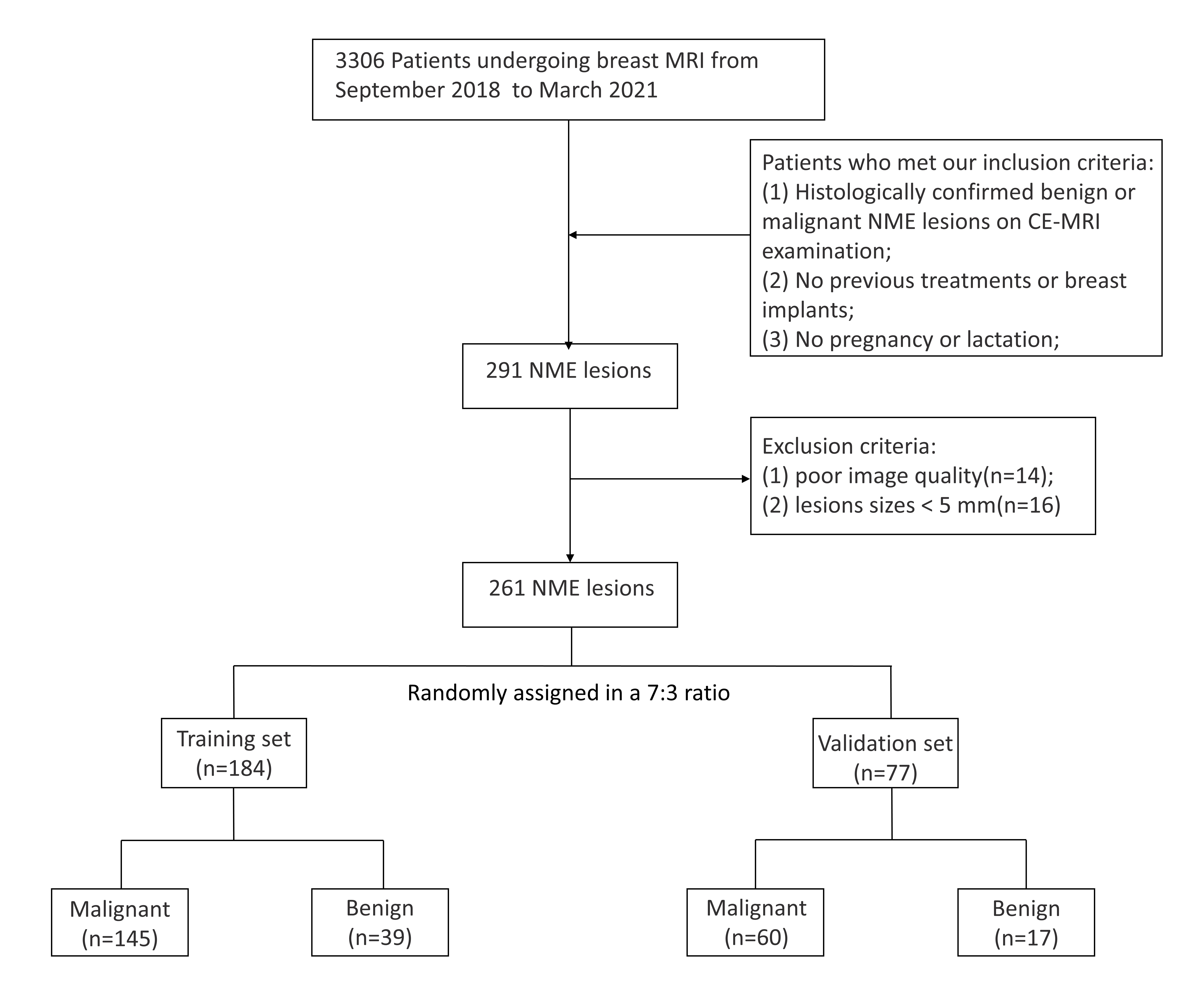
In this retrospective study, 261 NME lesions (184 for training and the following 77 for validation) on DCE-MRI with pathological results were enrolled. The apparent diffusion coefficient (ADC) model (ADC + morphology) and the TIC model (TIC + morphology) were constructed with binary logistic regression for NME lesions in the training set. The sensitivities, specificities, and area under the curve (AUC) were compared between the two models in the training cohort and the validation cohort.Results
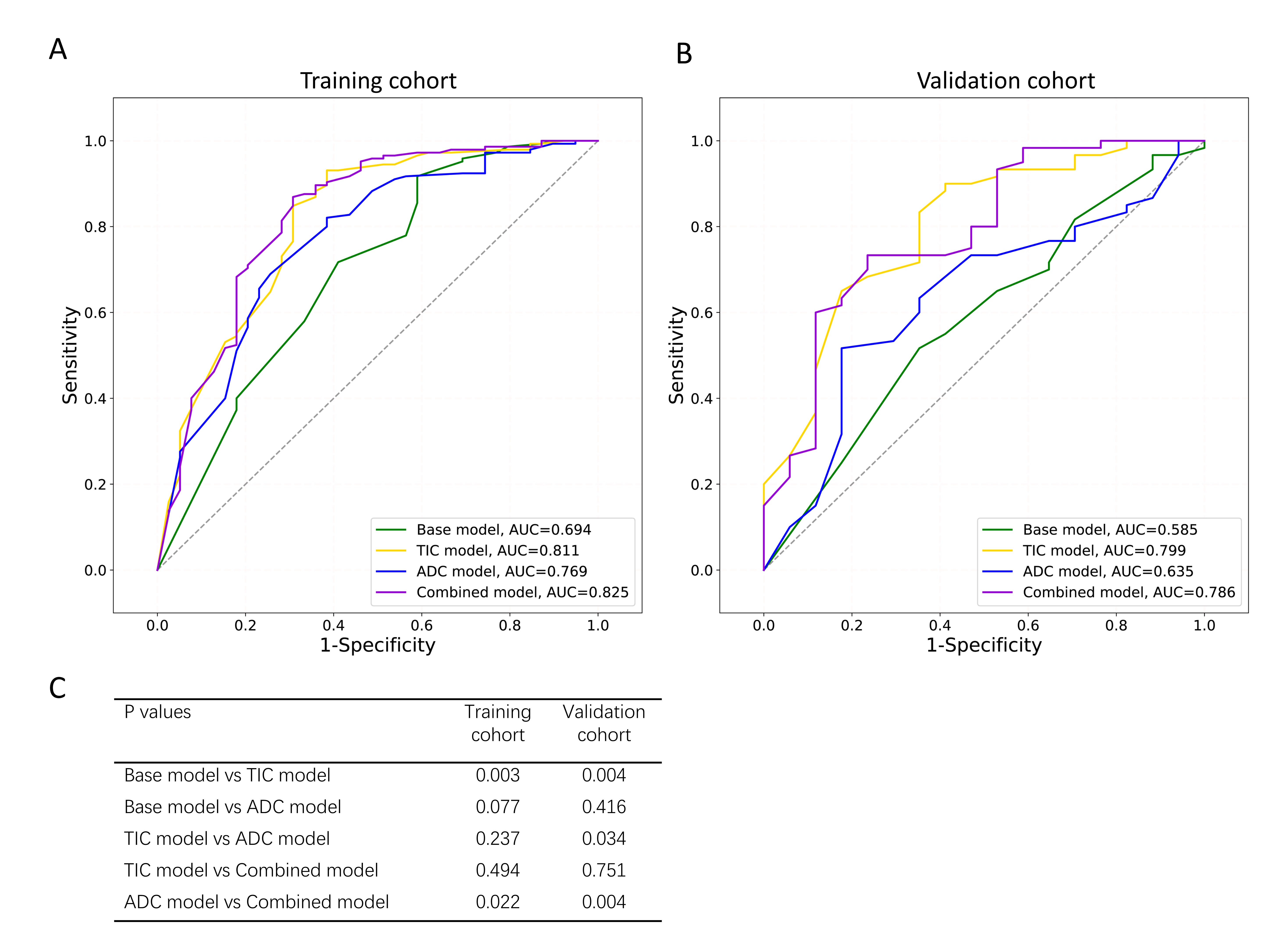
In the training cohort, the sensitivities, specificities, and AUCs of the TIC/ADC model was 0.924/0.814, 0.615/0.615, and 0.811(95%,0.727,0.894)/0.769(95%,0.681,0.856). The AUC of the TIC-ADC combined model was significantly higher than ADC model alone (P=0.022), while comparable with the TIC model (P=0.494). In the validation cohort, the AUCs of TIC/ADC model were 0.799/0.635 (P=0.034).Conclusion
Based on the morphologic analyses, the TIC model was validated superior than the ADC model for differentiating between benign and malignant NME lesions. A whole DCE-MRI scan for NMEs is recommended without the need to acquire additional DWI data.Acknowledgements
All authors have approved the abstract for submission and without any potential competing interestsReferences
1. ACR BI-RADS® ATLAS — BREAST MRI. https://www.acr.org/-/media/ACR/Files/RADS/BI-RADS/MRI-Reporting.pdf. 2013.
2. Baltzer PA, Benndorf M, Dietzel M, et al.: False-positive findings at contrast-enhanced breast MRI: a BI-RADS descriptor study. AJR Am J Roentgenol 2010, 194(6):1658-1663.
3. Torous VF, Resteghini NA, Phillips J, et al.: Histopathologic Correlates of Nonmass Enhancement Detected by Breast Magnetic Resonance Imaging. Arch Pathol Lab Med 2021, 145(10):1264-1269.
4. Lunkiewicz M, Forte S, Freiwald B, et al.: Interobserver variability and likelihood of malignancy for fifth edition BI-RADS MRI descriptors in non-mass breast lesions. European Radiology 2019, 30(1):77-86.
5. Clauser P, Krug B, Bickel H, et al.: Diffusion-weighted Imaging Allows for Downgrading MR BI-RADS 4 Lesions in Contrast-enhanced MRI of the Breast to Avoid Unnecessary Biopsy. Clin Cancer Res 2021, 27(7):1941-1948.
6. Sanaz A. Jansen, Xiaobing Fan, Gregory S. Karczmar, et al.: DCEMRI of breast lesions: Is kinetic analysis equally effective for both mass and nonmass-like enhancement? Medical Physics 2008, 35(7):3102-3109.
7. Avendano D, Marino MA, Leithner D, et al.: Limited role of DWI with apparent diffusion coefficient mapping in breast lesions presenting as non-mass enhancement on dynamic contrast-enhanced MRI. Breast Cancer Res 2019, 21(1):136.
8. Yabuuchi H, Matsuo Y, Kamitani T, et al.: Non-mass-like enhancement on contrast-enhanced breast MR imaging: lesion characterization using combination of dynamic contrast-enhanced and diffusion-weighted MR images. Eur J Radiol 2010, 75(1):e126-132.
9. Yang X, Dong M, Li S, et al.: Diffusion-weighted imaging or dynamic contrast-enhanced curve: a retrospective analysis of contrast-enhanced magnetic resonance imaging-based differential diagnoses of benign and malignant breast lesions. Eur Radiol 2020, 30(9):4795-4805.
10. Mori N, Sheth D, Abe H: Nonmass Enhancement Breast Lesions: Diagnostic Performance of Kinetic Assessment on Ultrafast and Standard Dynamic Contrast-Enhanced MRI in Comparison With Morphologic Evaluation. AJR Am J Roentgenol 2020, 215(2):511-518.
Figures