2724
When BOLD Optimized ME-EPI Echo Combination Isn't BOLD Optimized
R. Allen Waggoner1, Chisato Suzuki1, Ken-ichi Ueno1, and Keiji Tanaka1
1RIKEN Center for Brain Science, Wako-shi, Saitama, Japan
1RIKEN Center for Brain Science, Wako-shi, Saitama, Japan
Synopsis
Keywords: Artifacts, fMRI, Multi-Echo, Multi-Shot
In regions suffering from magnetic susceptibility effects, the use of BOLD optimized echo combination for averaging Single-Shot/Multi-Echo EPI data across echos, leads to voxels with improves signal but reduced BOLD sensitivity. The use of Multi-shot/Multi-Echo EPI can suppress the susceptibility effects allowing BOLD optimized echo averaging that retains BOLD sensitivity.Introduction
Multi-Echo EPI1 is a technique which is growing in popularity for improving fMRI sampling efficiency2 and for use in advanced denoising methods3. The images from different echo times can be combined in a weighted average which accounts for variations in optimum TE for BOLD contrast across the brain2. These BOLD optimized weights are obtained using the following equation,$$w(T_2^*)=\frac{TE_{n}\cdot\exp(-TE_{n}/T_{2(fit)}^*)}{\sum_n TE_{n}\cdot\exp(-TE_{n}/T_{2(fit)}^*)}\:\:\:\:Eq. 1$$
Using Equation 1 for combing data from voxels suffering from susceptibility artifacts, will bias the average for that voxel to data from the shortest echo time, which is least corrupted by the susceptibility effects. The weight obtained by Equation 1 biases the weighted average to data close to the measured T2* for each voxel. But in areas of the brain that suffer from susceptibility artifacts the BOLD effect is not the dominate mechanism for T2* relaxation. So, for such voxels the weighted average is optimized for signal but not BOLD sensitivity. Multi-Shot EPI is an alternative approach for minimizing the impact of susceptibility gradients on EPI data. With Multi-Shot EPI only a subset of k-space is acquired following the RF excitation, so multiple excitations are needed to acquire all of k-space. The multi-shot acquisition increases the bandwidth in the phase-encode direction, minimizing susceptibility effects. Combining Multi-Shot and Multi-Echo will enable regaining of BOLD sensitivity in regions suffering from susceptibility artifacts. The use of Multi-Shot/Multi-Echo EPI will also permit acquisitions with higher spatial resolution than can be achieved with conventional Single-Shot/Multi-Echo EPI, which will further suppress the susceptibility artifacts. We present data here that illustrates that the susceptibility effects weights obtain from Equation 1 and that Multi-Shot/Multi-Echo EPI can, to some extent, restore typical BOLD like T2* behavior in these regions, particularly at higher resolution.
Methods
Experiments were performed on a 3T MAGNETOM Prisma (Siemens Healthcare, Erlangen, Germany), using a Siemens 64-channel head/neck array and a prototype Multi-shot/Multi-Echo EPI sequence. For comparison, Single-shot/Multi-echo EPI data was also acquired for each subject using a customized multi-echo EPI sequence (CMRR C2P R2016a, University of Minnesota). The details of each acquisition are specified in Figure 1. The subject was a 37 yr. old male. To illustrate the impact of susceptibility artifacts on BOLD optimized echo combination weighting, three points were chosen from the example slice displayed in Figure 1. Point 1 was chosen in the location of a mild susceptibility artifact. Point 2, was located in a more severe susceptibility artifact. For comparison, point 3 was chosen in a location that did not suffer from susceptibility effects. For each acquisition strategy, the weights for BOLD optimized echo combination were determined using Equation 1.Results & Discussion
Point 1 is location in a region where susceptibility related signal loss is only observed in the single-shot case. For the single-shot case, the weights are biased toward shortest TE. For both the low- and high-resolution multi-shot data sets, the weights are biased toward the longer TEs. In these cases, the weights are consistent with the weights found for point 3, where there is no susceptibility effect. Point 1 illustrates that BOLD contribution to T2* has not changed in regions experiencing susceptibility effects. For point 2, where the susceptibility effect is observed for all acquisition strategies, the optimization weights are all biased toward the short TE.Conclusions
The BOLD optimized weights shown in Table 1, show that in locations where BOLD is not the dominate T2* relaxation mechanism, the resulting echo combination will have low BOLD sensitivity, even though there is image signal at that location in the combined image. Thus, to obtain data that has both image signal and BOLD sensitivity throughout the brain, the combination of high resolution and Multi-Shot/Multi-Echo EPI is more effective than Single-Shot/Multi-Echo EPI.Acknowledgements
This work was partially funded by a grant from the Brain/MINDS project.References
- Posse S, Weise S, Gembris D, et al. Enhancement of BOLD-contrast sensitivity by single-shot multi-echo functional MR imaging. Magn Reson Med. 1999;42:87-97.
- Poser BA, Versluis MJ, Hoogduin JM, et al. BOLD contrast sensitivity enhancement and artifact reduction with multiecho EPI: parallel-acquired inhomogeneity-desensitized fMRI. Magn Reson Med. 2006;55:1227-1235.
- Kundu P, Inati SJ, Evans JW, et al. Differentiating BOLD and non-BOLD signals in fMRI time series using multi-echo EPI. NeuroImage 2012;60:1759-1770.
Figures
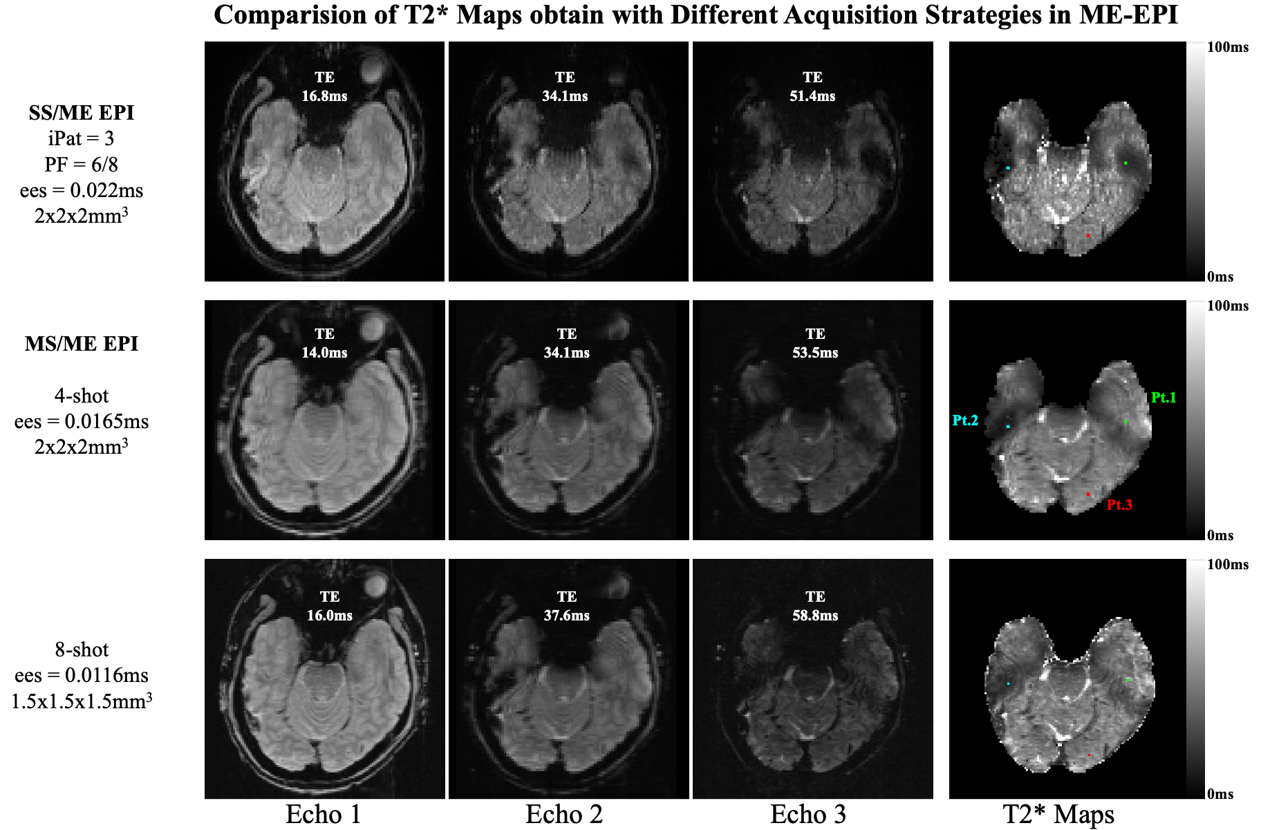
Figure 1. Multi-Echo EPI images from an example slice. The top row was acquired with a SS/ME EPI
sequence and a resolution of 2mm3.
The middle row was acquired with a 4-shot/ME EPI sequence and a
resolution of 2mm3. The
bottom row was acquired with an 8-shot/ME EPI sequence and a resolution of 1.5mm3. The right column shows the T2* maps
calculated for this slice from each acquisition. The green, cyan, and red dots, mark the locations
used for the weight calculations summarized in Table 1.
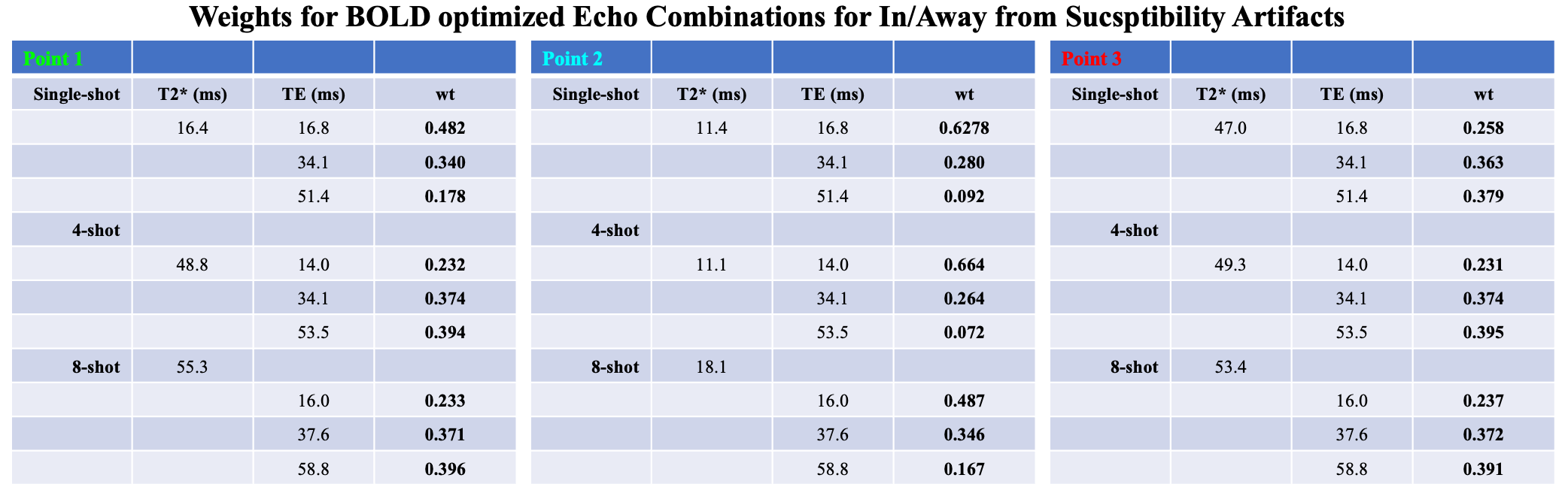
Table 1. For the 3 points highlighted in Figure 1, the
T2* values determined from the Multi-Echo EPI data. The right most column for each point shows
the weights determined using Equation 1, for a BOLD optimized echo combination.
DOI: https://doi.org/10.58530/2023/2724