2720
Where is each finger area in brain? Enhanced characterization of the somatosensory area using high-resolution EPIK at 3T1Medical Device Development Center, K-MEDI hub, Daegu, Korea, Republic of, 2Department of Rehabilitation Medicine, Yeungnam University, Daegu, Korea, Republic of, 3K-MEDI hub, Daegu, Korea, Republic of, 4Institute of Neuroscience and Medicine 4, INM-4, Forschungszentrum Juelich, Juelich, Germany, 5Institute of Neuroscience and Medicine 11, INM-11, Forschungszentrum Juelich, Juelich, Germany, 6JARA - BRAIN - Translational Medicine, Aachen, Germany, 7Department of Neurology, RWTH Aachen University, Aachen, Germany
Synopsis
Keywords: Data Acquisition, fMRI (task based)
The tactile stimulus of fingers in fMRI can effectively reveal the somatosensory functional region. However, this research requires a high-resolution fMRI technique, enabling a distinct delineation of functional areas for each finger. This work demonstrates the use of high-resolution EPIK (1.25 x 1.25 mm2) for enhanced characterization of somatosensory areas at 3T. EPIK provides improved spatial resolution than EPI (1.72 x 1.72 mm2) with increased brain coverage while keeping the same temporal resolution as EPI. The activation region from EPIK was shown to be more locally specific, thereby enabling a clearer recognition of functional areas for each finger.Introduction
The tactile stimulus of fingers in fMRI (functional MRI) can effectively reveal the functional areas for the somatosensory region. For this research, one attempted approach is to utilize an advanced stimulation paradigm to improve the detection sensitivity. Another methodological approach demanded in this investigation is to employ a high-resolution fMRI technique which enables the delineation of the functional area for each finger. In fMRI studies, EPI has been in widespread use due to its relatively high temporal resolution. However, EPI with high spatial resolution requires a greater number of phase encoding lines, leading to a significant increase in the minimum TE required.1 Thus, achieving the optimal TE for fMRI becomes increasingly difficult in high-resolution EPI. This issue can be mitigated by means of the multi-shot approach. However, the shot-to-shot instabilities arising from the multi-shot scheme can significantly magnify the physiological noise, consequently resulting in the significant reduction of detection sensitivity.2,3 In order to overcome this difficulty, an alternative method, EPI with Keyhole (EPIK), has been previously presented and its use for fMRI has been verified in a number of studies.3-6 The method was shown to provide a higher temporal resolution than single-shot EPI, while maintaining comparable performance in the detection of functional signals as EPI. This work aims at configuring 1) a high-resolution EPIK protocol dedicated for finger somatosensory fMRI and 2) examining a putative benefit of the superior resolution of EPIK in direct comparison to EPI at 3T.Methods
The tactile stimulus based on the block design was utilized as shown in Fig. 1. The blocks of rest and stimuli were alternated and each block time was 20 seconds. The order of stimulus was from thumb to little finger, successively. The group with these five fingers and corresponding rest blocks was repeated three times. Then, the last rest block was added. The total task time was 10 minutes and 20 seconds.The EPI and EPIK sequences were optimized to offer the highest possible resolution under a typical fMRI condition at 3T: TR/TE = 2000/28 msec. The achieved imaging parameters are shown in Table. 1. The nominal spatial resolution provided by EPI and EPIK was 1.72 × 1.72 mm2 and 1.25 × 1.25 mm2, respectively. The much smaller number of slices in EPI was due to the fact that the multiband option was not available in the sequence. T1 image was acquired using 3D MPRAGE: voxel size = 1.00 × 1.00 × 1.00 mm3.
Data sets from a healthy volunteer screened with a standard safety procedure were acquired using the EPI and EPIK protocols. fMRI experiments were performed at a Siemens MAGNETOM Skyra 3T scanner with a 64-ch head/neck coil. The preprocessing and of fMRI data involved realignment, co-registration, segmentation, normalization and smoothing in SPM12 and the activation region for the stimuli at each finger was obtained with an uncorrected p-value of 0.001 and an extended threshold of five voxels. The activated voxels are depicted on the MPRAGE scan individually normalized for each EPI and EPIK case.
Results
Figure 2 shows a reconstructed image from the EPI and EPIK scans at a nearly identical slice location. Although depicting lower SNR than the EPI scan, the EPIK scan shows a clearer delineation of anatomical structures around the boundaries between gray and white matters); the voxel volume of EPIK (1.95 mm3) is only nearly one-fifth of that of EPI (10.35 mm3).The activation regions are shown in Fig. 3. It is observed that the activated voxels from EPIK are more locally distributed around the cortical regions, whereas those from EPI are not as locally specific as EPIK. The results of EPIK further shows that the functional areas for each finger are more distinctly presented.
Conclusions and Discussion
This work demonstrates the use of high-resolution EPIK for enhanced characterization of somatosensory areas at 3T. As shown in this work, the EPIK method provides improved spatial resolution and also covers the whole brain while keeping the same temporal resolution as EPI. In addition, the activation region was shown to be more locally distributed around the cortical region when compared to EPI, thereby enabling a clearer recognition of functional areas for each finger. As a future work, a study with more number of subjects is planned, in which more reliable and robust detection of functional areas for each finger is expected by applying the 2nd level analysis to the group subject data.Acknowledgements
This research was supported by grants of the National Research Foundation of Korea (NRF) funded by the Ministry of Science & ICT (NRF-2020R1C1C1012230) and the National Information Society Agency (NIA) funded by the Ministry of Science & ICT (3-2. Establishment of child-adolescent psychological assessment and brain imaging data).References
[1] Yun SD, Shah NJ. Analysis of EPI phase correction with low flip-angle excitation to reduce the required minimum TE: Application to whole-brain, submillimeter-resolution fMRI at 3 T. Magn Reson Med. 2020 Sep;84(3):1416-1429. doi: 10.1002/mrm.28218. Epub 2020 Feb 21. PMID: 32086847.
[2] Lutti A, Thomas DL, Hutton C, Weiskopf N. High-resolution functional MRI at 3 T: 3D/2D echo-planar imaging with optimized physiological noise correction. Magn Reson Med. 2013;69(6):1657–1664. pmid:22821858
[3] Yun SD, Shah NJ. Whole-brain high in-plane resolution fMRI using accelerated EPIK for enhanced characterisation of functional areas at 3T. PLoS One. 2017 Sep 25;12(9):e0184759. doi: 10.1371/journal.pone.0184759. PMID: 28945780; PMCID: PMC5612468.
[4] Yun SD, Reske M, Vahedipour K, Warbrick T, Shah NJ. Parallel imaging acceleration of EPIK for reduced image distortions in fMRI. Neuroimage. 2013 Jun;73:135-43. doi: 10.1016/j.neuroimage.2013.01.070. Epub 2013 Feb 9. PMID: 23403182.
[5] Yun SD, Weidner R, Weiss PH, Shah NJ. Evaluating the Utility of EPIK in a Finger Tapping fMRI Experiment using BOLD Detection and Effective Connectivity. Sci Rep. 2019 Jul 29;9(1):10978. doi: 10.1038/s41598-019-47341-y. PMID: 31358817; PMCID: PMC6662889.
[6] Yun SD, Pais-Roldán P, Palomero-Gallagher N, Shah NJ. Mapping of whole-cerebrum resting-state networks using ultra-high resolution acquisition protocols. Hum Brain Mapp. 2022 Aug 1;43(11):3386-3403. doi: 10.1002/hbm.25855. Epub 2022 Apr 6. PMID: 35384130; PMCID: PMC9248311.
Figures
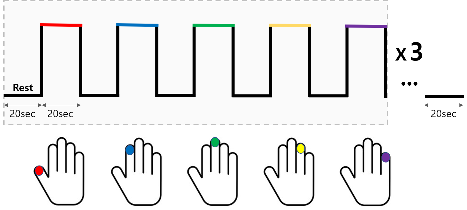
Figure 1. Tactile stimulus based on block design
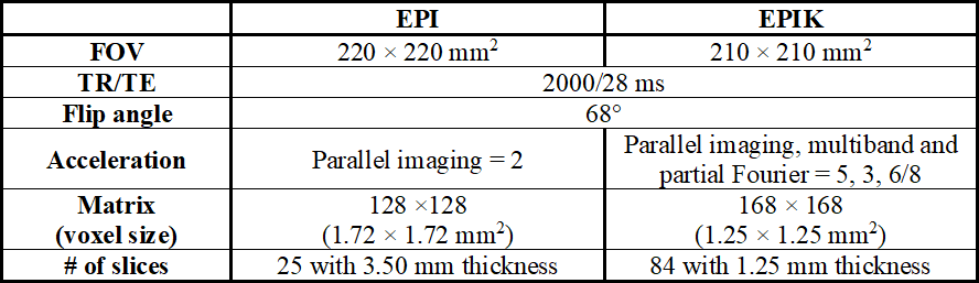
Table 1. Imaging parameters of EPI and EPIK
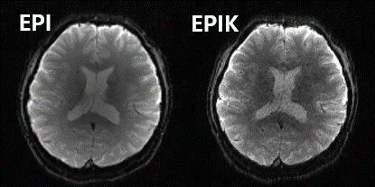
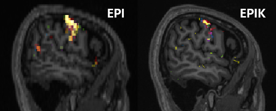
Figure 3. Activation regions from EPI (Left) and EPIK (Right), obtained with an uncorrected p-value of 0.001 and an extended threshold of five voxels