2717
BISEPI high-resolution fMRI at 7T with NORDIC-PCA
Guoxiang Liu1,2, Takashi Ueguchi1,2, and Seiji Ogawa1,3
1NICT, Osaka, Japan, 2Graduate School of Frontier Biosciences, Osaka University, Osaka, Japan, 3Tohoku Fukushi University, Sendai, Japan
1NICT, Osaka, Japan, 2Graduate School of Frontier Biosciences, Osaka University, Osaka, Japan, 3Tohoku Fukushi University, Sendai, Japan
Synopsis
Keywords: Image Reconstruction, fMRI (task based), Highe resolution
Recently, a new noise reduction technique NOise Reduction with Distribution Corrected (NORDIC) PCA has been proposed for High resolution fMRI, and the improvements in keys including temporal-SNR, BOLD detectability have been demonstrated using this technique. In this work, we applied this technique to 0.4-mm isotropic human fMRI data acquired with our Block-interleaved segmented EPI, and compare the BOLD detectability of the data with and without NORDIC-performed.Introduction
Submillimeter isotropic high-resolution functional MRI (fMRI) enables noninvasive studies on the responses of brain regions at the mesoscale of cortical columns and layers. However, the detectability of variations in task-elicited functional activity is low when using normal EPI owing to the reduced temporal SNR (tSNR). Some sub-millimeter fMRI sequences have been developed and applied towards advanced segmentation approaches [1,2,3,4]. We also developed a novel multi-shot EPI technique called block-interleaved segmented EPI (BISEPI) [5]. It realigns k-space trajectories according to the timing information of the stimuli originating from a block design paradigm during acquisition and reconstruction to maintain the temporal resolution and tSNR. In this work, we applied a recently proposed noise reduction technique, NORDIC (NOise Reduction with DIstribution Corrected) (NORDIC) PCA [6] to reduce noise in our 0.44-mm isotropic fMRI data with simple visual task stimuli. The activity maps of three subjects analyzed using general linear model showed that BISEPI with NORDIC PCA denoising is quite a one good combination for submillimeter high-resolution fMRI studies to detect BOLD responses.Materials & Methods
The fMRI data were acquired using a Siemens MAGNETOM 7T MRI scanner (Siemens, Erlangen, Germany) with a 32-channel phased-array head coil. A bite-bar stabilization system was used during scanning to minimize the subject head motion. In addition, an in-bore display was mounted on a table 50 cm away from the subject’s eyes to present the visual stimuli. The subjects were scanned under 18s/12s ON/OFF drafting gratings visual stimuli of total 16 minutes (Figure 1). Scanning was performed using an in-house 3D BISEPI sequence. All imaging parameters were the same for three subjects, including 0.4 mm isotropic voxel size, 16 slices per slab, TR=1500 ms, TE=31 ms, FOV of 77 mm, partial Fourier factor of 6/8, flip angle of 20°, bandwidth of 636 Hz/pixel, and oblique coronal slab orientation. The raw data from each coil channel were, first shot-wise separate motion-corrected in k-space [7], then Fourier transformed to complex image time-series. The reconstructed data from each channel were processed by NORDIC PCA (https://github.com/SteenMoeller/NORDIC_Raw). The pitch size of NORDIC of 5x5x3 is selected to fit the spatial and temporal dynamics of the acquisition request. Finally, sum-of-squire (SOS) was used to combine denoised data to one image series for the analysis. Using BrainVoyager, the data were then temporally filtered using a GLM Fourier basis set of three sine/cosines and temporal Gaussian smoothing with an FWHM kernel with two data points.Results and Discussion
We confirmed the performance improvement of our BISEPI with NORDIC performed at 0.4-mm isotropic resolution, compared the BOLD signal responses obtained from BISEPI with and without NORDIC-processed data. Figure 2 shows the tSNR maps for the data of one subject reconstructed directly, using k-space motion correction and k-space motion correction then NORDIC rrespectively. These data do not have any spatial smoothing just brain area masking applied to them. The tSNR is more than 4-fold larger for the data with NORDIC-performed compared to the standard or k-space motion corrected data. The bite bar system minimized the subject head motion, resulting little tSNR increased from standard to k-space motion corrected data. One possible reason for the significant increase in tSNR of NORIDC-performed data is, the thermal noise from coils located far away from the slice group (check slice location in Figure 1) were reduced very well by the coil channel separate NORDIC processing, while the standard SOS approach does not analyse the noise level of coil channel separately. Figure 3 shows the functional maps from three subjects (standard reconstruction in top row, NORDIC reconstruction in bottom row). The functional images (not struct images like MPRAGE images acquired additionally) were used in this figure to show the different noise levels between standard and NORDIC reconstruction. No activation at deep cortical could be detected with Standard reconstruction. Highly precise BOLD activation was observed for NORDIC images even at deep layers. Our fMRI experimental results show that stimulus-evoked signal changes in the fMRI time course at 0.4 mm isotropic level were obviously visible with the use of BISEPI and NORDIC at 7T.Summary of Main Findings
BISEPI with NORDIC PCA denoising can reach 0.4 mm isotropic high resolution fMRI studies at 7TAcknowledgements
We thank the members of CiNet/NICT for their continuous support and technical assistance in completing this study. We also thank the planning office of CiNet/NICT and the administrative staff for their support.
References
1. Berman AJL, Witzel T, Grissom WA, Park DJ, Setsompop K, Polimeni JR. High-resolution segmented-accelerated EPI using Variable Flip Angle FLEET with tailored slice profiles. Proc Intl Soc Mag Res Med. 2019:1169. doi:10.1109/42.75611
2. Huber L, Finn ES, Chai Y, et al. Layer-dependent functional connectivity methods. Prog Neurobiol. 2020:in print. Doi:j.pneurobio.2020.101835
3. Pais-Roldán P, Yun SD, Palomero-Gallagher N, Shah NJ. Cortical depth-dependent human fMRI of resting-state networks using EPIK. bioRxiv. 2020:1-26. doi:10.1101/2020.12.07.414144
4. Wang F, Dong Z, Wald LL, Polimeni JR, Setsompop K. Simultaneous pure T2 and varying T2′-weighted BOLD fMRI using Echo Planar Time-resolved Imaging (EPTI) for mapping laminar fMRI responses. bioRxiv. 2021:1-24.
5. Liu G, Shah A, Ueguchi T. Block-Interleaved Segmented EPI for voxel-wise high-resolution fMRI studies at 7T. Proc. Intl. Soc. Mag. Reson. Med. 26; 2018; 5450.
6. Vizioli, L., Moeller, S., Dowdle, L. et al. Lowering the thermal noise barrier in functional brain mapping with magnetic resonance imaging. Nat Commun 12, 5181 (2021). https://doi.org/10.1038/s41467-021-25431-8
7. Liu G, Shah A, Ueguchi T, Ogawa S. Shot-wise separate motion correction and ICA denoising for BISEPI high-resolution fMRI study at 7T. ISMRM 2021
Figures
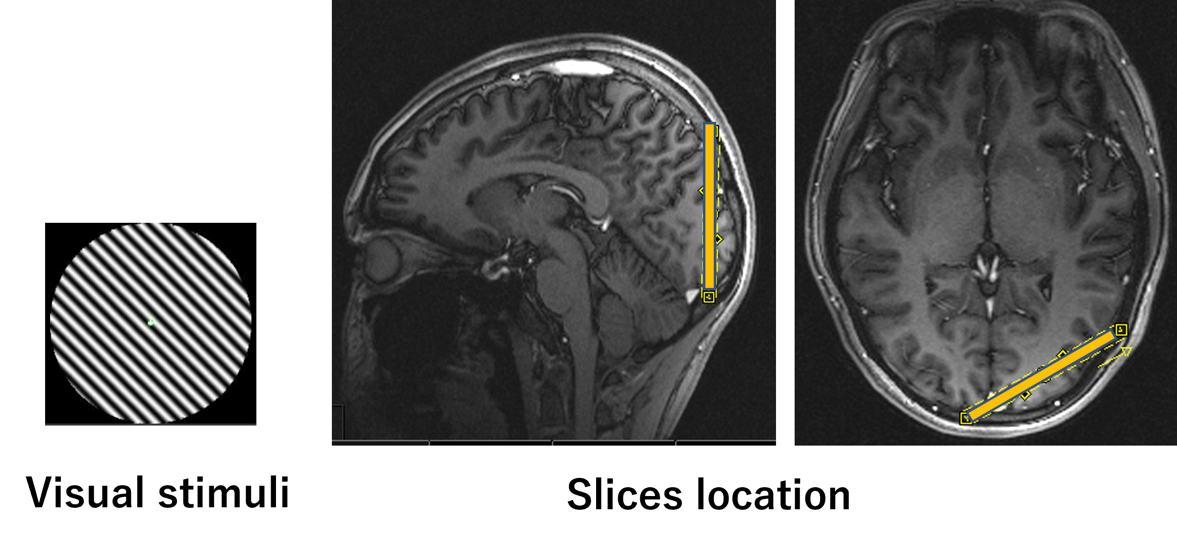
FIGURE 1: visual stimuli and slices location.
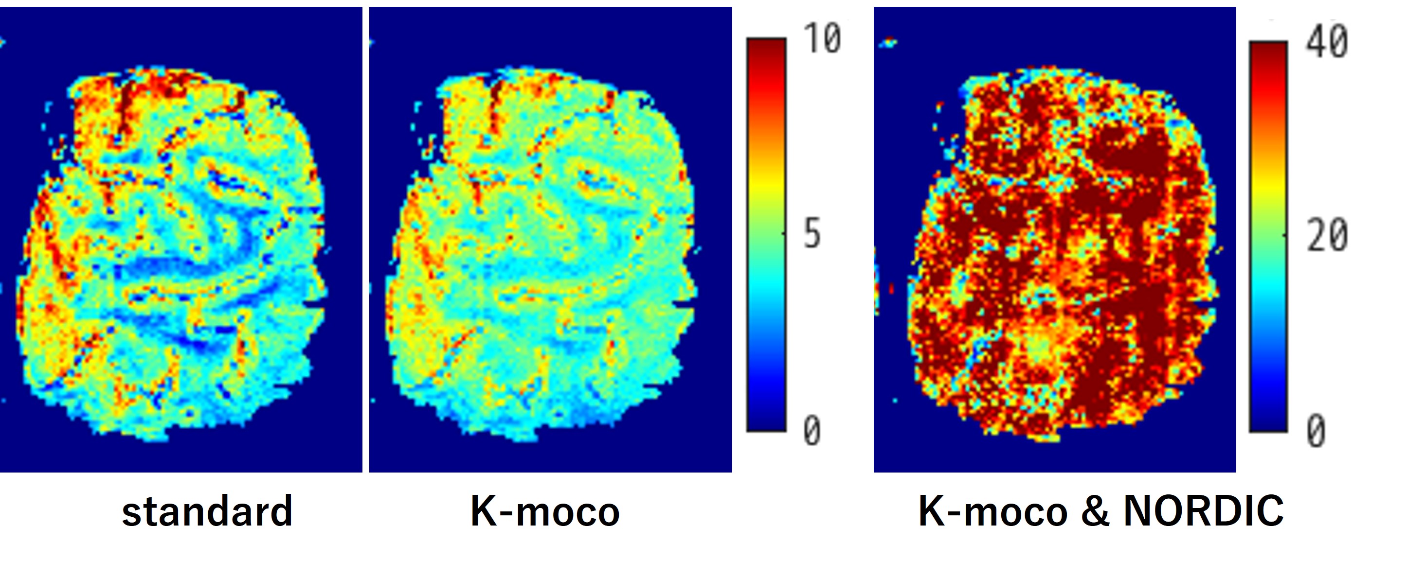
FIGURE 2: tSNR maps for different reconstruction approaches
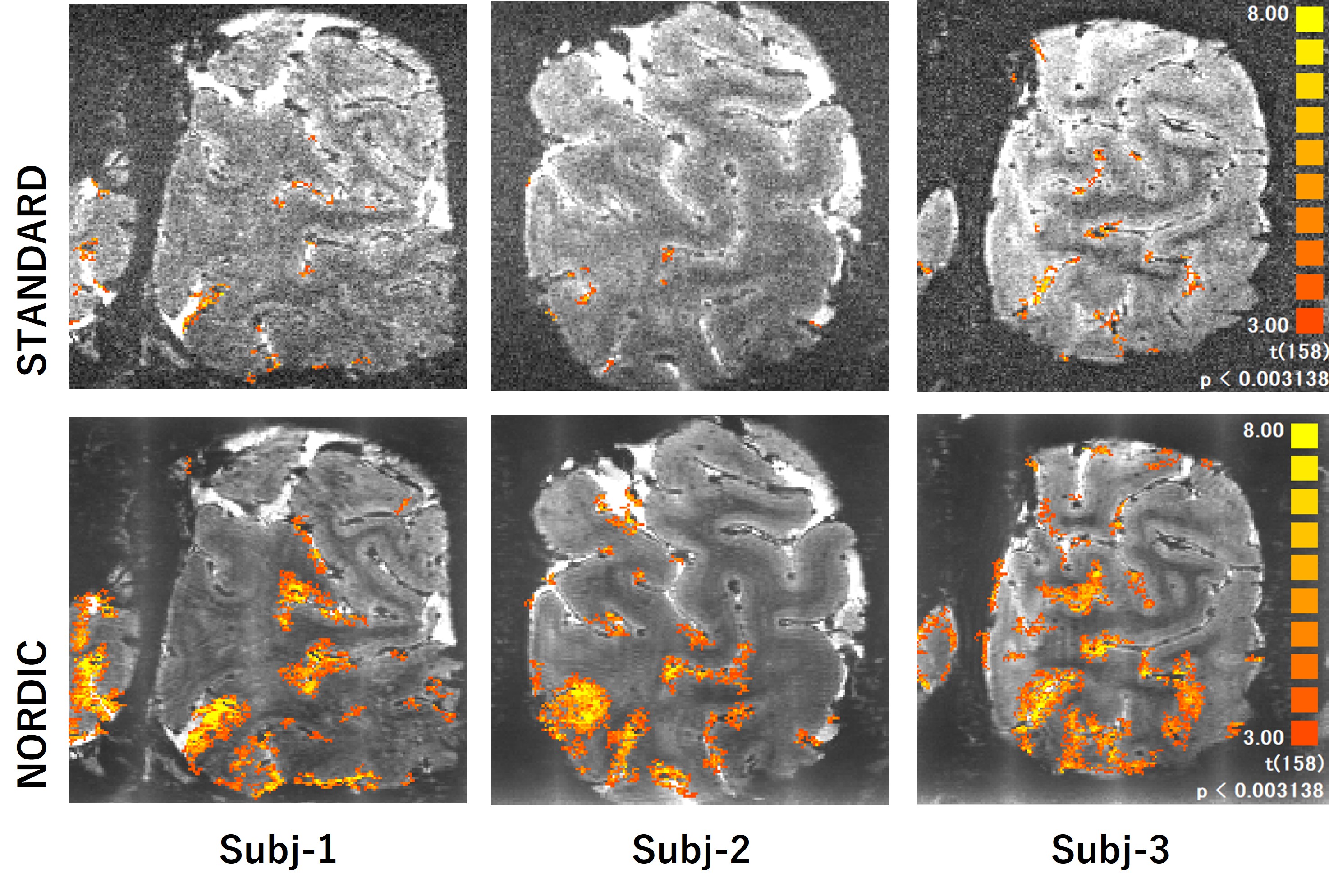
FIGURE 3: Detected fMRI activity during visual tasks of viewing flipping checkerboard using standard (top) and NORDIC (bottom) reconstruction.
DOI: https://doi.org/10.58530/2023/2717