2714
Joint Slice-Selective Pulse Design for Segmented FLEET-EPI using a Differential Bloch Simulator and Conjugate Gradient-Optimal Control
Imam Ahmed Shaik1, Mukund Balasubramanian2,3, Avery J.L. Berman4,5, Jonathan R. Polimeni2,6,7, and William Grissom1
1Institute of Imaging Science, Vanderbilt University, Nashville, TN, United States, 2Harvard Medical School, Boston, MA, United States, 3Boston Children's Hospital,, Boston, MA, United States, 4Carleton University, Ottawa, ON, Canada, 5University of Ottawa Institute of Mental Health Research, Ottawa, ON, Canada, 65Athinoula A. Martinos Center for Biomedical Imaging, Massachusetts General Hospital,, Charlestown, MA, United States, 7Harvard-MIT Division of Health Sciences and Technology,Massachusetts Institute of Technology, Cambridge, MA, United States
1Institute of Imaging Science, Vanderbilt University, Nashville, TN, United States, 2Harvard Medical School, Boston, MA, United States, 3Boston Children's Hospital,, Boston, MA, United States, 4Carleton University, Ottawa, ON, Canada, 5University of Ottawa Institute of Mental Health Research, Ottawa, ON, Canada, 65Athinoula A. Martinos Center for Biomedical Imaging, Massachusetts General Hospital,, Charlestown, MA, United States, 7Harvard-MIT Division of Health Sciences and Technology,Massachusetts Institute of Technology, Cambridge, MA, United States
Synopsis
Keywords: Data Acquisition, fMRI
Multishot EPI-based fMRI can achieve high spatial resolutions to resolve functional activity at the level of layers and columns, but suffers sensitivity to motion and phase changes between shots. Reordering the shots in a multislice stack so that each slice’s shots are acquired sequentially (VFA-FLEET-EPI) reduces this sensitivity but requires specialized RF pulses to maintain consistent signal between shots as longitudinal magnetization evolves. In this work we show that designing these pulses jointly using an autodiff optimal control algorithm yields more consistent signal across shots which reduces ghosting compared to VFA sinc and recursively designed SLR pulses.Intoduction
High-spatial resolution functional MRI (fMRI) is needed to resolve brain activity at fine spatial scales but requires long EPI readouts. For single shot EPI (SSH-EPI) this corresponds to long echo train lengths (ETL) and increased echo-spacing leading to severe image distortions. Multi-shot EPI (MSH-EPI) reduces the number of encoding lines per TR segment and hence reduces distortion. However, segmented EPI is sensitive to shot-to-shot signal changes due to sources such as respiratory and cardiac motion which causes ghosting when the data are combined in image reconstruction. Variable Flip-Angle (VFA-) FLEET (Fast Low-angle Excitation Echo-planar Technique) reorders multishot EPI segments to be acquired immediately sequentially for each slice and reduces the signal variation between segments [1]. To maintain consistent signal between shots despite variable longitudinal magnetization, a recursive Shinnar-Le Roux (SLR) radiofrequency (RF) pulse design (VFA-FLEET-SLR) algorithm was recently proposed which generates unique pulses for each shot of a VFA-FLEET-EPI scan that maintain constant signal. However, it required approximations and smoothing steps to stabilize the recursion which led to suboptimal slice profiles. In this work, we propose to instead jointly design pulses for VFA-FLEET-EPI using the optimal control algorithm [2] with autodifferentiation and a conjugate gradient solver, which improves slice sharpness for a given pulse duration with improves signal consistency between shots.Methods
An optimal control-based RF pulse design algorithm for multi-shot VFA FLEET acquisitions was developed that uses autodifferentiation in Julia (via ForwardDiff.jl) to jointly design a set of slice-selective RF pulses to achieve the same slice (Mxy) profile, even as longitudinal magnetization (Mz) evolves between pulses. The algorithm comprised a Bloch equation simulator to simulate each RF pulse across space given initial longitudinal magnetization and it minimized a cost function given by: err = w[ii] * ((Mx3 - Mxd[ii])^2 + (My3 - Myd[ii])^2 + (Mz3 - Mzd[ii])^2) + lamda * ((Mx1 - Mx3)^2 + (My1 -My3)^2) where Mx, My, Mz are actual magnetization & Mzd, Myd & Mzd are desired magnetization. where lambda balances each shot’s excitation error with the error between the shots’ Mxy profiles. The algorithm was used to design RF pulses for a 3-shot VFA-FLEET sequence with a time-bandwidth product of 4, and lambda =100 using the Optim.jl conjugate gradient solver. The RF pulses as well as time-bandwidth-matched sinc pulses were implemented in a multishot VFA-FLEET EPI pulse sequence on a Philips Elition X 3T scanner (Philips Healthcare, Best, Netherlands). To measure signal consistency between shots, images were acquired using a homogenous small phantom with a 32-channel head receive coil and a large FOV (300 mm) to resolve ghosting in individual-shot images. Data were acquired with TR = 80 ms, TE = 30 ms, shots = 3, matrix size = 144 x 144 with 2.08 mm in plane resolution, slice thickness = 4mm. FAs = 35, 45, 90, number of slices = 10, Volume TR = 2.4 s. Slice profiles were also acquired by moving the slice select gradient to the readout direction. No dummy excitations were performed so that Mz was fully relaxed prior to the first shot.Results
Figure 1 shows the waveforms and simulation results for scaled sinc and autodiff optimal control designs. The autodiff optimal control pulses Integrated Mxy signal is nearly constant across shots by varies more than 25% for sincs. Simulation results shown in figure 1, signifies that AOC method not only ensures lesser signal variation between shots but also matches the slice profile with more accuracy. It can be observed from Figure 2 that the slice profiles for each shot are more consistent in AOC method compared to Sinc profiles. FOV was selected to be thrice as big as phantom such that there is no fold over artefact and hence no overlapping aliases (Figure 3) in an individual shot image. Images were reconstructed without any navigator; hence significant ghosts are observed in previous methods while it is reduced in the proposed method but does not completely vanish (Figure 3). Average signal amplitude across shots indicates that OC method gives consistent signal.Discussion & Conclusion
We have presented a new algorithm to jointly design multishot slice-selective pulses for segmented VFA-FLEET EPI scans in fMRI. The algorithm is based on a differentiable Bloch equation solver and optimal control, and avoids recursions associated with a previous SLR-based approach [1]. The algorithm works by minimizing a cost function that balances excitation error of each pulse with errors between the shots’ excitation profiles. Simulations showed a large reduction in profile differences between consecutive shots, and experiments showed reduced ghosting compared to VFA sinc pulses. The pulses will next be directly compared to the SLR pulses and implemented in in vivo scans to assess ghosting and possible improvements in signal stability.Acknowledgements
This work was supported by NIH Grants R01 EB 016695 and R01 EB 019437References
[1] Berman AJL, Witzel T, Grissom WAG, Park D, Setsompop K, Polimeni JR. High-resolution segmented-accelerated EPI using Variable Flip Angle FLEET with tailored slice profiles. In: International Society of Magnetic Resonance in Medicine Annual Meeting. Vol. 27. Montreal, Canada; 2019. p. 1169. [2] S. Conolly, D. Nishimura, and A. Macovski. Optimal control solutions to the magnetic resonance selective excitation problem. IEEE Trans Med Imaging, 5:106–115, 1986.Figures
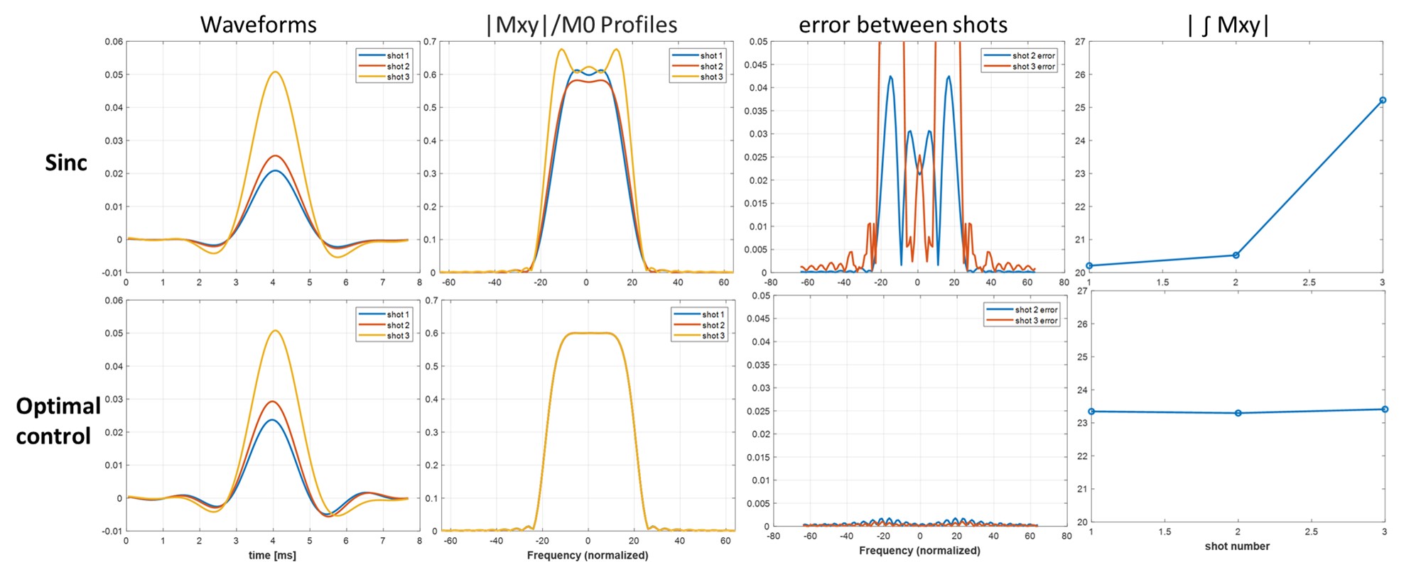
Sinc
and joint optimal control RF pulses (left column), Mxy profiles (second
column), errors between shots (third column), and integrated signal (fourth
column) for a 3-shot scan. Sinc pulses have the same waveform shape scaled to
different flip angles (35, 45, and 90 degrees). Due to varying Mz before each
pulse the sinc pulses produce inconsistent profiles between shots compared to
the optimal control pulses, as reflected by their varying integrated Mxy and
large errors between shots 2 and 3 versus shot 1.
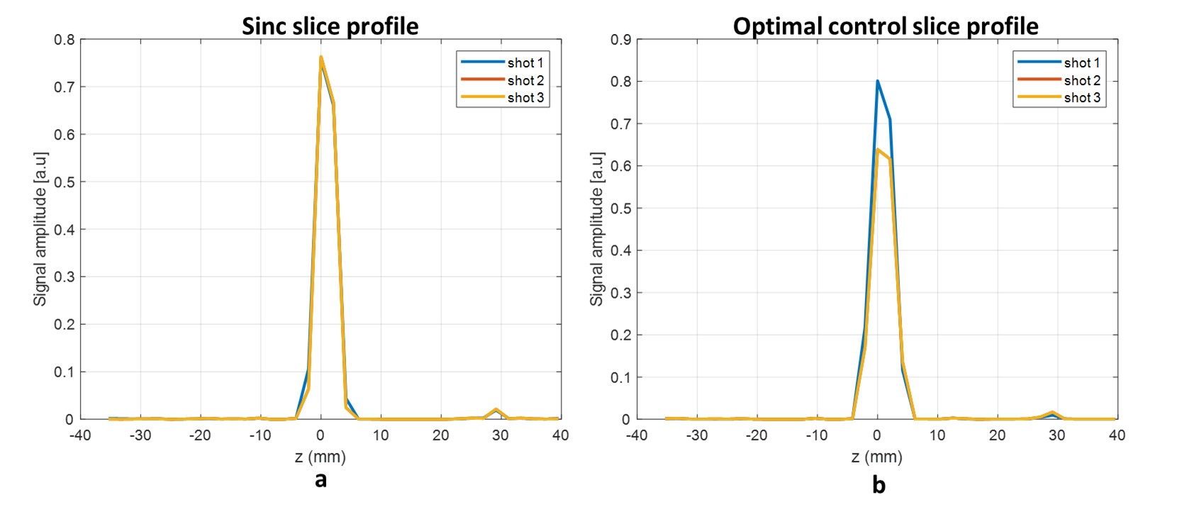
Measured slice profiles
for each shot in a phantom at 3T for sinc and optimal control pulses. The sinc
pulses produce varying signal amplitudes, with shot 3’s signal almost 25% lower
than shots 1 and 2. The optimal control pulses produced consistent slice
profiles and amplitudes for all shots.
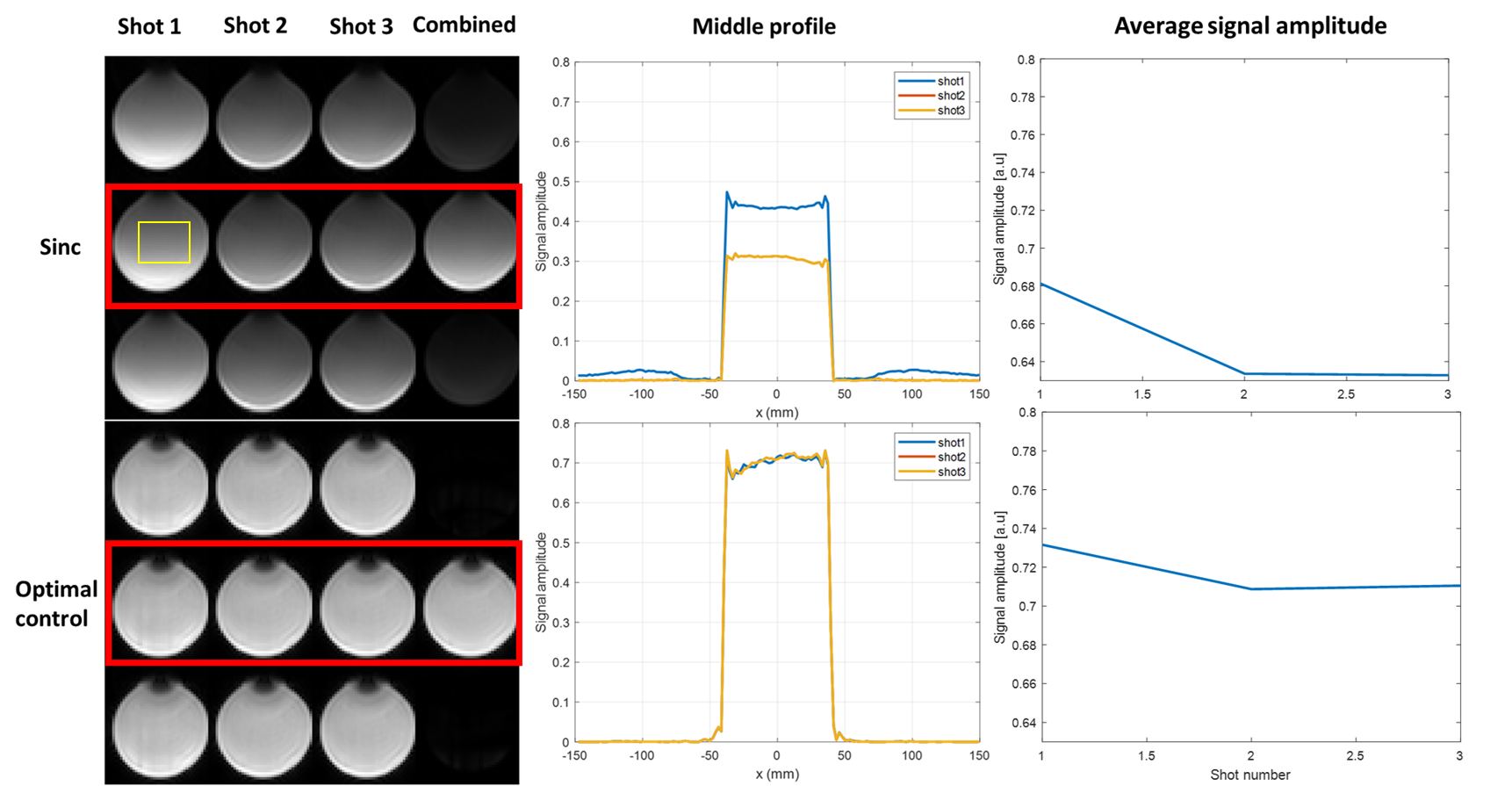
Shot-separated
multishot images of a phantom with diameter less than 1/3 the encoded FOV, to
measure aliased signals with sinc and optimal control pulses. Each individual
shot shows aliases above and below the central image (red rectangle) which
should be suppressed in the combined image. The first sinc slice had higher
signal amplitude than the other two shots which led to poor ghosting supression
in the combined image, and which are further reflected in the 1D profiles
across the images. Last column shows the
signal amplitude average of the selected region (yellow box) for all
shots.
DOI: https://doi.org/10.58530/2023/2714