2699
SAR and Temperature Safety Assessment on Patient Specific Models Generated with a Registration Method
Giuseppe Carluccio1,2, Eros Montin1, Christopher Michael Collins1,2, and Riccardo Lattanzi1
1Radiology, NYU Grossman School of Medicine, New York, NY, United States, 2Center for Advanced Imaging Innovation and Research (CAI2R), New York University, New York, NY, United States
1Radiology, NYU Grossman School of Medicine, New York, NY, United States, 2Center for Advanced Imaging Innovation and Research (CAI2R), New York University, New York, NY, United States
Synopsis
Keywords: Safety, Safety
Patient specific models are required to improve the accuracy of the simulations to perform safety assessment related to interaction of the RF fields with the body tissues. One registration method allows non-rigid transformations: this can be used to reshape a numerical body model to match the images of a patient, and generate a model similar to the shape of the patient. We performed SAR and temperature simulations to evaluate the ability of the registered model to perform a patient-specific safety assessment. The evaluation has been successful: the SAR and temperature patterns were corresponding to the ones of the target anatomy.Introduction
Interaction of radiofrequency (RF) electric fields with conductive tissues generate induced heat which can potentially cause discomfort in the patient or even tissue damage: therefore, limits are imposed on local and whole-body SAR and temperature. Usually, it is not practical to measure SAR or temperature in deep tissues, and RF safety assessment is performed through numerical simulations where a model of both the patient and the RF coil is used. Commonly, a patient-specific model is not available, and the patient is represented with one of the limited available body models. However, a patient-specific model can increase the accuracy of the simulations. A recently published method performs a novel registration between two models, able to perform not only rigid transformations such as translation and rotation, but also non-rigid operations that allow to match body models having different shapes1. Therefore, this method can be used to reshape a body model in order to match the images of a patient, and ultimately generate a model very similar to the shape of the patient. This model can be used to increase the accuracy of the safety assessment. In this work, we evaluate through simulations the accuracy of the estimation of SAR and temperature when a subject-specific model is generated with the non-rigid transformations registration.Methods
The registration method1 is used to transform the body model Norman2, by matching the simulated MRI images acquired with “Norman” with the simulated MRI images acquired with the body model “NLM Male” 3. The images are simulated with the PSUsoMRI MRI simulator4 emulating a 3T scanner, with a GRE sequence having TR=1s, TE=10 ms, 5mm resolution in each direction. Both uniform transmit and receive fields are assumed for the MRI simulations.The body models (Norman, NLM Male, and the new model obtained through registration) were then positioned within a 28 cm long, 15 cm radius, 16 leg, head-size birdcage coil to calculate SAR with commercial software (xFDTD; Remcom), and temperature with an in-house C++ software5 . Simulations were performed with a 5 mm isotropic resolution. The simulator provides a solution of the Pennes’ bioheat equation $$\rho c \frac{\partial T}{\partial t}=(\nabla (k\cdot \nabla (T)))-W_{bl}c_{bl}(T-T_{bl})+Q+\rho SAR$$where c is the heat capacity, W is the blood perfusion rate, k is the thermal conductivity, ρ is tissue mass density, the subscript bl indicates values for blood, Q is the heat generated by metabolism. The temperature simulations were performed for an imaging time of 30 minutes.Results and Discussion
Figure 3 provides the sagittal profile of the 10g average SAR and temperature increase for three body models: Norman, NLM Male, and the body model obtained with the registration method. The registration method is able to generate models more accurately matching the target anatomy. As a consequence, the accuracy of the safety assessment performed with the registered model increases accordingly. The images in Figure 3 clearly show SAR and temperature patterns closer to the target anatomy. Therefore, the registration method is robust enough to be used with less ideal conditions, and we expect to have similar performance with in vivo acquired images. In addition, in the future we plan to test its performance with lower SNR values, different field strengths, different image contrast between source model and target anatomy, different resolution, and non-homogeneous transmit and receive fields.Conclusions
We have presented a method based on a no-rigid transformations registration method. The method, operating on simulated MRI images, is able to generate a body model closer to the target anatomy. Therefore, it can be used to generate a patient-specific model to improve accuracy of safety assessment.Acknowledgements
This work was performed under the rubric of the Center for Advanced Imaging Innovation and Research (CAI2R, www.cai2r.net) at the New York University School of Medicine, which is an NIBIB Biomedical Technology Resource Center (NIH P41 EB017183).References
- Montin E, Belfatto A, Bologna M, Meroni S, Cavatorta C, Pecori E, Diletto B, Massimino M, Oprandi MC, Poggi G, Arrigoni F. A multi-metric registration strategy for the alignment of longitudinal brain images in pediatric oncology. Medical & Biological Engineering & Computing. 2020 Apr;58(4):843-55.
- Dimbylow PJ. “FDTD calculations of the whole-body averaged SAR in an anatomically realistic voxel model of the human body from 1 MHz to 1 GHz.” Physics in Medicine & Biology. 1997 Mar;42(3):479.
- Collins CM, Smith MB. “Calculations of B1 distribution, SNR, and SAR for a surface coil adjacent to an anatomically‐accurate human body model.” Magnetic Resonance in Medicine: An Official Journal of the International Society for Magnetic Resonance in Medicine. 2001 Apr;45(4):692-9.
- Cao Z, Oh S, Sica CT, McGarrity JM, Horan T, Luo W, Collins CM. Bloch‐based MRI system simulator considering realistic electromagnetic fields for calculation of signal, noise, and specific absorption rate. Magnetic resonance in medicine. 2014 Jul;72(1):237-47.
- Collins CM, Liu W, Wang J, Gruetter R, Vaughan JT, Ugurbil K, Smith MB. Temperature and SAR calculations for a human head within volume and surface coils at 64 and 300 MHz. Journal of Magnetic Resonance Imaging: An Official Journal of the International Society for Magnetic Resonance in Medicine. 2004 May;19(5):650-6.
Figures
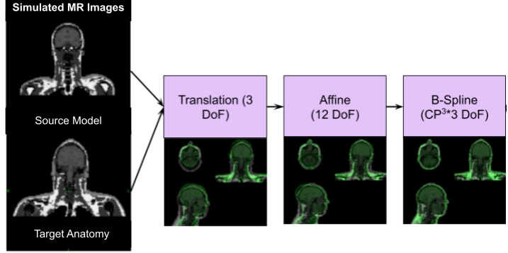
Figure1: The Registration Workflow: starting from MRI images generated from 2 body models (source model and target anatomy), by applying the registration transformations to the source it is possible to generate a body model similar to the target starting from the knowledge of the segmented source body model. The registration applies rigid and non-rigid transformations.
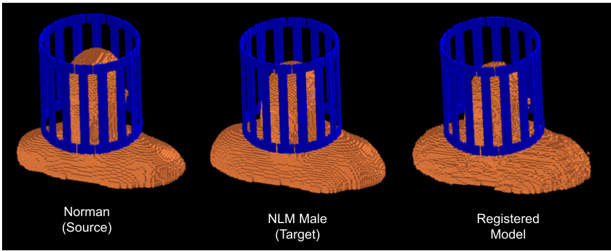
Figure 2: Geometry of the electromagnetic simulations with the three models in the birdcage coil: 1) Norman, corresponding to the source (left), 2) NLM Male, corresponding to the target (middle), 3) the registered model (right).
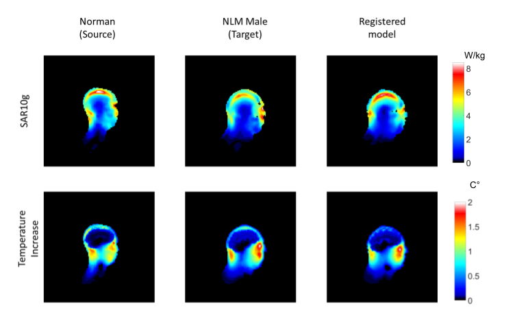
Figure 3: 10g average SAR (top row) and temperature increase (bottom row) profile for three models: Norman, the source model (left column), NLM Male, the target model (middle column), and the registered model with TE=10ms (right column).
DOI: https://doi.org/10.58530/2023/2699