2675
Task functional magnetic resonance imaging-guided individualized repetitive transcranial magnetic stimulation after a brief nap attenuates the sustained attention performance deterioration induced by sleep deprivation
Yuanqiang Zhu1, Fan Guo1, and Yingjuan Chang1
1Air Force Medical University, Xi'an, China
1Air Force Medical University, Xi'an, China
Synopsis
Keywords: Data Analysis, fMRI (task based)
Our pilot study validated the effectiveness of rTMS after a brief nap in terms of improving sustained attention in the context of SD. An investigation of the dynamic changes of PVT task-related cerebral responses across the three conditions showed that the middle frontal gyrus recovered least after the nap and was selected as the stimulation target. Through modulating the functional connectivity within the FPN and DMN, individualized, 10-Hz rTMS showed promise in terms of improving the vigilance of military academy cadets accepting real stimulation.Background: Military operations may require human functioning 24 hours a day and sleep deprivation (SD) is thus not uncommon[1]. However, in counteracting the effects of SD, the most effective method of long-time recovery sleep is not always feasible in military life. Although frequently used as a countermeasure, a brief nap is not enough to fully restore cognitive performance following SD[2]. Thus, targeted interventions after the brief nap, such as repetitive transcranial magnetic stimulation (rTMS), might better restore performance following SD. However, besides the stimulation frequency affecting the stimulation effects, one must be aware of the importance of the stimulation site. Even subtle changes in the stimulation target might lead to large changes in the rTMS effects[3]. Hence, determining the optimal rTMS target area is crucial to achieving maximal performance improvements.
Methods: Fifty military academy cadets were recruited and participated in two SD experiments. In experiment 1, each participant performed a psychomotor vigilance task (PVT) while undergoing functional magnetic resonance imaging (fMRI) for three conditions: (1) following a normal night of sleep (Resting wakefulness, RW), (2) following 30 hours of SD, and (3) after a subsequent 30-min nap. Dynamic changes of PVT outcomes and cerebral responses across the three conditions were analyzed to determine the optimal simulation target.In experiment 2, the same protocol (i.e., the three conditions of RW, 30 hours of SD and a 30-min nap) was adopted. In addition, after the nap, a 10-Hz, sham-controlled and individualized rTMS was administrated. Then, ANOVA analyses were conducted to investigate the stimulation effect for the improvement of the fastest PVT reaction times.
Results: Utilizing task-related fMRI, we first showed that the cerebral responses within the right middle frontal gyrus (MFG) recovered least after the nap and the right MFG was selected as the stimulation target. We next showed that the individualized 10-Hz rTMS over right MFG after the nap attenuated the sustained attention performance deterioration induced by SD.
Conclusion: We demonstrate that a nap combined with individualized rTMS may contribute to the recovery of the impaired sustained attention after SD by modulating the neural activity within functional brain networks.
Acknowledgements
This study was supported by the Key R&D Program Projects, National Science Foundation of China (Grant No.2016YFC1306900), the National Natural Science Foundation of China under Grant Nos. 81974215 and 81801772, Key R&D Program Projects of Shaanxi, China (No.2021SF-287 and 2022JM-575), Boost Program of Xijing Hospital (XJZT18ML84 and XJZT19ML56), China Postdoctoral Science Foundation (2019M653963), and Military Medical Science and Technology Youth Training Program (20QNPY049).References
1. Good CH, Brager AJ, Capaldi VF, Mysliwiec V. Sleep in the United States Military. Neuropsychopharmacology 2020; 45(1): 176-191.
2. Ong JL, Lau TY, Lee XK, van Rijn E, Chee MWL. A daytime nap restores hippocampal function and improves declarative learning. Sleep 2020; 43(9).
3. Siddiqi SH, Weigand A, Pascual-Leone A, Fox MD. Identification of Personalized Transcranial Magnetic Stimulation Targets Based on Subgenual Cingulate Connectivity: An Independent Replication. Biol Psychiatry 2021; 90(10): e55-e56.
Figures
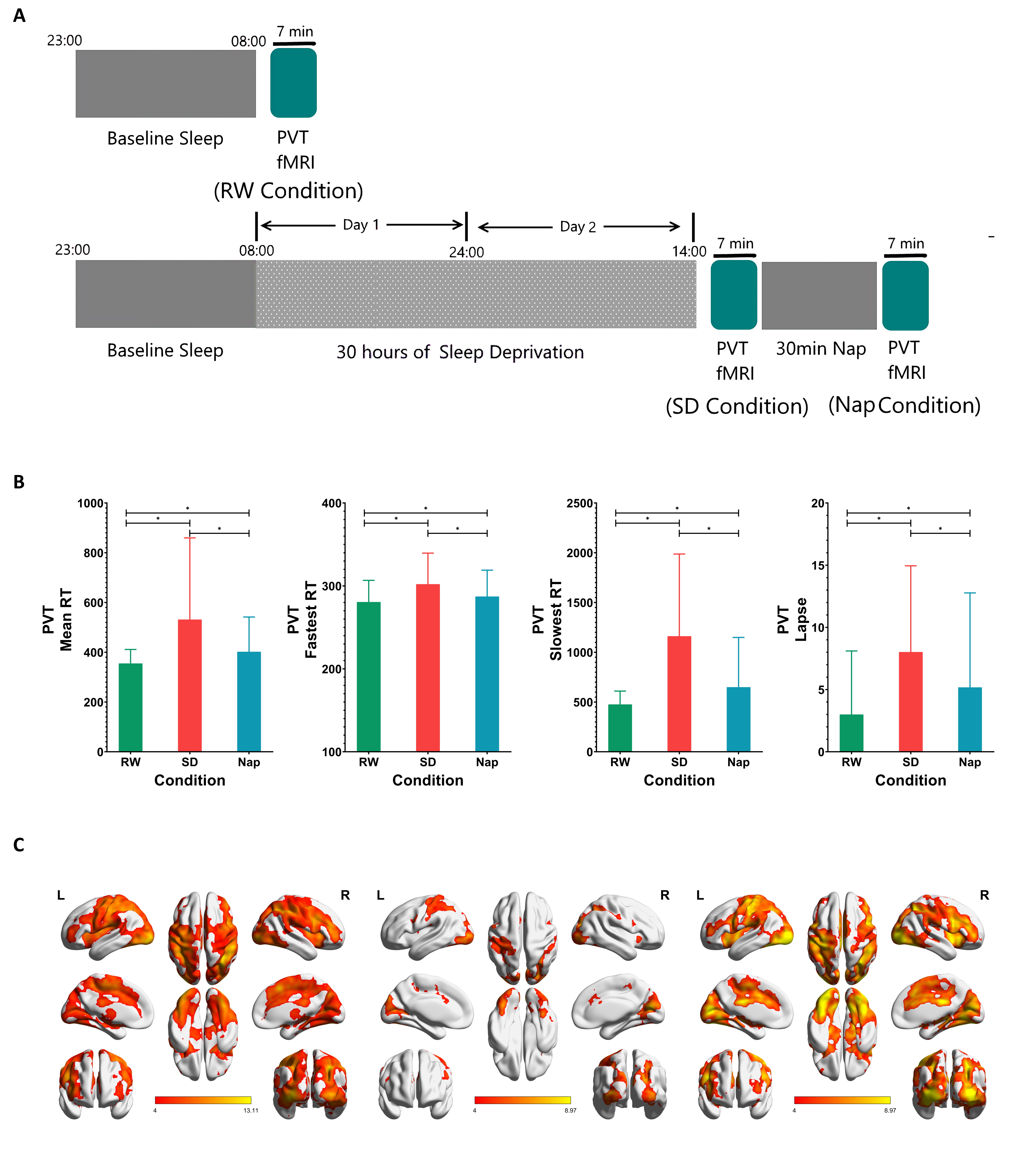
Experiment 1. A Participant performed a PVT task in the MR scanner after resting wakefulness (RW), 30 hours of SD and a subsequent 30 min nap. B Significant time effects (p < 0.001) for the mean RT, mean of the fastest RTs, mean of the slowest RTs and number of attention lapses. Posthoc analysis indicated significant differences in each pairwise comparison between the RW, SD and nap conditions (p < 0.01). C One-sample t test results of the cerebral response for the fastest RT under each condition.
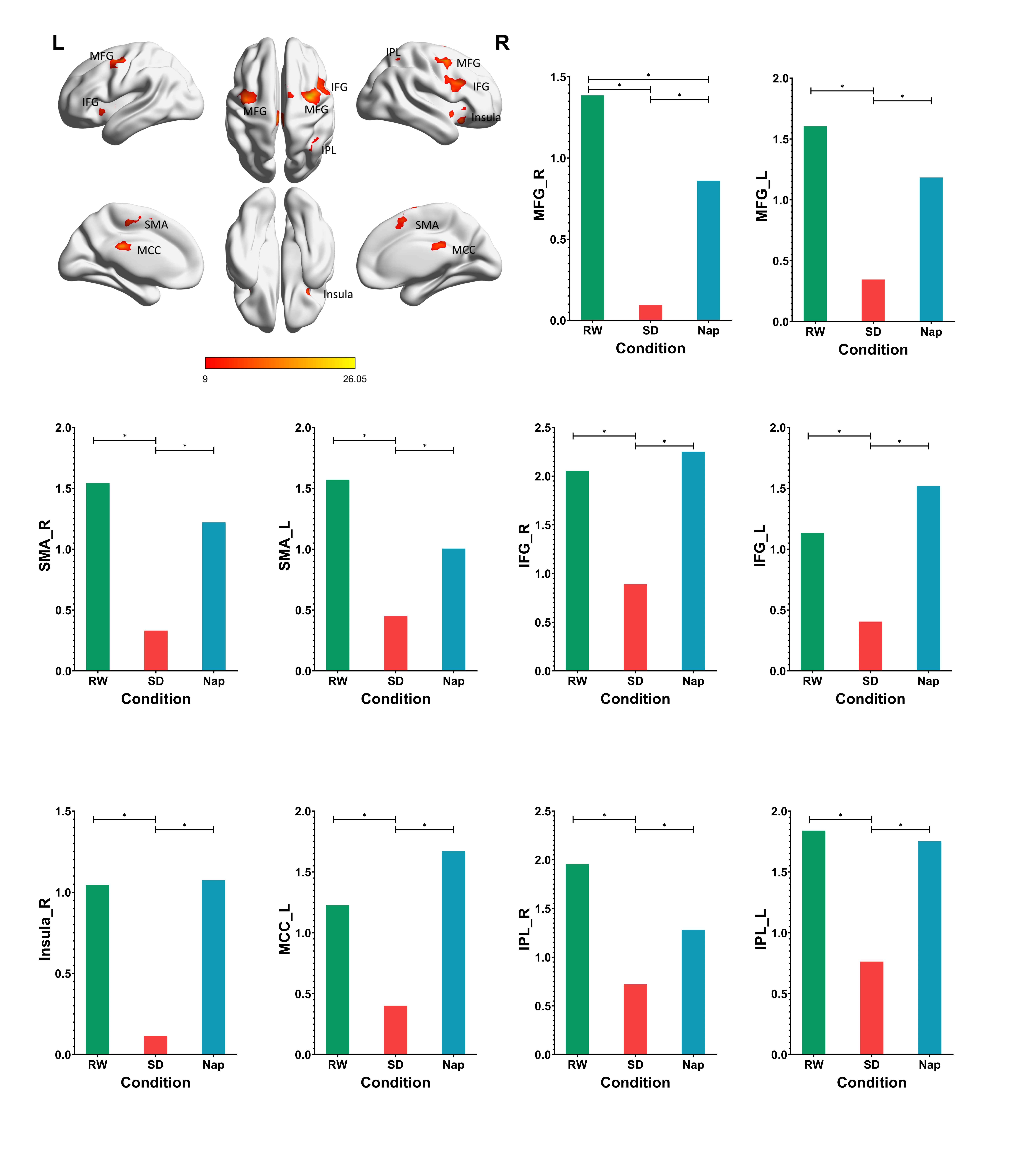
Experiment 1. One-way repeated-measures ANOVA results for the dynamic changes of PVT activation across the three conditions. Statistically significant changes were found in the MFG, SMA, IFG, right insula, left MCC, right IPL, and left inferior superior parietal gyrus. Note that only the pairwise differences in the right middle frontal gyrus were similar to the observed pairwise differences in the fastest PVT RT.
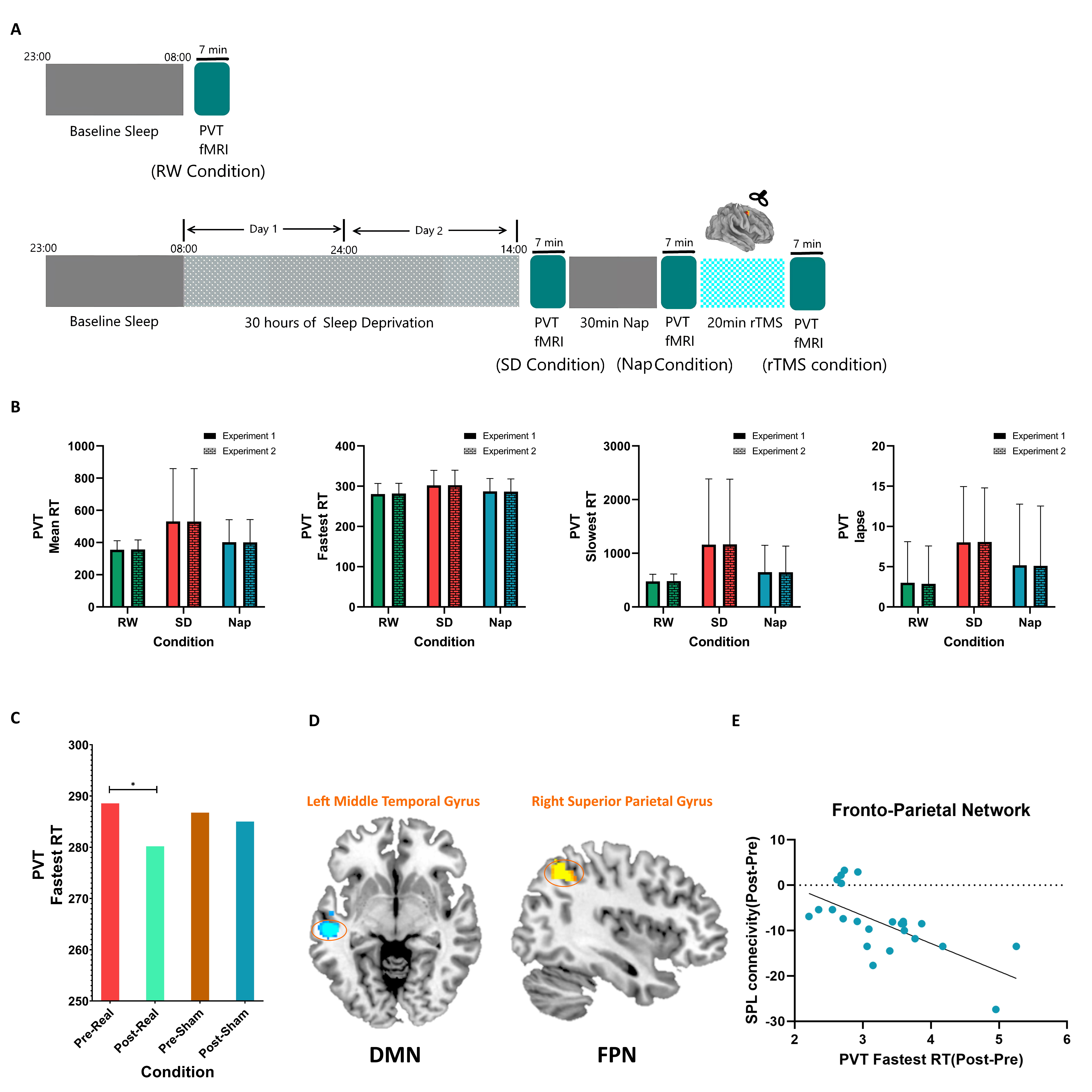
Experiment 2. A Participant performed a PVT task in the MR scanner after resting wakefulness (RW), 30 hours of SD, the 30 min nap and 20 min repetitive transcranial magnetic stimulation (rTMS). B No differences in PVT outcomes were found between experiments 1 and 2 for each condition (RW, SD and Nap). C The mixed two-way repeated-measures ANOVA revealed a significant main condition effect and a significant interaction effect. The reaction time was significantly reduced after rTMS administration, and the real rTMS most improved the processing speed. D For participants receiving real rTMS, the planned condition comparisons (pre-stimulation vs post stimulation) revealed increased functional connectivity in the frontoparietal network (FPN), mainly at the right superior parietal gyrus, and reduced FC within the default mode network (DMN), mainly at the medial frontal gyrus. E The increased functional connectivity in the right superior parietal gyrus after stimulation was negatively correlated with a reduction in the fastest RT for participants receiving real rTMS.
DOI: https://doi.org/10.58530/2023/2675