2673
Neural perspective on observational drawing: A longitudinal resting state functional connectivity study using 7T MRI
Apoorva Safai1, Jeffrey Katz2,3,4,5, Barbara Bondy6, and Gopikrishna Deshpande2,3,4,5,7,8,9,10
1Symbiosis Centre for Medical Image Analysis, Symbiosis International University, Pune, India, 2Department of Psychological Sciences, Auburn University, Auburn, AL, United States, 3Department of Electrical & Computer Engineering, Auburn University, Auburn, AL, United States, 4Alabama Advanced Imaging Consortium, Birmingham, AL, United States, 5Center for Neuroscience, Auburn University, Auburn, AL, United States, 6Department of Art and Art History, Auburn University, Auburn, AL, United States, 7Key Laboratory for Learning and Cognition, Capital Normal University, Beijing, China, 8Department of Psychiatry, National Institute of Mental Health and Neurosciences, Bangalore, India, 9Centre for Brain Research, Indian Institute of Science, Bangalore, India, 10Department of Heritage Science and Technology, Indian Institute of Technology, Hyderabad, India
1Symbiosis Centre for Medical Image Analysis, Symbiosis International University, Pune, India, 2Department of Psychological Sciences, Auburn University, Auburn, AL, United States, 3Department of Electrical & Computer Engineering, Auburn University, Auburn, AL, United States, 4Alabama Advanced Imaging Consortium, Birmingham, AL, United States, 5Center for Neuroscience, Auburn University, Auburn, AL, United States, 6Department of Art and Art History, Auburn University, Auburn, AL, United States, 7Key Laboratory for Learning and Cognition, Capital Normal University, Beijing, China, 8Department of Psychiatry, National Institute of Mental Health and Neurosciences, Bangalore, India, 9Centre for Brain Research, Indian Institute of Science, Bangalore, India, 10Department of Heritage Science and Technology, Indian Institute of Technology, Hyderabad, India
Synopsis
Keywords: Brain Connectivity, Neuroscience, Neuroplasticity
Observational drawing is representation of observed 3D real-life objects into 2D drawings on paper, which involves engaging complex skills like visual perception, spatial encoding, memory and decision making. This study investigates longitudinal functional connectivity (FC) in art students who underwent a 16-week long drawing course, in comparison to students who took unrelated courses. The art group showed improved drawing skills and demonstrated enhanced FC between DMN and salience network and in cerebellar regions post-art class, in comparison, with healthy non-art students. These findings imply positive changes in brain functioning on learning and practicing art, thereby highlighting its potential therapeutic applications.Body of Abstract:
Introduction: Observational drawing involves creatively producing 2D drawings of 3D objects that are directly observed in real life, by synchronously developing and engaging cognitive skills like visual perception, spatial encoding, top-down control, memory and decision making [1,2]. Neuroimaging studies assessing neural correlates of observed drawing have demonstrated altered structural organization [3] and functional activations [2,4] in cortical and cerebellar regions in art students compared to non-art students. Varied visual art forms have also shown an involvement of the default mode network (DMN) using task-based fMRI [5,6]. In this study we perform a longitudinal exploratory assessment of whole brain resting state functional connectivity in art students who attended observational drawing class and non-art students. The observational drawing class provided training on basic drawing techniques such as tonal variations in light, effect of shadows and linear perspectives for volume modelling. Understanding the effect of art training on brain functional connectivity patterns and network structure is important particularly for its pedagogy and its therapeutic application in mental health.Methods: Dataset for this study consisted of 51 participants, among which 32 were categorized as the experimental art group who were enrolled in an observational drawing class and 19 as the healthy control (HC) group of non-art undergraduate students, without any previous drawing experience or enrollment in art classes. Inclusion of HC group ensured that neural changes obtained pre and post class were due to the drawing course. Drawing skills of both groups were assessed both pre and post course, using a composite art score based on a three-part questionnaire, a freehand charcoal drawing task (experimental group only), and task-based fMRI[2]. A written consent was obtained from all participants along with research approval by the Institutional Review Board at Auburn University. All participants were scanned on a Siemens 7T MAGNETOM MR Scanner using 32-channel head coil at the Auburn University MRI Research Center. T1 weighted MPRAGE scan was acquired with 256 slices, voxel size=0.6 mm3, TR/TE 2200/3.05ms, flip angle=7◦. Resting state fMRI scan was acquired with a TR/TE=1000/22ms, no. of volumes=600, slice thickness=2x2x2.2mm, FOV=100x100x51. These MRI scans were acquired pre as well as post art class for the experimental and pre/post non-art class for the HC group, which are further referred as pre-art, post-art, pre-HC and post-HC groups. The detailed demographics as well as scores of all participants/groups used in further analysis is given in Figure-1. Resting state fMRI data was pre-processed using the standard pre-processing pipeline. Additionally, we deconvolved the time series to remove the confounding effect of the variable HRF[7,8,9] , followed by band pass filtering in 0.01 to 0.1 Hz range. Mean fMRI time series were extracted for 272 regions of interest (ROIs) which included cortical, subcortical and cerebellar regions from the Power, Harvard-Oxford and Buckner atlases, respectively. Resting state functional connectivity (FC) was obtained by computing pairwise Pearson’s correlation between the mean time series of all ROIs. A set of statistical t-tests were performed on Fisher’s normalized FC matrix, in order to investigate the intrinsic functional connectivity changes in experimental group post art class, in comparison with their pre art class performance and with HC group. Specifically, FC that showed non-significant (p>0.05) baseline difference in pre-art vs pre-HC, pre vs post HC groups, and significant (p<0.05) difference in post vs pre-art group were considered.
Results and Discussion: The interaction effect analysis revealed increased FC in post-art vs pre-art and post-HC groups. Within cerebellar FC was most significantly higher, followed by increased FC within sections of cingulum and frontal regions, comprising of DMN and salience network, along with higher FC between sensory motor network (SMN) and cerebellum, as shown in Figure-2 and Figure-3. Figure-4 depicts the higher FC in post-art group compared to pre-art and post-HC groups, with similar baseline patterns in experimental and HC group. These findings indicate that learning and continued practice of observational drawing enhances connectivity within and between cerebellar, DMN and salience networks, which are generally involved in complex motor and cognitive tasks.
Conclusion: Longitudinal assessment of the effect of learning and practicing observational drawing in art students demonstrated enhanced FC in cerebellar regions as well as between DMN and salience network regions. These findings suggest that visual art method of observational drawing enhances neuroplasticity, thereby providing support for its therapeutic applications.
Acknowledgements
No acknowledgement found.References
1. Kozbelt, A. (2001). Artists as experts in visual cognition. Visual cognition, 8(6), 705-723. 2. Katz, J. S., Forloines, M. R., Strassberg, L. R., & Bondy, B. (2021). Observational drawing in the brain: A longitudinal exploratory fMRI study. Neuropsychologia, 160, 107960. 3. Schlegel, A., Alexander, P., Fogelson, S. V., Li, X., Lu, Z., Kohler, P. J., ... & Meng, M. (2015). The artist emerges: Visual art learning alters neural structure and function. NeuroImage, 105, 440-451. 4. Lin, C. S., Liu, Y., Huang, W. Y., Lu, C. F., Teng, S., Ju, T. C., ... & Hsieh, J. C. (2013). Sculpting the intrinsic modular organization of spontaneous brain activity by art. PLoS One, 8(6), e66761. 5. Bolwerk, A., Mack-Andrick, J., Lang, F. R., Dörfler, A., & Maihöfner, C. (2014). How art changes your brain: Differential effects of visual art production and cognitive art evaluation on functional brain connectivity. PloS one, 9(7), e101035. 6. Vessel, E. A., Starr, G. G., & Rubin, N. (2012). The brain on art: intense aesthetic experience activates the default mode network. Frontiers in human neuroscience, 6, 66. 7. Rangaprakash, D., Wu, G. R., Marinazzo, D., Hu, X., & Deshpande, G. (2018). Hemodynamic response function (HRF) variability confounds resting‐state fMRI functional connectivity. Magnetic resonance in medicine, 80(4), 1697-1713 8. Yan, W., Palaniyappan, L., Liddle, P. F., Rangaprakash, D., Wei, W., & Deshpande, G. (2022). Characterization of Hemodynamic Alterations in Schizophrenia and Bipolar Disorder and Their Effect on Resting-State fMRI Functional Connectivity. Schizophrenia bulletin, 48(3), 695-711. 9. Yan, W., Rangaprakash, D., & Deshpande, G. (2018). Aberrant hemodynamic responses in autism: implications for resting state fMRI functional connectivity studies. NeuroImage: Clinical, 19, 320-330.Figures
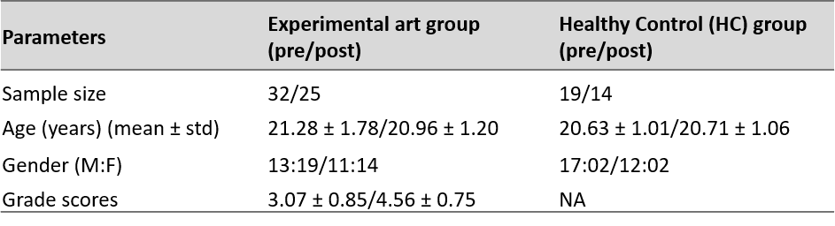
Figure-1: Detailed demographics of experimental
art group and HC group.
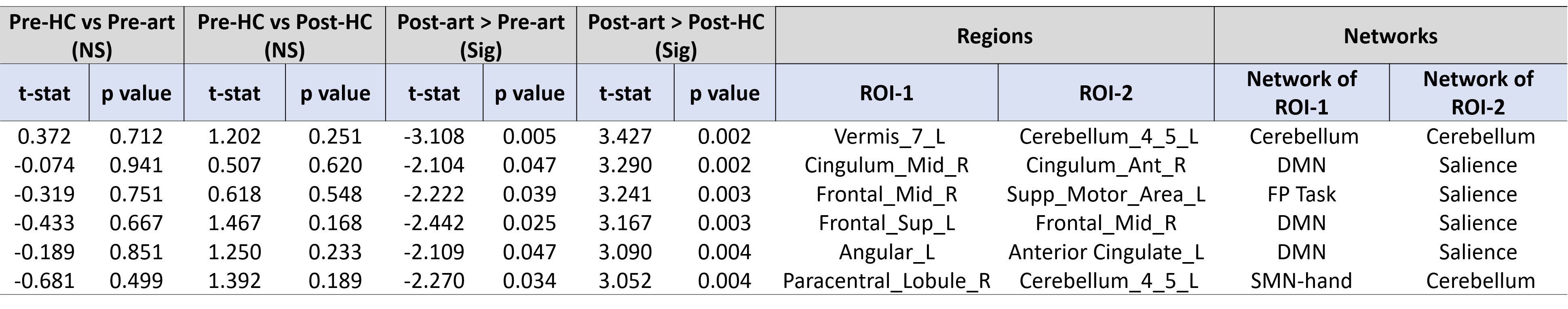
Figure-2: Significantly (uncorrected p<0.005)
increased FC in post-art compared to pre-art and HC groups, with interaction
effect– pre-HC vs pre-art (NS) ∩ pre-HC vs post-HC (NS) ∩ post-art > pre-art
(Sig) ∩ post-art > post-HC (Sig). The connections are visualized in Figure.3
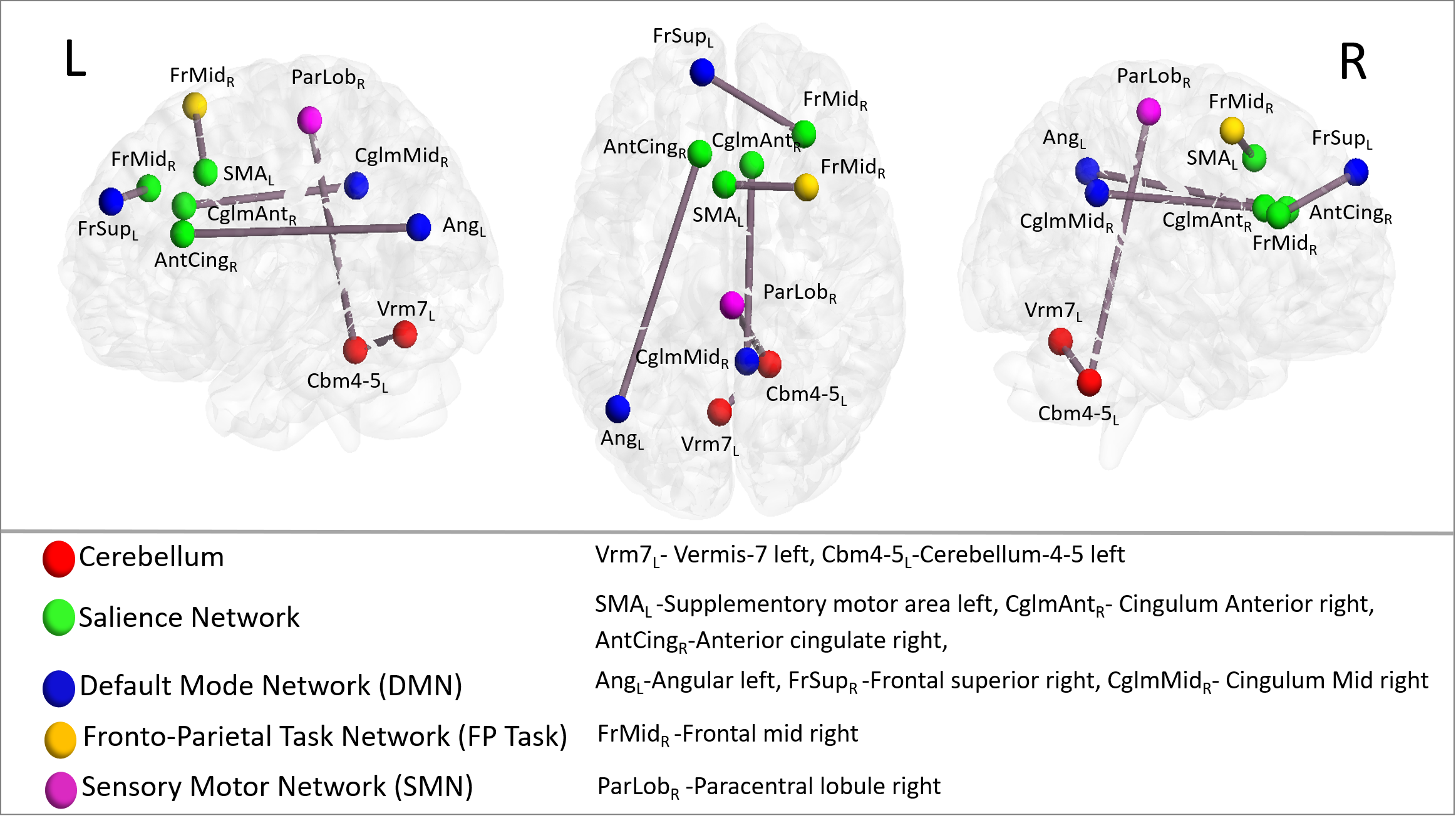
Figure-3: Visualization of connections
with increased FC in post art group compared to pre art and HC groups.
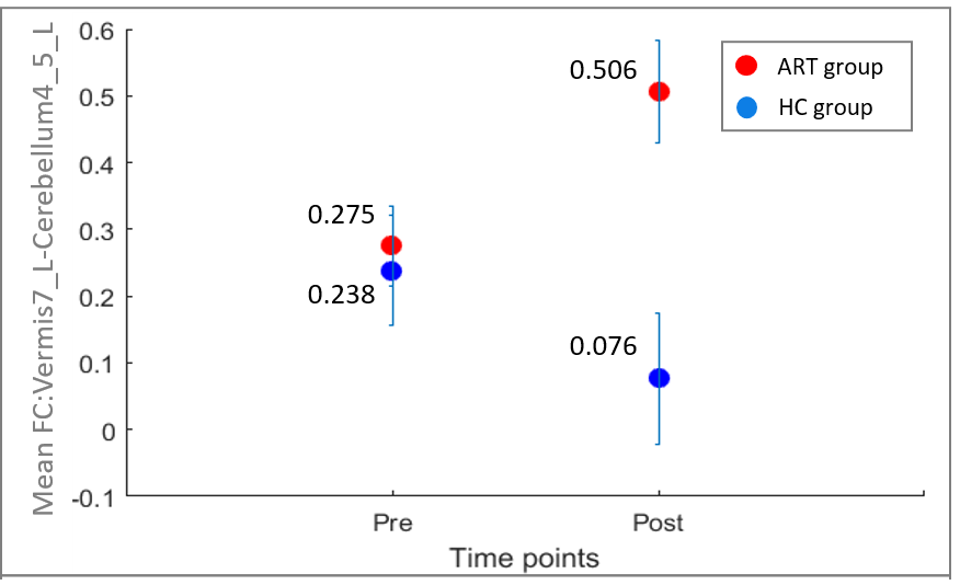
Figure-4: Most significantly
altered FC pattern of vermis-7_L and cerebellum_4_5_L, showing increased FC in
post art group compared to pre art and HC groups. (Values on error bars
indicate mean group FC values)
DOI: https://doi.org/10.58530/2023/2673