2667
Comparing the efficacy of data-driven noise regression techniques in preserving age-related resting-state connectivity information1University of Calgary, Calgary, AB, Canada, 2Rotman Research Institute at Baycrest, Toronto, ON, Canada, 3Department of Biophysics, University of Toronto, Toronto, ON, Canada
Synopsis
Keywords: Brain Connectivity, Artifacts
Data-driven denoising methods (global-signal regression (GS), white matter and CSF (cerebrospinal fluid) regression, anatomical and temporal CompCor, ICA AROMA) have been shown to remove cardiac and respiratory contributions from the fMRI signal. In this study, we compared the effectiveness of these methods in preserving the signals associated with age-related brain connectivity changes. We show that GS and AROMA resulted in diminished age-related brain connectivity differences, aCompCor and tCompCor retained the most connectivity differences while denoising effectively.Introduction
Physiological contributions from cardiac and respiratory frequencies are a major source of noise in fMRI data1. As these physiological signals are often not recorded, it is common to use data-driven denoising methods to estimate and remove physiological noise from the fMRI signal. In theory, to investigate the behaviors of these denoising techniques, it is necessary to faithfully sample the cardiac and respiratory signals, which requires acquiring a dataset with high temporal resolution. In this study we used fMRI data with high temporal resolution with whole-brain coverage to evaluate the performance of several data-driven denoising methods. In our previous work, we demonstrated differences in the extent to which each method removed cardiac, respiratory and low-frequency signal content2. In this work, we related the previous findings with the extent to which the output of each denoising technique can preserve age-related functional-connectivity (fcMRI) differences. It is hypothesized that methods that removed more low-frequency signal power also resulted in a loss of sensitivity to age-related fcMRI differences.Method
18 healthy young subjects (age=26.7 ± 6.5 years) and 18 healthy older subjects (age=74.2 ± 7.0 years) were scanned using a Siemens TIM Trio 3T scanner. rs-fMRI scans were acquired using SMS-EPI BOLD (TR/TE = 380/30 ms, FA = 40°, 20 5-mm slices, 64x64 matrix, 4x4x5 mm voxels, multiband factor = 3, 1,950 volumes). The rs-fMRI preprocessing was implemented through fMRIPrep, and includes motion correction, spatial smoothing (5mm FWHM), high-pass filtering (>0.01 Hz) and brain extraction. This is a common processing pipeline applied prior to all physiological denoising methods.Physiological denoising: Five data-driven methods of noise correction are included in the comparison, as implemented through fMRIPrep: global signal regression (GS), white matter and CSF (cerebrospinal fluid) signal regression (WM-CSF), anatomical CompCor (aCompCor)3, temporal CompCor (tCompCor)3, and ICA AROMA4. All methods were also compared to the case of no physiological denoising (“no correction”).
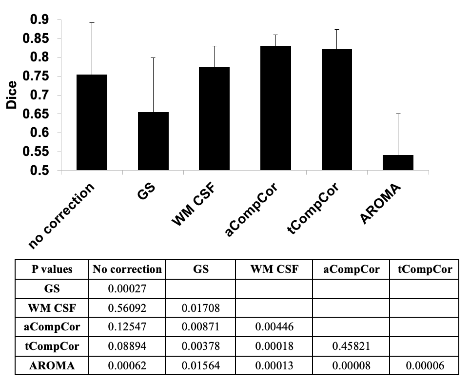
Evaluation metrics: For each processed fMRI dataset, template-based rotation (TBR)5 is used to generate resting-state connectivity (rs-connectivity) maps using Yeo’s seven resting-state network (rs-network) templates6. For each network, voxel-wise 2-factor repeated measure ANOVA is used with processing method and age as the two factors. F-map associated with the age factor is considered as a pseudo-ground truth, as it shows fcMRI differences between age groups regardless of the denoising method. Next, for each network and denoising method a difference map between the two age groups is generated using voxel-wise unpaired t-tests. Performance of the noise-removal methods are evaluated by comparing the difference t-map created by each method with the ground-truth F-map. Two parameters are used for this comparison. First, for each of the 7 networks, correlation between the F-map and the absolute value of the t-map is calculated. The correlation values are then compared using paired t-test across the 7 networks. Next, F-maps and t-maps are thresholded (p = 0.01 and Gaussian random field correction of p = 0.05). Dice coefficients between the thresholded F-map and the t-map of each method are calculated. The Dice coefficients are then compared between pairs of methods across the 7 networks using paired t-test.
Results
Figure 1 shows the spatial correlation between the pseudo ground-truth F-map and the t-map for different processing methods averaged across the networks. The Dice coefficient between the thresholded F-map and the t-maps for different methods are presented in Figure 2. Both figures showed that the difference maps generated from AROMA and GS are less similar to the ground-truth, indicating these denoising methods also remove meaningful fcMRI differences between age groups. In contrast, aCompCor and tCompCor create maps that have the highest similarity with the pseudo-ground-truth map.Discussion
As we showed in our previous work2, the performances of different denoising methods vary by age. We previously showed that AROMA and GS consistently remove more heart-beat and respiratory frequencies but also the most low-frequency signals. However, we were uncertain how this affects our ability to distinguish age-related differences in functional connectivity from changes in the fMRI signal due to physiological noise. Here we confirmed that the removed low-frequency signals are in-fact signals that contain information about brain connectivity. aCompCor and tCompCor seem to offer a good compromise in removing physiological signals while preserving fcMRI information.Conclusion
The findings of this study directly influence the choice of rs-fMRI denoising methods. The results suggest that aCompCor and tCompCor remove physiological noise while retaining neuronal information better than global-signal regression and ICA AROMA.Acknowledgements
No acknowledgement found.References
1. Liu, T. T. Noise contributions to the fMRI signal: An overview. Neuroimage 143, 141–151 (2016).
2. Golestani A.M., Chen J.J., Comparing the efficiency of data-driven noise regression in removing heartbeat and respiratory signals from resting-state fMRI data: Difference across age groups. ISMRM (2022).
3. Behzadi, Y., Restom, K., Liau, J. & Liu, T. T. A component based noise correction method (CompCor) for BOLD and perfusion based fMRI. Neuroimage 37, 90–101 (2007).
4. Pruim, R. H. R. et al. ICA-AROMA: A robust ICA-based strategy for removing motion artifacts from fMRI data. Neuroimage 112, 267–277 (2015).
5. Schultz, A. P., Chhatwal, J. P., Huijbers, W., Hedden, T., van Dijk, K. R. A., McLaren, D. G., et al. Template based rotation: a method for functional connectivity analysis with a priori templates. Neuroimage 102(Pt 2), 620–636 (2014).
6. Yeo, B. T. T., Krienen, F. M., Sepulcre, J., Sabuncu, M. R., Lashkari, D., Hollinshead, M., et al. The organization of the human cerebral cortex estimated by intrinsic functional connectivity. J. Neurophysiol. 106, 1125–1165 (2011).
Figures