2653
Functional and Structural Connectivity in the TgF344-AD Transgenic Rat Model of Alzheimer's Disease using rs-fMRI & DTI at 21.1 T
Jenna M. Radovich1,2, Jordan Ogg3, Zachary Baty1,2, Aaron Wilber3, and Samuel Colles Grant1,2
1National High Magnetic Field Laboratory, Florida State University, Tallahassee, FL, United States, 2Chemical & Biomedical Engineering, FAMU-FSU College of Engineering, Tallahassee, FL, United States, 3Psychology, Florida State University, Tallahassee, FL, United States
1National High Magnetic Field Laboratory, Florida State University, Tallahassee, FL, United States, 2Chemical & Biomedical Engineering, FAMU-FSU College of Engineering, Tallahassee, FL, United States, 3Psychology, Florida State University, Tallahassee, FL, United States
Synopsis
Keywords: Alzheimer's Disease, Brain Connectivity, Graph theory, Progressive Degeneration
A hallmark of Alzheimer's Disease (AD) is spatial disorientation, such as getting lost in new locations. A potential cause is disrupted exchange between egocentric and allocentric reference frames, in which the parietal and retrosplenial cortex have roles. This study aimed to examine rs-fMRI and DTI in relationship with coordination between reference frames in a transgenic AD rat model. Behavioral and DTI data suggest that both age and genotype lead to declines in action-orientation performance with alterations appearing at 5 mon, and at 6 mon structurally with profound changes developing longitudinally across multiple ROI.INTRODUCTION
A hallmark of preclinical Alzheimer’s Disease (AD) is spatial disorientation, such as getting lost in new locations. One potential cause is disrupted exchange between egocentric and allocentric reference frames. Both the parietal (PC) and retrosplenial cortices (RSC) have garnered attention for their roles in encoding and transforming information between these reference frames, as well as the hippocampus (HPC) due to its role in learning and memory. The RSC and PC also are earlier sites of dysfunction in humans with AD and rodents modeling aspects of AD. This study aimed to examine resting-state functional MRI (rs-fMRI) and diffusion tensor imaging (DTI) in relationship with coordination between reference frames in the TfF334-AD rat model. We hypothesized that pathology development in transgenic AD rats leads to brain network dysfunction, which causes impaired coordination between reference frames.METHODS
In double transgenic TgF344-AD rats and littermate controls, longitudinal MRI data acquired in vivo at 21.1 T to assess functional and structural connectivity alterations by rs-fMRI and structural DTI using the application of graph theory. Under light anesthesia (1-2% isoflurane), full brain rs-fMRI datasets were acquired at 250x250x1500 mm using 300 repetitions without stimulation, with a TE/TR of 20 ms/5 s, 23 slices, and a 4-segment spin-echo EPI. rs-fMRI was followed by acquisition of an 18-direction spin-echo EPI DTI dataset at 200x200x1000 mm, with 23 slices, 4 averages, and 6 segments. Rats were scanned at 2, 4, 6, 10 & 12 mons.MR data were compared to behavioral tasks in which reference frame coordination was assessed. Using an action-oriented spatial navigation task, rats were required to associate action of removing one of two objects (left versus right) with locations. Day to criterion were defined as a scored of 85% correct out of 40 trials for 3 d in a row, with a maximum of 6 d. Testing stopped at 35 d maximum if criterion not met to allow for MR imaging at intended time points.
RESULTS
DTI data were analyzed via DSI Studio, with the software aligning the Waxholm-Ferris Brain Atlas to define the ROI. Particular ROI of note included the PC, RSC, HPC, temporal (TEa), piriform (PIR) and cerebellar cortex (CBX). The PC, RSC, and HPC were selected based on the stated hypothessi while the TEa, PIR and CBX were reported due to major differences seen between Tg and WT at the 12-mon timepoint.At 6 mo, Tg rats had consistently higher weighted degree values than WT in all ROI. There was little change to 10 mo with Tg rats having higher values than WT for all but the PIR. By 12 mo, WT had much higher values than Tg.
Behavioral data indicated significant deficits emerging in the location-action task average in both groups at 3.5- and 8.5-mon compared to 5 mon. Tg rats performed significantly better compared to WT at 9 mo. However, head scanning was significantly reduced in Tg at 5 mon compared to WT.
DISCUSSION
Higher values of Tg at 6 mon is not unexpected, as previous studies have shown higher connectivity at 6 mon and was hypothesized to be a compensatory mechanism as the AD pathology begins to develop. ROI may be connected to more nodes at this point in time, but it is likely that the connections are beginning to degrade. At 10 mon, WT values begin to meet and overtake Tg, as their brains are still developing and able to make new connections over time. By 12 mon, WT specimens have much higher values than Tg as the ROI connections to other nodes have degraded and a more complete, full brain AD pathology has developed. This result matches previous studies.CONCLUSIONS
Data suggest that both age and genotype lead to declines in action-orientation performance, with differences in genotype emerging at 5 mon of age and increasing at older timepoints. These alterations were reflected in the structural networks in all ROI by 12 mon, with more profound changes developing longitudinally. These findings highlight a new focus for understanding cognitive deficits in AD by using allocentric and egocentric coordination as a novel predictor of early declines in AD.Acknowledgements
All work has been done in accordance with the Florida State University Animal Care and Use Committee. This work was supported by the Keck Foundation, US National High Magnetic Field Laboratory, which is supported by the National Science Foundation (DMR-1644779) and the State of Florida, and the NIH (R00 AG049090 and R01 AG070094). Research also funded by the Florida Department of Health (20A09).References
No reference found.Figures
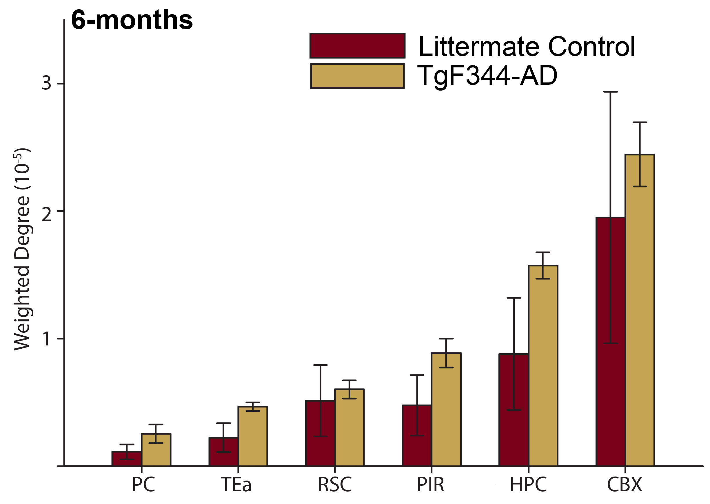
The weighted degree at 6 mon of the Tg is consistently higher than WT for all ROI. The higher values at this time point could be related to previous reports of hyperactivity and overcompensation for appearing brain pathology in AD rodents at 6 mon.
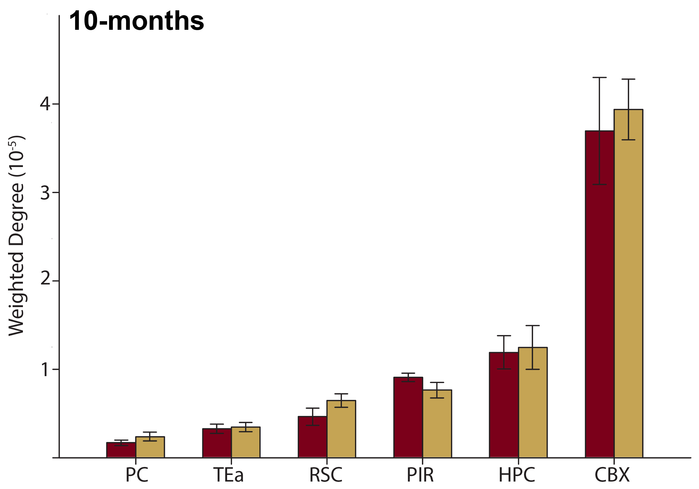
The weighted degree at 10 mon of the Tg is consistently higher than the WT for all ROI, with the exception of the piriform region.
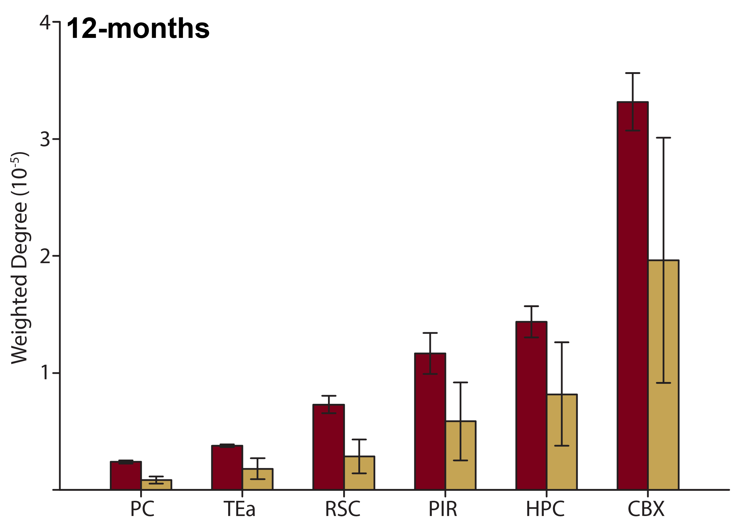
The weighted degree at 12 mon of the Tg is consistently lower than WT for all ROI. ROI of Tg animals have fewer connections to other nodes than WT at this timepoint, reflecting later stages of pathology related to emerging hypoactivity within the brain.
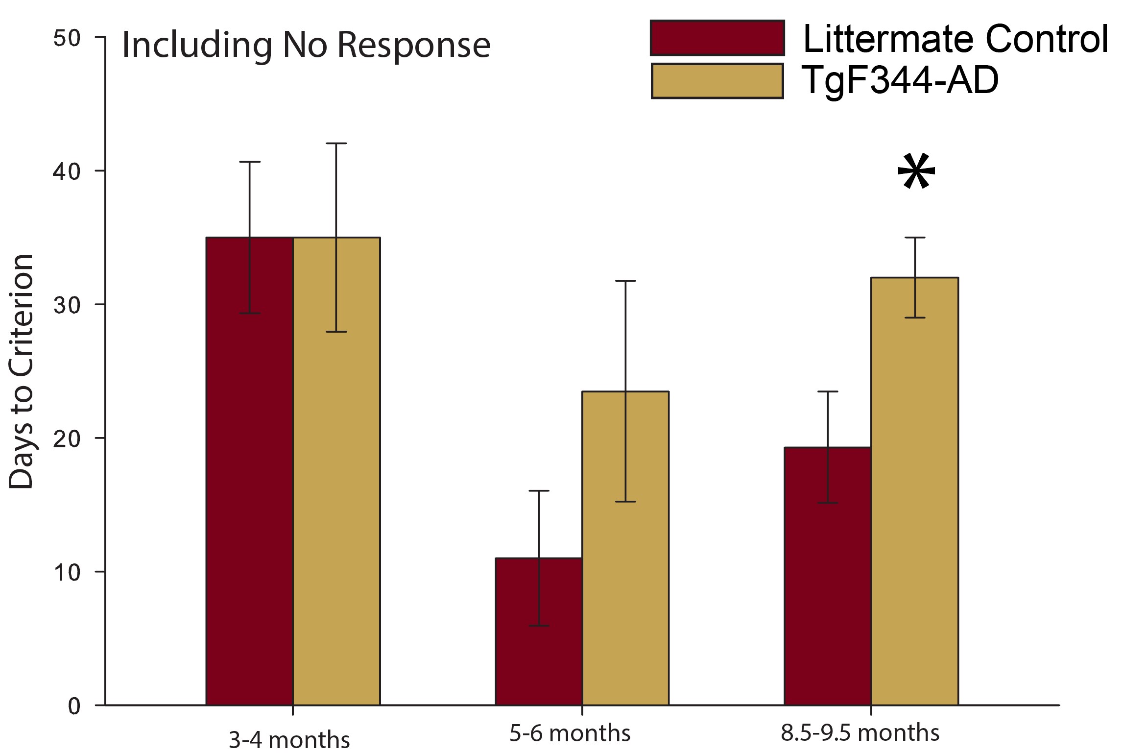
Animals demonstrated worse average performance in both groups at 3 and 8.5 mon, compared to 5 mon.
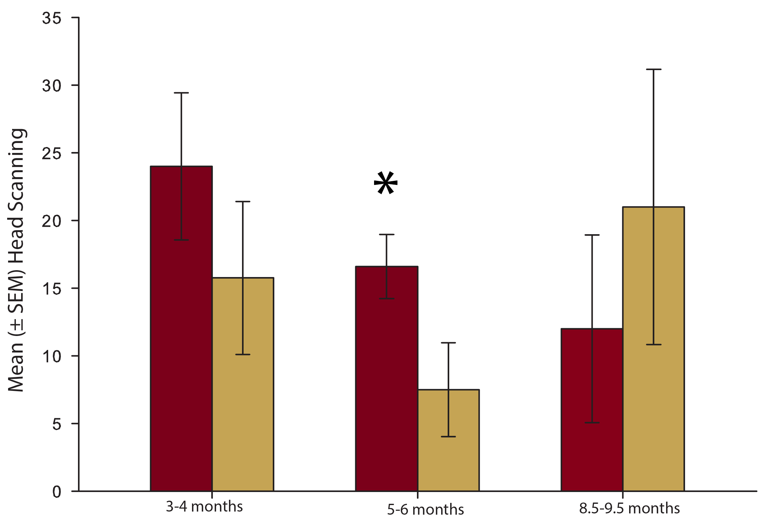
Head scanning is significantly reduced at 5 mon in Tg animals. A head scan is defined as when an animal exits the arm of the plus maze and looks in a particular direction (either left or right).
DOI: https://doi.org/10.58530/2023/2653