2643
Two clinically feasible myelin water imaging methods (MCR-DIMWI and METRICS) can differentiate patients with a leukodystrophy from controls.1Department of Pediatric Neurology, Amsterdam UMC, Amsterdam, Netherlands, 2Donders Institute for Brain, Cognition and Behaviour, Radboud University, Nijmegen, Netherlands, 3Department of Physics and Astronomy, University of British Columbia, Vancouver, BC, Canada, 4Department of Radiology and Nuclear Medicine, Amsterdam UMC, Amsterdam, Netherlands
Synopsis
Keywords: White Matter, Genetic Diseases, Leukodystrophy
Imaging biomarkers are needed for studying white matter (WM) diseases. Myelin water imaging (MWI) uses multi-compartment relaxometry to estimate myelin content, and is promising for use in leukodystrophies. We applied MCR-DIMWI and METRICS, two novel whole-brain MWI techniques, to a cohort of 9 leukodystrophy patients and 15 controls. Myelin water fractions (MWFs) from both techniques correlated well. In patients, MWF was decreased. For both techniques region-specific MWFs and relaxation metrics could differentiate patients from controls. They are promising for use in the context of leukodystrophies; additional studies are required to further explore potential clinical application.
INTRODUCTION
Leukodystrophies are a heterogeneous group of genetic diseases selectively affecting the white matter (WM) of the central nervous system. MRI has a central role in both diagnosing and monitoring patients. However, imaging biomarkers are scarce and rarely optimised in the leukodystrophy setting. Recently, two myelin water imaging (MWI) techniques were described, providing 3D images within clinically feasible acquisition times: multi-spin echo T2 relaxation imaging with compressed sensing (METRICS)1, and multi-compartment relaxometry with diffusion-informed MWI (MCR-DIMWI)2. METRICS is based on multi-spin echo MWI3, separating myelin water from intra/extracellular and free water based on T2 values. MCR-DIMWI relies on the short apparent T1 and T2* of myelin water, as well as its frequency offset as predicted by the hollow cylinder model2,4. We applied both techniques in a cohort of patients with a leukodystrophy and controls.METHODS
9 patients with several different leukodystrophies (median age (in years) 9.2; range 0.4-62.4) and 15 control subjects (23.5; 2.3-61.3) were included.The MRI acquisition (3T, Philips) and analysis protocol consisted of:
a) 3D-T1 MPRAGE, segmented into WM and (deep) GM using SynthSeg5.
b) 3D-FLAIR, to identify WM-abnormalities.
c) Multi-shell DWI: 9b0, 29b1000, 59b2000 + reversed phase-encode references (TR=3750ms, TE=95ms, SENSE=2x1.5, MB factor=2, 6min36s acquisition time). Analyzed with NODDI6 and FSL-BEDPOSTX. Results were used in MCR-DIMWI model. Single-shell tensor data were used to register WM lobes and tract-based ROIs from JHU- and IIT-atlases with DTI-TK7.
d) MCR-DIMWI: multi-echo gradient echo (TR=46ms, 12 echoes, TE=2.15-35.7ms, ΔTE=3.05ms, 7 flip angles 5-70°, SENSE=1.4x1.5, 10min5s). Analysis as described2.
e) METRICS: multi-echo spin echo (TR=1066ms, 56 echoes, TE=7-392ms, ΔTE=7ms, compressed SENSE factor 10, 9min26s), as described1. Analysis as described, assuming myelin-water T2 10-40ms, IET2 40-200ms1, while including spatial correlations8.
Conventional images (a and b) have 1 mm isotropic resolution. Quantitative methods (c,d,e) have 2.5 mm isotropic resolution to ensure high SNR within clinically feasible acquisition times.
MCR-DIMWI and METRICS myelin water fractions (MWFs), relaxation times, and quality control parameters were extracted per ROI using FSL and analyzed using RStudio.
RESULTS
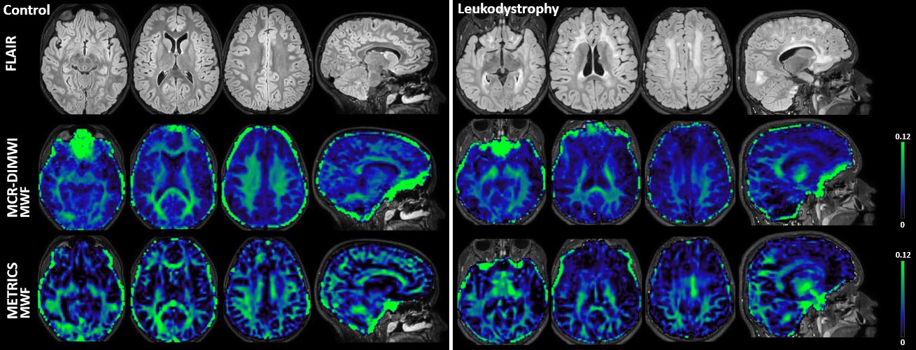
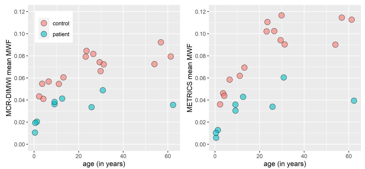
Examples of MWF maps of a control subject and a patient are shown in Figure 1. Good quality MWF maps were obtained throughout the brain. Artefactually high MWF is commonly observed in orbitofrontal regions in MCR-DIMWI due to susceptibility artefacts, and in METRICS in areas with CSF or venous blood flow and in deep GM areas.Both techniques detect lower MWF of cerebral WM in patients than in controls (Figure 2). An increase of MWF with age is also seen, corresponding to myelination during development.
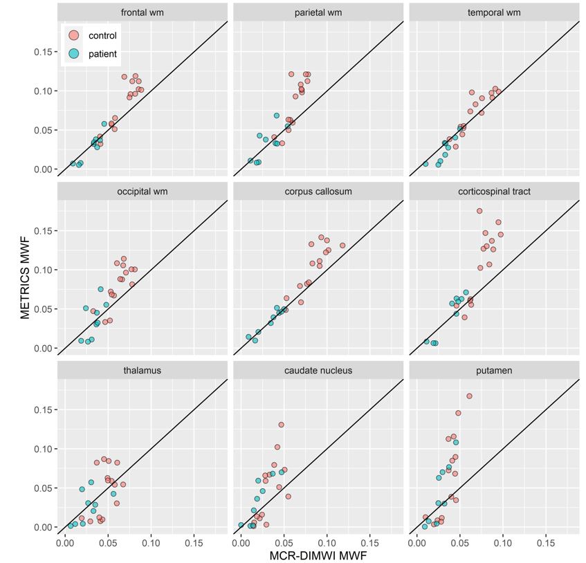
The techniques correspond well regarding MWFs in most ROIs, although METRICS tends to yield higher values (Figure 3).
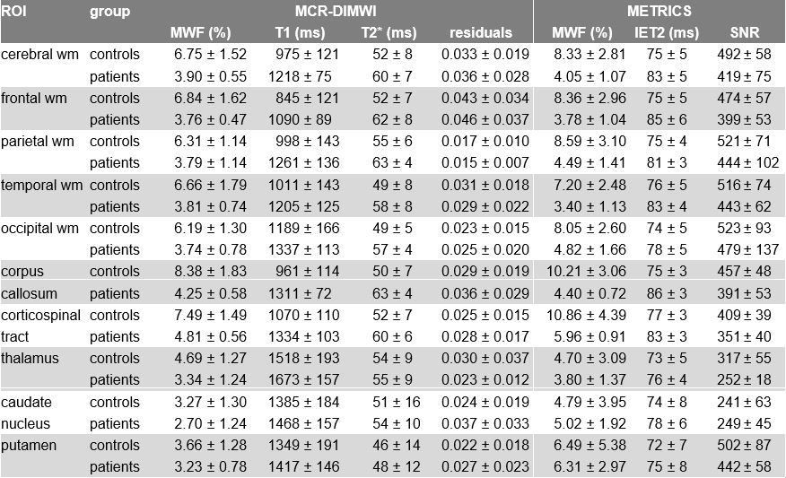
Table 1 shows mean MWF values per region obtained with both techniques for all subjects above the age of 2 years (thus excluding the 3 youngest patients). Differences in MWF between patients and controls are most prominent in WM ROIs. The coefficient of variation is larger for METRICS than for MCR-DIMWI, both within subjects (variation within ROIs, data not shown) and between subjects. Table 1 also shows single-compartment relaxation times from MCR: T1 (from TE=2.15ms images), and T2* (from FA=10° images), and from METRICS the geometrical mean of intra/extracellular water T2 (IET2). Quality control parameters for both methods are also shown. All relaxation times are higher in patients than controls.
DISCUSSION
Both MCR-DIMWI and METRICS provide significantly different results between leukodystrophy patients and control subjects. The lower MWF values in the patient group were expected, as all patients had varying degrees of pathologically affected WM. This was also the reason for the increased relaxation times in patients, as healthy myelin plays an important role in accelerating the relaxation process. Very short T2 relaxation times (for instance in deep GM) may mistakenly be modelled as myelin water and overestimate METRICS-MWF. Differences between both techniques regarding homogeneity of MWF within regions will be explored in future studies. Furthermore, control values of subjects under 2 years of age should be obtained.MCR-DIMWI has limited value in areas with susceptibility artefacts (which will be less severe at higher spatial resolution). In MCR-DIMWI, motion correction is possible between volumes acquired with variable flip angles. With METRICS, no motion correction is currently possible. Both techniques can be further accelerated9,10.
CONCLUSION
Both MCR-DIMWI and METRICS provide whole brain MWF-maps and are able to distinguish leukodystrophy patients from controls. A relatively low spatial resolution ensures a high SNR and short acquisition times, and is sufficient for globally affected WM disorders. Future studies incorporating clinical measures from leukodystrophy patients can further explore the potential use of these MWI techniques in monitoring disease progression, as well as treatment effects.Acknowledgements
No acknowledgement found.References
1 Dvorak, A. V. et al. Multi-spin echo T2 relaxation imaging with compressed sensing (METRICS) for rapid myelin water imaging. Magn Reson Med, doi:10.1002/mrm.28199 (2020).
2 Chan, K. S. & Marques, J. P. Multi-compartment relaxometry and diffusion informed myelin water imaging - Promises and challenges of new gradient echo myelin water imaging methods. Neuroimage 221, 117159, doi:10.1016/j.neuroimage.2020.117159 (2020).
3 MacKay, A. et al. In vivo visualization of myelin water in brain by magnetic resonance. Magn Reson Med 31, 673-677, doi:10.1002/mrm.1910310614 (1994).
4 Wharton, S. & Bowtell, R. Fiber orientation-dependent white matter contrast in gradient echo MRI. Proc Natl Acad Sci U S A 109, 18559-18564, doi:10.1073/pnas.1211075109 (2012).
5 Billot, B. et al. SynthSeg: Domain Randomisation for Segmentation of Brain MRI Scans of any Contrast and Resolution. arXiv preprint arXiv:2107.09559 (2021).
6 Zhang, H., Schneider, T., Wheeler-Kingshott, C. A. & Alexander, D. C. NODDI: practical in vivo neurite orientation dispersion and density imaging of the human brain. Neuroimage 61, 1000-1016, doi:10.1016/j.neuroimage.2012.03.072 (2012).
7 Zhang, H., Yushkevich, P. A., Alexander, D. C. & Gee, J. C. Deformable registration of diffusion tensor MR images with explicit orientation optimization. Med Image Anal 10, 764-785, doi:10.1016/j.media.2006.06.004 (2006).
8 Kumar, D. et al. Using 3D spatial correlations to improve the noise robustness of multi component analysis of 3D multi echo quantitative T2 relaxometry data. Neuroimage 178, 583-601, doi:10.1016/j.neuroimage.2018.05.026 (2018).
9 Chan, K.-S., Chamberland, M. & Marques, J. P. On the performance of multi-compartment relaxometry for myelin water imaging-Intra-subject and inter-protocol reproducibility. bioRxiv (2022).
10 Dvorak, A. V. et al. The CALIPR framework comprehensively improves acquisition, reconstruction & analysis of multi-component relaxation imaging. ISMRM abstracts 2851 (2022).
Figures