2636
Bright light therapy increases myelin density in posterior thalamic radiation in young adults with subthreshold depression: An ihMT MRI study1First Affiliated Hospital of Jinan University, Guangzhou, China, 2MR Research, GE Healthcare, Beijing, China
Synopsis
Keywords: White Matter, Magnetization transfer
Subthreshold depression (SD) is a significant risk indicator of major depressive episodes. This study used the inhomogeneous magnetization transfer (ihMT) technique to probe myelin abnormalities and its response to bright light therapy (BLT) in SD. The findings of this study suggest the macromolecular disruption of myelin in the posterior thalamic radiation, sagittal stratum, and uncinate fasciculus in the early stages of depression. Furthermore, the myelin impairments in the PTR could be reversed by BLT, thus suggesting they might be used as the potential neural target for BLT in SD.Background
Subthreshold depression (SD) is highly prevalent in the general population, particularly in adolescence and early adulthood [1]. As a significant risk indicator of later major depressive episodes [2], SD is associated with an increased burden of disease and suicide risk among adolescents [3]. However, the pathophysiological mechanism underlying SD remains unclear, and more attention should be paid to the treatment of SD and prevention of MDD. The inhomogeneous magnetization transfer (ihMT) is an emerging MRI technique that may offer high specificity for myelinated tissue [4]. Quantitative measures such as pseudo-quantitative ihMT (qihMT) and ihMT ratio (ihMTR) can be derived from ihMT data [5], while good inter- and intra-scanner reliability and reproducibility of ihMT measurements were reported by a test-retest, multi-site study [6-8]. Bright light therapy (BLT) is considered a cost-effective, well-tolerated treatment with fewer side effects compared with medications, thus holding considerable promise as a treatment for seasonal affective disorder [9] and non-seasonal affective disorders such as MDD [10]. In this study, we used ihMT technique to investigate myelin integrity in a relatively large sample size of university students with SD. Part of the SD participants underwent eight weeks of BLT. We hypothesized that SD might show reduced ihMT values in some WM fibers such as PTR, IFOF, and UF compared to controls at baseline. In addition, we assumed that the myelin density of certain WM fibers might be changed in SD participants after treatment.Methods
A total of 104 college students with SD and 91 age- and sex-matched healthy controls (HCs) were included. All participants underwent ihMT imaging, and pseudo-quantitative ihMT (qihMT), and ihMT ratio (ihMTR) were obtained. The qihMT and ihMTR values of 50 white matter (WM) fibers were compared between SD and HCs. Thirty-one SD participants underwent eight weeks of BLT, after which we observed the effect of treatment on WM fibers with abnormal qihMT and ihMTR values at baseline in SD. Also, the psychological variables were compared before and after BLT in SD.Results
Differences in IhMT measurements between SD and HCs groupCompared with the HCs group, qihMT values in the bilateral PTR (including optic radiation) (right PTR: t = -3.402, p < 0.001; left PTR: t = -3.314, p = 0.001), right sagittal stratum (SS) (including ILF and IFOF) (t = -3.445, p < 0.001), and right UF (t = -3.334, p = 0.001) were significantly decreased in the SD group. Also, the SD group had significantly decreased ihMTR values in the left PTR (including optic radiation) (t = -3.395, p = 0.001) (Table 3 and Figure 1). No other WM fiber tracts were found to have significantly different qihMT and ihMTR values between the SD group and HCs group (p > 0.001). There were no significant correlations between the qihMT values in the above WM fiber tracts and any mood scale in the SD group (p > 0.05). With reference to DTI measures, there were no significant differences in FA, MD, RD, and AD between SD and HCs groups (p > 0.001).
Differences in IhMT measurements in SD after BLT
After 8 weeks of BLT, we focused on the bilateral PTR (including optic radiation), right SS (including ILF and IFOF), and right UF, finding that compared with the qihMT values of the right PTR (including optic radiation) at baseline, the qihMT values of the right PTR (including optic radiation) were increased in SD after treatment (t = 2.258, p = 0.032) (Figure 2A). In addition, the qihMT values of the right SS (including ILF and IFOF) increased in post-treatment SD compared with pre-treatment SD (t = 1.949, p = 0.061); the p-value was indicative of marginal significance. No significant differences of qihMT values in other WM fiber tracts and ihMTR values were found between before and after treatment in SD (p > 0.05) (Table 2). No significant differences of FA, MD, AD, and RD values were found between before and after treatment in all observed WM fiber tracts in SD (p > 0.05) (Table S1).
For correlation analysis, the changes of qihMT values in the right PTR (including optic radiation) had a significant negative correlation with the changes in PANAS-negative scores of SD (r = -0.421, p = 0.026) (Figure 2B).
Conclusion
This study provides preliminary evidence of macromolecular disruption of myelin in several WM tracts, including the PTR, IFOF, ILF, and UF in SD. Decreased density of the WM myelin may have an important role in the neural pathology of SD and may occur early in the initial stages of depression. Furthermore, the myelin impairments in the PTR could be reversed by BLT, which represents the potential neural targets for BLT in SD. The ihMT technique may become a valuable and sensitive screening method for evaluating stages of depression and assessing treatment responses.Acknowledgements
The study was supported by grants from the National Natural Science Foundation of China (81671670, 81971597, and 82172530); National Key Research and Development Project (2020YFC2005700); Key-Area Research and Development Program of Guangdong Province (2020B1111100001). The funding organizations play no further role in study design, data collection, analysis and interpretation and paper writing.
References
1. Bertha, E.A. and J. Balázs, Subthreshold depression in adolescence: a systematic review. Eur Child Adolesc Psychiatry, 2013. 22(10): p. 589-603.
2. Tuithof, M., et al., Course of subthreshold depression into a depressive disorder and its risk factors. J Affect Disord, 2018. 241: p. 206-215.
3. Balázs, J., et al., Adolescent subthreshold-depression and anxiety: psychopathology, functional impairment and increased suicide risk. J Child Psychol Psychiatry, 2013. 54(6): p. 670-7.
4. Malik, S.J., et al., Steady-state imaging with inhomogeneous magnetization transfer contrast using multiband radiofrequency pulses. Magn Reson Med, 2020. 83(3): p. 935-949.
5. Varma, G., et al., Magnetization transfer from inhomogeneously broadened lines: A potential marker for myelin. Magn Reson Med, 2015. 73(2): p. 614-22.
6. Zhang, L., et al., Reproducibility of inhomogeneous magnetization transfer (ihMT): A test-retest, multi-site study. Magn Reson Imaging, 2019. 57: p. 243-249.
7. Zou, K.H., et al., Statistical evaluations of the reproducibility and reliability of 3-tesla high resolution magnetization transfer brain images: a pilot study on healthy subjects. Int J Biomed Imaging, 2010. 2010: p. 618747.
8. McHinda, S., et al., Whole brain inhomogeneous magnetization transfer (ihMT) imaging: Sensitivity enhancement within a steady-state gradient echo sequence. Magn Reson Med, 2018. 79(5): p. 2607-2619.
9. Pjrek, E., et al., The Efficacy of Light Therapy in the Treatment of Seasonal Affective Disorder: A Meta-Analysis of Randomized Controlled Trials. Psychother Psychosom, 2020. 89(1): p. 17-24.
10. Lam, R.W., et al., Efficacy of Bright Light Treatment, Fluoxetine, and the Combination in Patients With Nonseasonal Major Depressive Disorder: A Randomized Clinical Trial. JAMA Psychiatry, 2016. 73(1): p. 56-63.
Figures
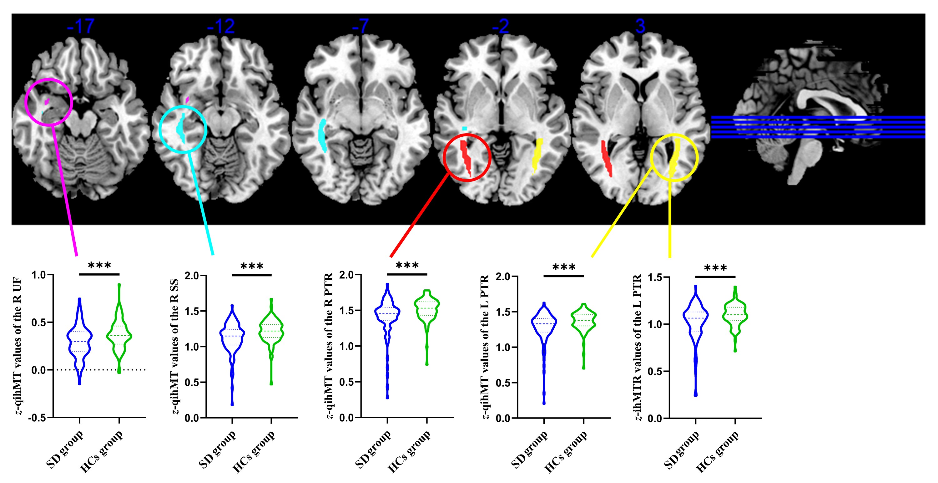
Figure 1. Significant different qihMT and ihMTR values between the SD group and HCs group. qihMT, pseudo-quantitative inhomogeneous magnetization transfer; ihMTR, inhomogeneous magnetization transfer ratio; SD, subthreshold depression; HCs, healthy controls; PTR, posterior thalamic radiation (include optic radiation); SS, sagittal stratum; UF, uncinate fasciculus; L, left; R, right; ***, p < 0.001.
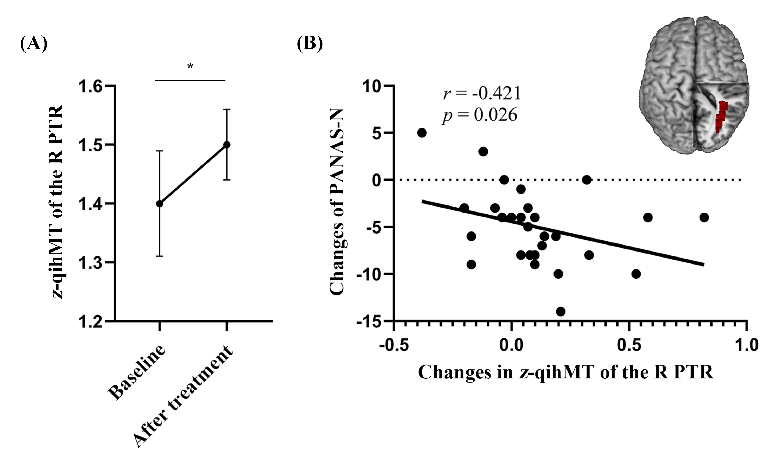
Figure 2. Differences of IhMT measurements of SD before and after BLT, and correlations between changes of qihMT values and changes of PANAS-N scores in SD. SD, subthreshold depression; BLT, bright light therapy; PANAS-N, negative affect schedule; qihMT, pseudo-quantitative inhomogeneous magnetization transfer; PTR, posterior thalamic radiation (include optic radiation); R, right; *, p < 0.05.