2632
Cortical thickness and white matter integrity abnormalities in dysthyroid optic neuropathy
Mengsha Zou1, Hongzhang Zhu1, Yunzhu Wu2, and Zhiyun Yang1
1Department of Radiology, The First Affiliated Hospital of Sun Yat-Sen University, 58th, The Second Zhongshan Road, Guangzhou, China, Guangzhou, China, 2MR Scientific Marketing, Siemens Healthineers Ltd., Shanghai, China, Shanghai, China
1Department of Radiology, The First Affiliated Hospital of Sun Yat-Sen University, 58th, The Second Zhongshan Road, Guangzhou, China, Guangzhou, China, 2MR Scientific Marketing, Siemens Healthineers Ltd., Shanghai, China, Shanghai, China
Synopsis
Keywords: White Matter, Brain
A pilot study to examine the structural MRI features of patients with dysthyroid optic neuropathy (DON) as compared to normal controls, and correlate pathologic MRI changes to clinical symptoms of the disease. DON patients show reduced cortical volume in precuneus and occipital pole, decrease area in precuneus and precentral gyrus, and widespread decreased FA value, mainly in genu of corpus callosuma and body of corpus callosum. Meanwhile, the FA of was associated with visual acuity. Our study provides a new insight of pathological mechanism of DON.Introduction
Dysthyroid optic neuropathy (DON) is a serious complication of Graves orbitopathy (GO) that may result in permanent loss of vision. DON shows ischemia and axonal loss of the optic nerve that could be identified by measuring the diffusion tensor imaging[1]and T2 mapping[2]. However, the degree to which dysthyroid optic neuropathy has effects in the brain beyond the eye and the visual pathways is unclear. This study aimed to explore the morphological and microstructural changes of grey and white matter in DON patients.Methods
We acquired magnetic resonance imaging (MRI) scans from 42 DON patients and 42 well-matched healthy controls (HCs). GO diagnosis was based on the diagnostic criteria postulated by Bartley GB et al. [3]. DON diagnosis was based on at least two of the following signs: the deterioration of VA (< 1.0), loss of color vision, optic disc swelling, and relative afferent pupillary defect [4]. Exclusion criteria were: 1) no history of neurological, psychiatric, major-medical conditions, or substance abuse; 2) no intracranial or other intra-orbital lesions on routine MR images. Scanning was performed on a 3T MRI system (MAGNETOM Prisma, Siemens Healthcare, Erlangen, Germany) using a 64-channel head coil. The protocol included high-resolution T1-weighted MR images and q-space acquisition mode diffusion weight image (DWI) data. T1-weighted magnetization prepared rapid acquisition gradient echo (MPRAGE) sequence scans were acquired at a resolution of 0.8 mm3, 224 slices, TR/TE = 2300 ms/2.43 ms, scan time = 6 min. DWI data was acquired using a 7 min spin-echo echo-planar imaging sequence with a half coverage Cartesian q-space grid scheme. the DWI parameters: TR/TE = 3700 ms/72 ms, FOV = 220 × 220 mm2, voxel size = 2 × 2 × 2 mm3, in-plane acceleration factor=2, slice acceleration factor=2. Multiple b-values with the maximum 3000 s/mm2 along 99 diffusion gradient directions were sampled on the grid points in the 3D q-space. DTI parameters were calculated using NeuDiLab software developed in-house with Python, which is based on an open-resource tool DIPY (Diffusion Imaging in Python). Cortical thickness, cortical volume and area were measured using the surface-based morphometry (SBM) approach. We also evaluated DTI-metrics (fractional anisotropy (FA), mean diffusivity (MD), axial diffusivity (AD) and radial diffusivity (RD)) derived from DWI data using tract-based spatial statistics (TBSS). For those brain regions exhibiting altered structure, correlations between alterations and visual acuity were analyzed in all patientsResults:
Brain region of significant change in grey matter and white matter between DON patients and healthy controls revealed by SBM and TBSS were summarized in Table 2. Compared with controls, DON patients exhibited decrease cortical volume in precuneus and occipital pole (Fig1A-B). DON patients exhibited decrease cortical area in precuneusand precentral gyrus (Fig1C-D). However, cortical thickness showed no significance between DON patents and HC. DON exhibited widespread decreased FA value (Genu of corpus callosum, body of corpus callosum, anterior corona radiata superior corona radiata, posterior corona radiata and superior longitudinal fasciculus) (Fig2). MD, AD and RD showed no significant difference between DON patents and HC. In patient group, Pearson partial correlation showed no significant correlation between the change of cortical volume in precuneus and visual acuity (r = 0.143, P = 0.387), the change of cortical volume in occipital pole showed no significant correlation with visual acuity (r = 0.252, P = 0.121). the FA value in the body of corpus callosum was positively correlated with visual acuity (r = 0.446, P = 0.015). (Fig. 3)Discussion
To the best of our knowledge, this is the first study to investigate brain morphometric alterations in DON patients using multimodal neuroimaging measures including cortical thickness, cortical volume and area, as well as white matter integrity. The use of multimodal in brain imaging analysis allows us to conduct a more comprehensive understanding in the neuropathology of DON. Additionally, we analyzed correlation between visual acuity and altered brain structure. The main findings of our study are that DON patients have significantly reduced cortical volume and area structural alteration of precuneus, and cortical volume change in occipital pole. And lower FA in brain white matter compared with HC. Especially, the FA value in the body of corpus callosum was positively correlated with visual acuity.Conclusions
DON patients demonstrated reduced cortical volume and area structural alteration of precuneus, and cortical volume change in occipital pole. Microstructural alterations in brain white matter. Combined multimodal neuroimaging methods may provide a more comprehensive perspective to clarify the neuropathology of DON patient.Acknowledgements
No acknowledgementsReferences
[1] B. Özkan, Y. Anik, B. Katre, Ö. Altintaş, M. Gençtürk, N. Yüksel, Quantitative Assessment of Optic Nerve With Diffusion Tensor Imaging in Patients With Thyroid Orbitopathy, Ophthalmic Plastic and Reconstructive Surgery 31(5) (2015) 391-395.[2] M. Zou, D. Wu, H. Zhu, X. Huang, X. Zhao, J. Zhao, W. Fu, R. Li, B. Li, P. Wan, S. Hong, Y. Li, H. Xiao, Z. Yang, Multiparametric quantitative MRI for the evaluation of dysthyroid optic neuropathy, Eur Radiol 32(3) (2022) 1931-1938.
[3] L. Barrett, H.J. Glatt, R.M. Burde, M.H. Gado, Optic nerve dysfunction in thyroid eye disease: CT, Radiology 167(2) (1988) 503-7.
[4] B. Rutkowska-Hinc, E. Maj, A. Jablonska, J. Milczarek-Banach, T. Bednarczuk, P. Miskiewicz, Prevalence of Radiological Signs of Dysthyroid Optic Neuropathy in Magnetic Resonance Imaging in Patients with Active, Moderate-to-Severe, and Very Severe Graves Orbitopathy, Eur Thyroid J 7(2) (2018) 88-94.
Figures
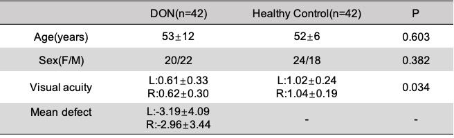
Table1 Demographic characteristic and visual function of DON patients and healthy control groups (P< 0.05)
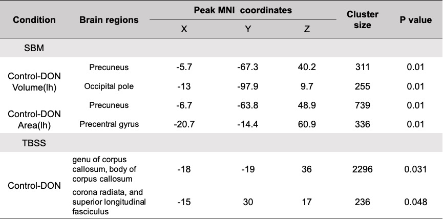
Table 2. Brain region of significant change in grey matter and white matterbetween DON patients and healthy controls revealed by SBM and TBSS(P < 0.05)
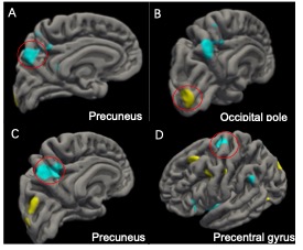
Figure 1 Cortical regions exhibiting significant gray matter change in dysthyroid optic neuropathy patients compared to normal controls. Data are rendered on a standard cortical surface. Significant reduction of gray matter volume was observed in the Precuneus(A), Occipital(B). Significant reduction of gray matter area was observed in the Precuneus(C), Precentral(D). Locations and sizes of all clusters are listed in Table 2.
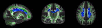
Figure 2 Representative sections of voxel-wise TBSS analysis show reduced FA in DON patients than in healthy controls. The voxels with blue or light blue color represent those with significant reduction of FA in DON patients relative to healthy controls (P<0.05, threshold-free cluster enhancement family-wise error corrected). The significant regions are thickened for better visibility. The white matter skeleton is shown in green (FA>0.2). The background image is the mean FA map derived from all subjects.
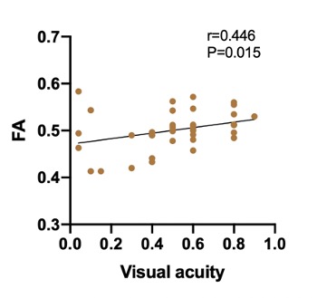
Figure 3 Scatter diagrams showed the significant correlation between visual acuity and FA value in the body of corpus callosum in DON group. FA was positively correlated with visual acuity (r = 0.446, P = 0.015).
DOI: https://doi.org/10.58530/2023/2632