2621
Cortical demyelination in chronic post-traumatic stress disorder: A study of World Trade Center responders
Juin W. Zhou1, Chuan Huang1,2, Kritikos Minos3, Sean A.P. Clouston3, Megan K. Horton4, Roman Kotov5, Roberto G. Lucchini6,7, Evelyn J. Bromet5, and Benjamin J. Luft8,9
1Biomedical Engineering, Stony Brook University, Stony Brook, NY, United States, 2Radiology, Renaissance School of Medicine, Stony Brook, NY, United States, 3Family, Population, and Preventative Medicine, Renaissance School of Medicine, Stony Brook, NY, United States, 4Environmental Medicine and Public Health, Icahn School of Medicine at Mount Sinai, New York, NY, United States, 5Psychiatry, Renaissance School of Medicine, Stony Brook, NY, United States, 6Environmental Health Sciences, Florida International University, Miami, FL, United States, 7Medical and Surgical Specialties, Radiological Sciences, and Public Health, University of Brescia, Brescia, Italy, 8Medicine, Renaissance School of Medicine, Stony Brook, NY, United States, 9Stony Brook WTC Wellness Program, Renaissance School of Medicine, Stony Brook, NY, United States
1Biomedical Engineering, Stony Brook University, Stony Brook, NY, United States, 2Radiology, Renaissance School of Medicine, Stony Brook, NY, United States, 3Family, Population, and Preventative Medicine, Renaissance School of Medicine, Stony Brook, NY, United States, 4Environmental Medicine and Public Health, Icahn School of Medicine at Mount Sinai, New York, NY, United States, 5Psychiatry, Renaissance School of Medicine, Stony Brook, NY, United States, 6Environmental Health Sciences, Florida International University, Miami, FL, United States, 7Medical and Surgical Specialties, Radiological Sciences, and Public Health, University of Brescia, Brescia, Italy, 8Medicine, Renaissance School of Medicine, Stony Brook, NY, United States, 9Stony Brook WTC Wellness Program, Renaissance School of Medicine, Stony Brook, NY, United States
Synopsis
Keywords: Neurodegeneration, Psychiatric Disorders
World Trade Center (WTC) responders are at mid-life, with 23% presenting with chronic post-traumatic stress disorder (PTSD). Epidemiologic studies suggest that chronic PTSD is associated with psychomotor slowing and physical functional limitations consistent with intracortical neuroinflammation. Prior neuroimaging has suggested that chronic PTSD is associated with glial activation and reduced cortical complexity, as well as neurodegeneration in the hippocampus and anterior cingulate, suggestive of PTSD-induced cortical neuropathology. Hypothesizing that these results might reduce intercortical density, we evaluated intercortical demyelination in WTC responders with chronic PTSD. We used gray-white contrast purported to measure cortical demyelination and, by extension, interneuronal health.Introduction
Search-and-rescue operations following the collapse of the World Trade Center (WTC) exposed responders to extraordinary physical and psychological experiences. More than two decades later, approximately 23% of this population presents with symptoms of chronic post-traumatic stress disorder (PTSD).1 Prior neuroimaging studies in WTC responders with PTSD have identified increased neuroinflammation2, reduced cortical complexity3, and demyelination in key brain regions.Recent work has suggested that the presence of these factors might increase cortical demyelination and are potentially indicative of poor interneuronal health. The present study used gray-white matter contrast (GWC)—a purported surrogate biomarker for intracortical myelin health, previously used in studies of patients with mild cognitive impairment (MCI)4, schizophrenia5, Parkinson’s disease6, or autism7—to investigate differences between WTC responders with or without PTSD. In this study of GWC in a PTSD-afflicted population, we hypothesized that we would observe reduced intracortical myelination in cortical regions of interest (ROIs) previously identified in studies of PTSD. A second aim was to determine the extent of regional overlap between GWC and both previously reported cortical complexity and neuroinflammation.
Methods
In total, 102 responders aged 44-65 were included in this study (44 with chronic PTSD) as diagnosed through Structured Clinical Interviews for DSM-IV. All subjects were scanned on a 3T Siemens Biograph mMR scanner using MPRAGE sequence (TR/TE/TI = 1900, 2.49, 900 ms, flip angle = 9o, acquisition matrix = 256 x 256, voxel resolution = 0.89 x 0.89 x 0.89 mm3). Subjects’ images were segmented into white (WM) and gray matter (GM). To calculate GWC, GM and WM signal intensities were measured at 50% of the cortical thickness above and 1 mm below the gray-white matter interface. Subjects’ GWC surfaces were transformed onto the default template brain, averaged, and then smoothed at 25 mm FWHM. Analyses were performed using Freesurfer version 7.2.0.8 Associations between PTSD and GWC were estimated using general linear model, adjusting for voxel-wise CT, APOE phenotype, age, sex, and cognitive status. The model was also corrected for intracranial volume (ICV)-adjusted hippocampal volume to account for the volume reduction associated with PTSD.9 Multiple comparison correction was performed using permutation testing (n=10,000).Results
Responders were aged 57 on average, and the majority were male. GWC was found to be significantly reduced (p≦0.05) in WTC responders with PTSD in ROIs spanning the frontal, temporal, parietal, and occipital lobes (Figure 1). Table 1 shows ROIs affected in the present study and two prior WTC neuroimaging measures that used measures of cortical complexity3 and neuroinflammation2 to identify overlapping regions including the pars triangularis. However, there are also several regions that are unique to GWC, particularly in the frontal lobe.Discussion
Individuals with chronic PTSD suffer from ongoing stressful symptoms that can be debilitating, and those limitations are associated with more rapid onset of diseases of old age. This is the first study of GWC in individuals with chronic PTSD. The present study expands the literature, by identifying intracortical demyelination consistent with reduced interneuronal density in WTC responders with PTSD, in both previously identified and unique ROIs.The link with neurodegeneration is important since prior PTSD studies have suggested that PTSD can cause neurodegeneration in the insula, superior frontal gyrus, insula, middle and inferior temporal gyri, anterior cingulate, rostral anterior cingulate, orbitofrontal cortex, hippocampus, and amygdala.9 Prior results from our group note that physical functional decline are consistent with intraneuronal atrophy, and show that neurodegeneration is consistently accompanied by cognitive decline and impairment among responders with chronic PTSD. The present study expands on prior work to show that chronic PTSD is marked by the presence of poorer intracortical demyelination which may suggest a unique neurobiological signature
Acknowledgements
We would like to thank all of the participants of this study. This work was supported by the Centers for Disease Control and Prevention (CDC/NIOSH U01 OH011314), the National Institute on Aging (NIH/NIA P50 AG005138) and (NIH/NIA R01 AG049953), as well as the WTC Health and Wellness Program (CDC 200-2011-39361).
References
- Clouston, S. A. et al. Cognitive impairment among World Trade Center responders: Long-term implications of re-experiencing the 9/11 terrorist attacks. Alzheimers Dement (Amst) 4, 67-75 (2016). https://doi.org:10.1016/j.dadm.2016.08.001
- Deri, Y. et al. Neuroinflammation and Mild Cognitive Impairment in World Trade Center responders at midlife: A pilot study using [18F]-FEPPA PET imaging. Brain, Behavior, & Immunity-Health, 100287 (2021).
- Kritikos, M. et al. Cortical complexity in world trade center responders with chronic posttraumatic stress disorder. Translational Psychiatry 11, 597 (2021). https://doi.org:10.1038/s41398-021-01719-74
- Jefferson, A. L. et al. Gray & white matter tissue contrast differentiates Mild Cognitive Impairment converters from non-converters. Brain Imaging Behav 9, 141-148 (2015). https://doi.org:10.1007/s11682-014-9291-25
- Jørgensen, K. N. et al. Increased MRI-based cortical grey/white-matter contrast in sensory and motor regions in schizophrenia and bipolar disorder. Psychol Med 46, 1971-1985 (2016). https://doi.org:10.1017/s00332917160005936
- Uribe, C. et al. Gray/White Matter Contrast in Parkinson's Disease. Front Aging Neurosci 10, 89 (2018). https://doi.org:10.3389/fnagi.2018.00089
- Bezgin, G., Lewis, J. D. & Evans, A. C. Developmental changes of cortical white-gray contrast as predictors of autism diagnosis and severity. Transl Psychiatry 8, 249 (2018). https://doi.org:10.1038/s41398-018-0296-28
- Fischl, B. FreeSurfer. Neuroimage 62, 774-781 (2012). https://doi.org:10.1016/j.neuroimage.2012.01.0219
- Bromis, K., Calem, M., Reinders, A., Williams, S. C. R. & Kempton, M. J. Meta-Analysis of 89 Structural MRI Studies in Posttraumatic Stress Disorder and Comparison With Major Depressive Disorder. Am J Psychiatry 175, 989-998 (2018). https://doi.org:10.1176/appi.ajp.2018.17111199
Figures
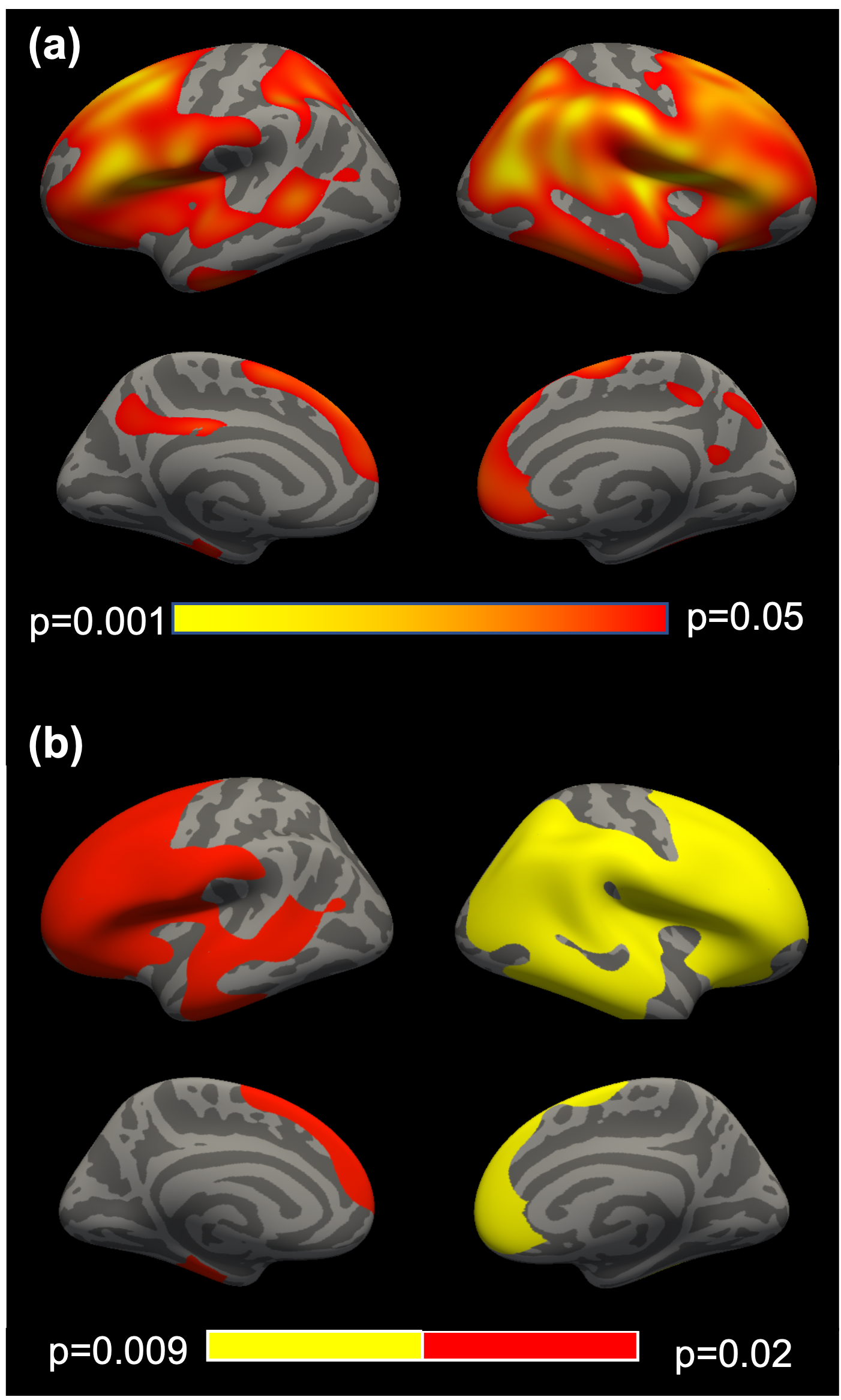
Figure 1. Cortical mapping showing regional
measures of reduced gray-white matter contrast among World Trade Center responders
with chronic post-traumatic stress disorder. (a) Results mapped to the surface of the brain
indicate regions with reduced gray-white matter contrast are expressed as
p-values. (b) Clusters survive multiple comparison corrections.
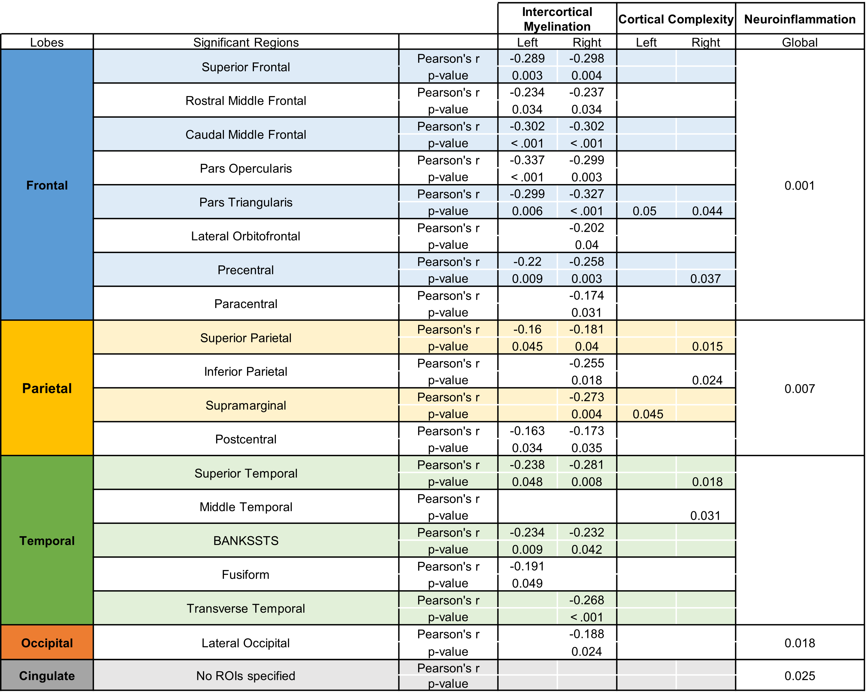
Table 1. Regional comparisons between measures of intracortical demyelination, cortical complexity3, and neuroinflammation2 among World Trade Center responders.
DOI: https://doi.org/10.58530/2023/2621