2619
Enlarged choroid plexus volume in multiple sclerosis is related with disability, cognition and brain atrophy1MS Center Amsterdam, Anatomy and Neurosciences, Vrije Universiteit Amsterdam, Amsterdam Neuroscience, Amsterdam UMC location VUmc, Amsterdam, Netherlands, 2Department of Molecular Cell Biology and Immunology, Vrije Universiteit Amsterdam, Amsterdam Neuroscience, Amsterdam UMC location VUmc, Amsterdam, Netherlands, 3MS Center Amsterdam, Radiology and Nuclear Medicine, Vrije Universiteit Amsterdam, Amsterdam Neuroscience, Amsterdam UMC location VUmc, Amsterdam, Netherlands, 4Institutes of Neurology and Healthcare Engineering, UCL London, London, United Kingdom, 5MS Center Amsterdam, Neurology, Vrije Universiteit Amsterdam, Amsterdam Neuroscience, Amsterdam UMC location VUmc, Amsterdam, Netherlands
Synopsis
Keywords: Multiple Sclerosis, Neuroinflammation
Enlargement of the choroid plexus (ChP) has been recently suggested in multiple sclerosis (MS), but relations with clinical and MRI outcome measures remain unclear. In this study, we compared automated segmentation approaches to assess ChP volume on 3D-T1 to manual outlines. Next, ChP volume was assessed in 327 patients with MS and 78 healthy controls. Gaussian Mixture Modelling (GMM)-based segmentation showed best agreement with manual segmentations in MS and controls. Enlargement of ChP was observed in MS compared to controls, and was associated with worse physical disability and cognitive impairment and more severe brain, cortical and thalamic atrophy.
Introduction
The choroid plexus (ChP) is a structure located in the ventricles of the brain which produces cerebrospinal fluid (CSF) and controls the trafficking of immune cells between the blood and CSF. In multiple sclerosis (MS), post-mortem brains showed inflammatory changes in the ChP1, 2. Recent studies have shown that in vivo quantifications of ChP changes are feasible on MRI3. Also it was observed that ChP volume seems larger in MS compared with healthy controls and was associated with inflammatory activity in the form of clinical relapses and brain atrophy4, 5. In one study, ChP volume correlated with disability and was predictive of future disease worsening6, a finding that still needs replication. As such, the number of MS studies using this novel MRI biomarker remains very limited and correlations with clinical outcome measures and other MRI markers are largely unknown. Therefore, we aimed to validate automated ChP segmentation in MS and explore clinical and radiological relations with ChP volumes. First, performance of automated ChP segmentation approaches were compared in MS and controls. Second, ChP volumes were compared between MS phenotypes and associations with physical disability, cognition and volumetric MRI measures were assessed.Methods
In this study, 3D-T1 and 3D-FLAIR weighted images of 327 MS patients and 78 healthy controls (HC) were retrospectively analyzed7. Clinical assessments included Expanded Disability Status Scale and an expanded Brief Repeatable Battery of Neuropsychological Tests. Manual reference segmentations were generated with 3D Slicer on 10 randomly selected MS subjects and 10 HC. Manual segmentations were then compared with two automated approaches: FreeSurfer (version 7.1.1) and a recently proposed light weight segmentation algorithm based on GMM3. The best performing segmentation approach (i.e. Dice score closest to manual) was used to segment ChP in the whole cohort. Structural brain volumes were quantified on lesion-filled T1-weighted images with FreeSurfer and all volumes (including ChP) were corrected for head size using total intracranial volume (TIV). Subsequently, ChP volume (ChPV) was compared between HC and MS, across disease phenotypes (relapse remitting MS (RRMS) vs. progressive MS (PMS), MS patients with low disability (EDSS<4) vs. high disability and cognitively preserved (CP) vs. cognitively impaired (CI) MS patients. Group comparisons were made with ANCOVA models, corrected for age and sex. Finally, the association between ChPV and clinically relevant MRI volume measures (brain, cortex, ventricle, thalamus, T2 lesions) was investigated with linear regression analysis, with age and sex as covariates. P-values were corrected for multiple comparisons with Holm-Bonferroni and corrected p-values< 0.05 were considered statistically significant.Results
Demographic and clinical variables of the studied cohort are presented in Table 1. Figure 1 shows ChP segmentation performed manually and automatically by FreeSurfer and GMM in a representative MS subject. Compared to manual segmentations, the GMM-based segmentation had a higher spatial overlap compared with FreeSurfer (median DC=0.60 vs. DC=0.33, p<0.001), see Figure 2A. ChPV was larger in MS compared with HC (see Figure 2B and Table 2, p<0.001, η2=0.06), but did not differ between RRMS and PMS (p=0.990, η2=0.00). MS patients with high disability had higher ChPV than those with low disability (p=0.001, η2=0.04), with similar effects for MS patients with versus without cognitive impairment (p=0.003, η2=0.03). In HC, ChPV was only related to ventricular size (std. β=0.52, p<0.001) and not with brain, cortical and thalamic volumes (Figure 3). In MS, larger ChPV was associated with lower brain (std. β=-0.52, p<0.001), higher ventricular (std. β=0.52, p<0.001), lower cortical (std. β=-0.33, p<0.001), lower thalamic (std. β=-0.32, p<0.001) and higher T2 lesion volume (std. β=0.39, p<0.001).Discussion
In this study, automated ChP segmentation approaches were compared with manual reference segmentations and associations between ChP volumes and clinical and radiological outcome measures were assessed in a large cohort of MS patients. Manual segmentation of ChP is still considered the golden standard in neuroimaging studies, but is not practical for application in large-scale studies. Here, we found that automated GMM-based ChP segmentation outperformed FreeSurfer, which was also seen in previous work in Alzheimer’s disease3. Moreover, we confirmed that choroid plexus volume was enlarged in patients with MS compared to controls and related to a broad scale of clinical and radiological measures. Interestingly, ChP volume was not different between relapsing remitting and progressive phase MS patients. In previous work, enlargement of ChP volume was already observed in the earliest clinical phases of MS8, possibly indicating that increased volumes remain constant towards the progressive phases of the disease. Direct clinical relevance of enlarged ChP volume has been debated recently, as this was shown by Fleisher et al.6, but not by Ricigliano et al.4. Our study did show clear relations with both disability and cognition, in addition to neurodegenerative and inflammatory MRI markers, supporting ChPV as an upcoming MRI biomarker of disease severity in MS.Conclusion
Choroid plexus volume can be automatically measured in vivo on MRI using Gaussian Mixture Modeling-based segmentation and is a clinically relevant new biomarker in MS. Relations were seen with physical disability and cognitive impairment, as well as global and regional atrophy and lesion volumes. As automated are now feasible, larger-scale studies can now further study the value of this new marker for monitoring disease activity and treatment effects.Acknowledgements
No acknowledgement found.References
1. Vercellino M, Votta B, Condello C, et al. Involvement of the choroid plexus in multiple sclerosis autoimmune inflammation: a neuropathological study. Journal of neuroimmunology 2008;199:133-141.
2. Rodríguez-Lorenzo S, van Olst L, Rodriguez-Mogeda C, et al. Single-cell profiling reveals periventricular CD56bright NK cell accumulation in multiple sclerosis. Elife 2022;11:e73849.
3. Tadayon E, Moret B, Sprugnoli G, et al. Improving choroid plexus segmentation in the healthy and diseased brain: Relevance for Tau-PET imaging in dementia. Journal of Alzheimer's Disease 2020;74:1057-1068.
4. Ricigliano VA, Morena E, Colombi A, et al. Choroid plexus enlargement in inflammatory multiple sclerosis: 3.0-T MRI and translocator protein PET evaluation. Radiology 2021;301:166-177.
5. Müller J, Sinnecker T, Wendebourg MJ, et al. Choroid plexus volume in multiple sclerosis vs neuromyelitis optica spectrum disorder: a retrospective, cross-sectional analysis. Neurology-Neuroimmunology Neuroinflammation 2022;9.
6. Fleischer V, Gonzalez-Escamilla G, Ciolac D, et al. Translational value of choroid plexus imaging for tracking neuroinflammation in mice and humans. Proceedings of the National Academy of Sciences 2021;118:e2025000118.
7. Eijlers AJ, Meijer KA, van Geest Q, Geurts JJ, Schoonheim MM. Determinants of cognitive impairment in patients with multiple sclerosis with and without atrophy. Radiology 2018;288:544-551.
8. Klistorner S, Van der Walt A, Barnett MH, et al. Choroid plexus volume is enlarged in clinically isolated syndrome patients with optic neuritis. medRxiv 2022.
Figures
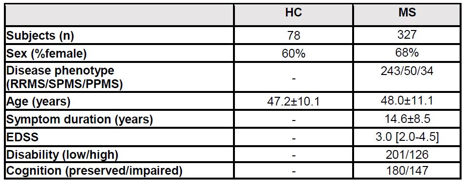
Table 1. Demographic and clinical variables of healthy controls (HC) and multiple sclerosis (MS) patients. MS=Multiple Sclerosis; Scale; RR=relapse remitting; SP=secondary progressive; PP=primary progressive; EDSS=Expanded Disability Status
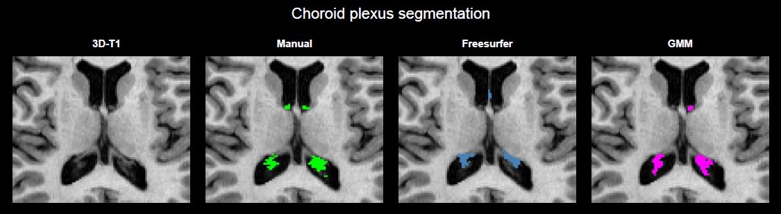
Figure 1. Comparison manual and automated segmentation approaches of choroid plexus.
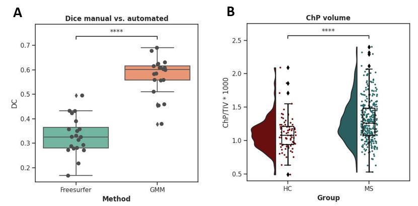
Figure 2. A) Dice (DC) score between manual reference and automated choroid plexus segmentation by using Freesurfer and Gaussian Mixture Models (GMM). B) Difference in ChP volume between HC and MS.
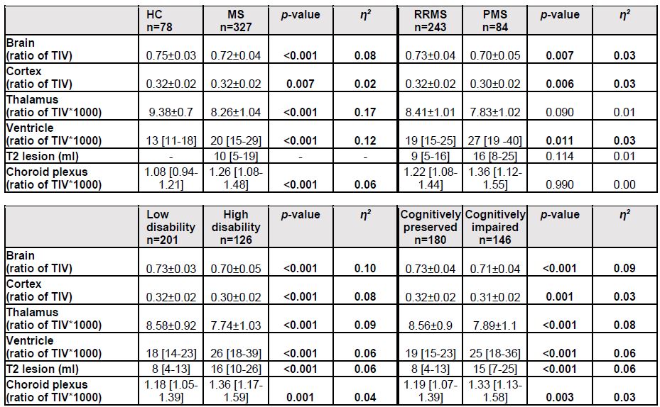
Table 2. Differences in MRI volumes (normalized by total intracranial volume) between HC vs. MS, RRMS vs. PMS, low disability vs. high disability and cognitively preserved vs. cognitively impaired MS patients.
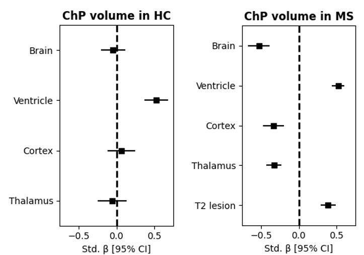
Figure 3. Correlation between choroid plexus volume and other MRI volumes within HC and MS, corrected for age and sex.