2605
A pilot study of quantitative T2* mapping in intervertebral lumbar disc patients with lower back pain
Adiraju Karthik1, Apoorwa Devappa2, Aakaar Kapoor3, Dharmesh Singh4, and Dileep Kumar4
1Department of Radiology, Sprint Diagnostics, Jubilee Hills, Hyderabad, India, 2Department of Radiology, Mahadevappa Rampure Medical College, Kalaburagi, India, 3Department of Radiology, City X-Rays Scan & Clinical Private Limited, New Delhi, India, 4Central Research Institute, Global Scientific Collaborations, United Imaging Healthcare, New Delhi, India
1Department of Radiology, Sprint Diagnostics, Jubilee Hills, Hyderabad, India, 2Department of Radiology, Mahadevappa Rampure Medical College, Kalaburagi, India, 3Department of Radiology, City X-Rays Scan & Clinical Private Limited, New Delhi, India, 4Central Research Institute, Global Scientific Collaborations, United Imaging Healthcare, New Delhi, India
Synopsis
Keywords: Skeletal, Spinal Cord, Intervertebral lumbar disc patients , T2* mapping
The human lumbar spine is comprised of multiple tissue components that serve to offer structural stability as well as optimal nutrition. Traditional magnetic resonance (MR) imaging techniques have been beneficial in assessing intervertebral disc (IVD). Quantitative T2* mapping has recently been used to diagnose and characterize abnormalities associated with IVD, particularly water content variations. However, this approach remains in research settings, and its clinical application has not been thoroughly investigated. This study quantitatively evaluate the clinical value of T2* mapping of intervertebral lumbar disc patients with lower back pain using 3T magnetic resonance imaging (MRI).Introduction
Low back pain (LBP) is one of the most prevalent musculoskeletal disorders. Intervertebral disc (IVD) degeneration is responsible for 41.8% of LBP patients1. IVD has a complicated structure whereby a central nucleus pulposus (NP) is surrounded by layers of annulus fibrosus (AF). Several studies have used biochemically sensitive MRI (chemical exchange saturation transfer, T1rho, sodium, and T2 mapping) to explore the qualitative and quantitative features of the IVD2,3. T2* mapping is an MR-based imaging technique that has gained popularity recently due to its advantages of fast imaging, high resolution, and three-dimensional evaluation of IVD homogeneity4. T2* appears to be an appropriate diagnostic approach that may be applied to a clinical MR procedure for determining the beginning of degeneration at the earliest possible stage, which is critical for the efficacy of regenerative IVD treatment. The goal of this study was to quantify the T2* values of IVD patients with LBP and investigate the clinical utility of T2* mapping.Methods
Patients with a history of low back pain (15 normal, 13 mild, and 8 severe IVD) were enrolled in this prospective study. T2* maps were obtained by scanning on a 3T MR system (United Imaging Healthcare Co. Ltd. Shanghai) with a multi-echo gradient recalled echo and 5 incremental echo time (TE - 6.71 ms) values. T2* maps were generated and analyzed on an uWS workstation, and T2* values were calculated by placing a region of interest (ROI) in the corresponding disc areas. An example of a patient with T2* map overlay is shown in figure 1.Results
A routine sequence (T2 weighted, T1 weighted, and STIR acquired in sagittal, coronal and axial plane) of 36 discs (L2 to L5) was evaluated morphologically. T2* maps were produced for the whole lumber spine, and by putting an ROI, T2* values were acquired in discs exhibiting normal, mild, and severe conditions. Mean T2* values were 73.80 ± 8.27, 41.55 ± 10.58, and 23.72 ± 7.0 for normal, mild, and severe discs, respectively.Discussion
MRI remains the gold standard for IVD evaluation in the current period. Biochemically sensitive approaches having the ability to detect disc degeneration early. It might increasingly lead to modifications in diagnosis and therapeutic management procedures. One of these techniques is T2* mapping, which has the advantages of fast acquisition, high sensitivity, and 3D visual interpretation without additional hardware modification. In this study, we quantitatively investigated the clinical value of T2* mapping of intervertebral lumbar disc patients with LPB using 3T MRI. The T2* values obtained in this investigation are consistent with other studies5. In comparison to sagittal-oriented imaging approaches, axial mapping allows for the study of a larger disc region and has been demonstrated to be a valuable grading instrument, particularly during the early stages of disc degeneration6. As a result, our hypothesis that axial T2 imaging could be a promising approach for detecting early stages of disc degeneration in all regions of the IVD. The degradation of cartilage is the primary cause of IVD degeneration, although the change in T2* value is caused by a change in water content, collagen fibre disorder, and the loss of certain biochemical macromolecules such as proteoglycans. Therefore, detection of IVD conditions (normal, mild and severe) using imaging can result in early IVD degeneration diagnosis, therapy, and prevention.Conclusion
T2* mapping is a viable approach for diagnosing IVD stages and predicting the degree of lumbar disc degeneration. Additionally, this technology can be used to establish the diagnostic criteria for IVD degeneration. This suggests that the T2* sequence could be useful in daily clinical practice.Acknowledgements
Authors would like to acknowledge the technical support of staff members at Sprint Diagnostics Private Limited and MR Application team members of United Imaging Healthcare for protocols optimization.References
- DePalma MJ, Ketchum JM, Saullo T. What is the source of chronic low back pain and does age play a role? Pain Med. 2011;12:224–33
- Muller-Lutz A, Schleich C, Schmitt B, et al. Gender, BMI and T2 dependencies of glycosaminoglycan chemical exchange saturation transfer in intervertebral discs. Magn Reson Imaging. 2016; 34:271–275.
- Schleich C, Muller-Lutz A, Eichner M, et al. Glycosaminoglycan chemical exchange saturation transfer of lumbar intervertebral discs in healthy volunteers. Spine. 2016; 41:146-152.
- Morgan P, Spiridonov S, Goebel R, et al. MR imaging with T2∗ mapping for improved acetabular cartilage assessment in FAI-a case report with arthroscopic correlation. Orthop Traumatol Surg Res. 2014;100: 971-973.
- Huang L, Liu Y, Ding Y, et al. Quantitative evaluation of lumbar intervertebral disc degeneration by axial T2* mapping. Medicine (Baltimore). 2017; 96(51):e9393.
- Hoppe S, Quirbach S, Mamisch TC, et al. Axial T2 mapping in intervertebral discs: a new technique for assessment of intervertebral disc degeneration. Eur Radiol. 2012;22:2013–9
Figures
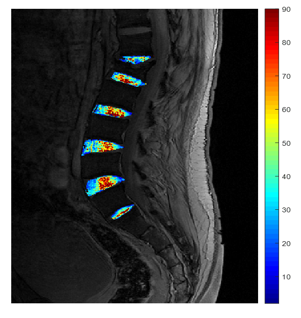
Figure 1: An example of a patient with T2* map
overlay
DOI: https://doi.org/10.58530/2023/2605