2604
Deep learning system for automated detection of posterior ligamentous complex injury in patients with thoracolumbar fracture on magnetic resonance imaging
Sang Won Jo1, Eun Kyung Khil1, Seun Ah Lee1, Jihe Lim1, Jae Hyeok Lee2, and Yu Sung Yoon3
1Radiology, Hallym University Dongtan Sacred Heart Hospital, Hwaseong, Korea, Republic of, 2Deepnoid, Seoul, Korea, Republic of, 3Radiology, Soonchunhyang University Bucheon Hospital, Bucheon, Korea, Republic of
1Radiology, Hallym University Dongtan Sacred Heart Hospital, Hwaseong, Korea, Republic of, 2Deepnoid, Seoul, Korea, Republic of, 3Radiology, Soonchunhyang University Bucheon Hospital, Bucheon, Korea, Republic of
Synopsis
Keywords: MSK, Machine Learning/Artificial Intelligence, thoracolumbar fracture
This study was aimed to develop a deep learning algorithm for automated detection and localization of posterior ligamentous complex (PLC) injury on magnetic resonance imaging and evaluate its diagnostic performance. The sensitivity, the specificity and AUC of inception-ResNet V2 architecture of second step were 88%, 82% and 0.928, for the internal test set and 86%, 74% and 0.916 for the external test set, respectively. A deep learning algorithm detected PLC injury in patients with thoracolumbar fracture with a high diagnostic performance which was validated using external data set.Purpose: To develop a deep learning algorithm for automated detection and localization of posterior ligamentous complex (PLC) injury on magnetic resonance imaging and evaluate its diagnostic performance.
Methods and Materials: In a retrospective and multicenter study, a midline sagittal fat-suppressed T2-weighted MR image with PLC injury based on radiological reports were extracted. The deep learning algorithm development in this study was conducted through two major steps. The first step was to train the deep learning algorithm (attention U-net) to segment the entire soft tissue area (back ground anatomy) including two PLCs (normal or damaged PLC) above and below the spinous process of thoracolumbar fracture. The second step was to have the deep learning algorithm detect and classify the PLC injury area expressed as T2 high signal intensity lesion in the segmented image by the attention U-net. To carry out the first and second steps, spine MR images were randomly divided into two data sets: training set of 300 examinations with thoracolumbar fracture (150 PLC injury group and 150 PLC normal group) and internal test set of 100 examinations with thoracolumbar fracture (50 PLC injury group and 50 PLC normal group). External test data set also consisted of 100 examinations (50 PLC injury group and 50 PLC normal group) with thoracolumbar fracture, which were extracted based on radiological reports from a different institution. After manual ground truth segmentation of thoracolumbar fracture, background anatomy, PLC injury, a deep learning algorithm based on inception-ResNet V2 architecture was established with the training set. Its dice score, sensitivity, specificity and AUC were evaluated in the internal and external test sets.
Results: The dice score of attention U-net of first step was 0.849 for the internal test set and 0.773 for the external test set, respectively. The sensitivity, the specificity and AUC of inception-ResNet V2 architecture of second step were 88%, 82% and 0.928, for the internal test set and 86%, 74% and 0.916 for the external test set, respectively.
Conclusions: A deep learning algorithm detected PLC injury in patients with thoracolumbar fracture with a high diagnostic performance which was validated using external data set.
Clinical Relevance/Application: PLC injury detection on MRI is a field highly dependent on the judgment of radiologists, and it is expected to play a very important role in determining the direction of treatment and providing practical help to the clinician rather than the detection of fractures. In addition, the development of such a system is expected to be in high demand in the clinical field. It will be used as an auxiliary means to help an accurate diagnosis of the radiologist
Acknowledgements
NoneReferences
1. Bharti et al.Traumatic Thoracolumbar Spine Injuries: What the Spine Surgeon Wants to Know. RadioGraphics 2013; 33:2031–2046
2. Vaccaro AR, Lehman RA, Jr., Hurlbert RJ, Anderson PA, Harris M, Hedlund R, et al. A new classification of thoracolumbar injuries: the importance of injury morphology, the integrity of the posterior ligamentous complex, and neurologic status. Spine (Phila Pa 1976). 2005;30(20):2325-33
3. Vaccaro AR, Lee JY, Schweitzer KM, Jr., Lim MR, Baron EM, Oner FC, et al. Assessment of injury to the posterior ligamentous complex in thoracolumbar spine trauma. Spine J. 2006;6(5):524-8.
Figures
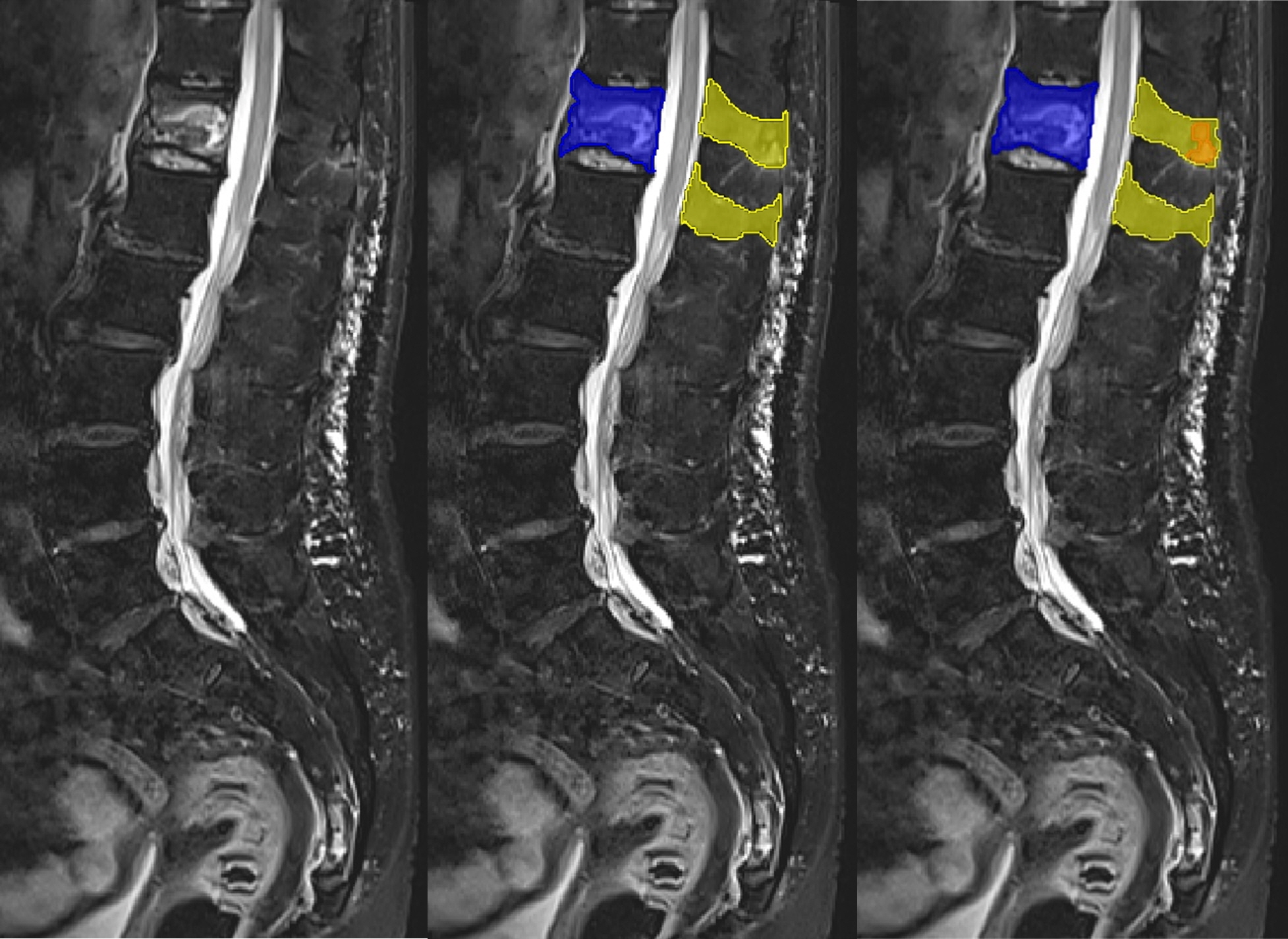
Figure 1. Representative case of manual segmentation of vertebral fracture, background anatomy, and posterior ligament injury.
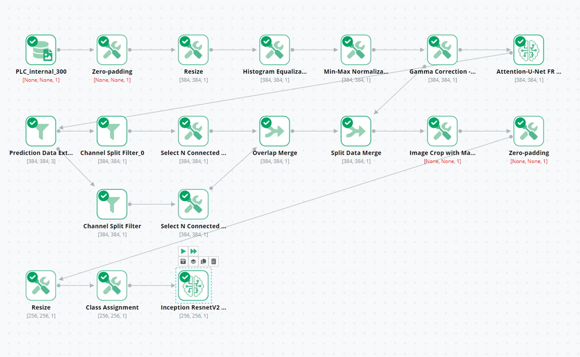
Figure 2. Overall deep learning training steps
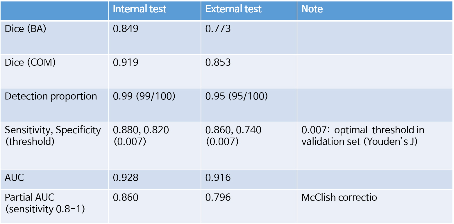
Table 1. Results summary of 1st Step and 2nd step of this study
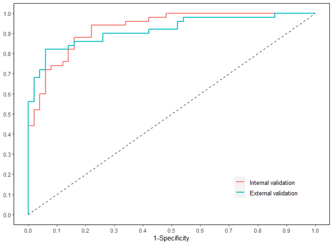
Figure 3. Receiver operator curves for diagnosing the posterior ligament complex injury
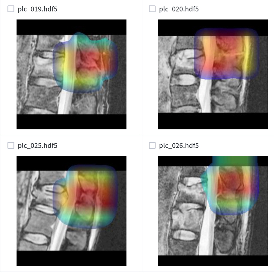
Figure 4. Representative case of GRAD cam
DOI: https://doi.org/10.58530/2023/2604