2603
Deep Learning Reconstructed T2-weighted Dixon Imaging of the Spine: Impact on Acquisition Time and Diagnostic Performance1Radiology, University Medical Center Freiburg, Freiburg, Germany, 2EMEA Scientific Partnerships, Siemens Healthcare GmbH, Erlangen, Germany, 3MR Application Predevelopment, Siemens Healthcare GmbH, Erlangen, Germany
Synopsis
Keywords: MSK, Machine Learning/Artificial Intelligence
Magnetic resonance imaging of the spine is considered one of the most commonly performed examinations in clinical routine. The raising demand for high quality imaging of the spine creates the need for tailored examination protocols, especially with regard to increasingly limited scanner capacities. Deep Learning based imaging reconstruction has emerged as promising novel technique to accelerate MR imaging while maintaining image quality. This study analyzed a novel deep learning accelerated T2-weighted Dixon sequence of the spine in terms of diagnostic performance. The results suggest that the here presented sequence is feasible with a diagnostic performance comparable to standard imaging.Introduction
Magnetic resonance imaging (MRI) of the spine is among the most frequently performed examinations in clinical routine and considered the standard imaging modality for the workup of lower back pain [1]. Given the high demand, the need for cost and time effective MRI examinations to overcome limited scanner capacities is increasing. Sagittal T2-weighted TSE Dixon imaging is replacing standard protocols due to the advantage of being able to provide different contrasts (including fat suppressed fluid sensitive and fat specific imaging) in a single acquisition [2]. Deep learning (DL) components involving neural networks in imaging reconstruction have recently been introduced as a novel technique, providing high acceleration factors while maintaining image quality [3]. The aim of this study was to compare a DL accelerated T2-weighted Dixon sequence (T2DL) with conventional T2-weighted TSE Dixon (T2std) imaging serving as reference standard. We hypothesized that T2DL is feasible with image quality and diagnostic performance comparable to T2std while allowing for a significant reduction of acquisition time.Methods
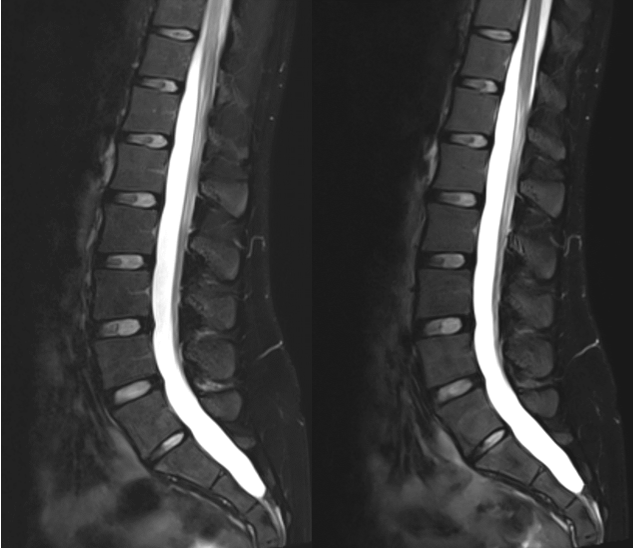
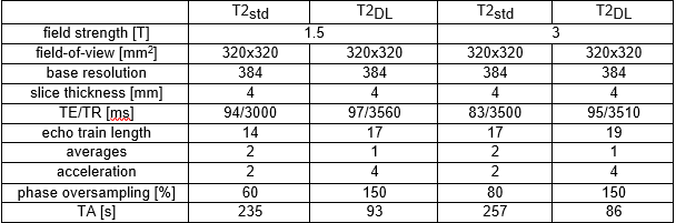
This prospective, single center study was approved by the local institutional review board. Written informed consent was obtained from all participants. N=18 consecutive patients with a clinical indication for lumbar MRI at our university radiology department between September 2022 and October 2022 were included in this study. MRI examinations were performed on 1.5-T and 3-T scanners using dedicated spine coils (MAGNETOM Aera and Vida; Siemens Healthcare, Erlangen, Germany). The MR study protocol consisted of our standard clinical protocol (including T2std) and the additional T2DL.The T2DL acquisition used a conventional sampling pattern with a higher parallel acceleration factor. The individual contrasts acquired for Dixon water-fat separation were then reconstructed using a research application as used and detailed in Refs. [4,5]. After reconstruction of the contrast images from k-space data, a conventional water-fat separation was performed to provide derived water images. Two readers with 6 (C.W.) and 4 (S.W.) years of experience in interpreting MSK imaging, respectively, analyzed the images in a randomized fashion. Readers were fully blinded to patients’ clinicopathological data, sequence type and each other’s rating. regarding Overall image quality, banding artifacts, artifacts, sharpness, noise, and diagnostic confidence were analyzed using a 5-point Likert scale (1 = non-diagnostic; 2 = low image quality; 3 = moderate image quality; 4 = good image quality; 5 = excellent image quality). For quantitative evaluation of image quality, the coefficient of variation (CV) was calculated and reported in %. Imaging quality scores, as well as differences in the CV were analyzed using the Wilcoxon signed-rank test. Inter-reader agreement was assessed via weighted Cohen’s kappa statistics. Analyses were performed using two-tailed testing and p-values below 0.05 were considered to denote a significant difference.Results
A total of 18 patients (median age 57 years, [IQR: 51,57], male sex: 33.3%) were prospectively included. Sixteen examinations were performed on 1.5-T and 2 examinations on 3-T- scanners. A sequence was successfully acquired in all patients. The total acquisition time of T2DL was 93 seconds at 1.5-T and 86 seconds at 3-T, compared to 235 seconds, and 257 seconds, respectively for T2std (reduction of acquisition time: 60.4% at 1.5-T, and 66.5% at 3-T; p< 0.01). Overall image quality was rated equal for both sequences (median 4.5, [IQR 4,5]; p = 0.27). T2DL showed significantly reduced noise levels compared to T2std (median 4.5, [IQR 4.5,5] versus median 4, [IQR 3.5,4.5]; p < 0.01). In contrast, sharpness was rated to be significantly higher in T2std (median 4.75, [IQR 4.5,5] versus median 4, [IQR 4,4.5]; p = 0.01). The range of artifacts was found to be comparable between T2std and T2DL (T2std: median 4.75, [IQR 4.13,5] and T2DL: median 4.5, [IQR 4,5]; p = 0.66), although T2DL displayed significantly more banding artifacts (median 4.3, [IQR 4,4.5] versus median 5, [IQR 4.5, 5], p < 0.01). However, no significant impact on the readers diagnostic confidence between sequences was noted (T2std: median 4.75, [IQR 4,5] and T2DL: median 4.5, [IQR 4.13,4.5]; p = 0.1). Mean CV values were 8,8% for T2std and 10.3% for T2DL, respectively (p = 0.03). Inter-reader agreement ranged from almost fair (κ for sharpness: 0.16) to substantial (κ for artifacts: 0.78).Discussion
The present study investigated the feasibility and diagnostic performance of a novel DL accelerated T2-weighted chemical-shift sequence compared to standard TSE imaging. The results indicate that T2DL has the potential to significantly reduce acquisition time while maintaining high image quality and diagnostic confidence. DL based imaging reconstruction has emerged as a promising new method to reduce acquisition time while overcoming the drawbacks of other acceleration techniques [6,7]. The results of this study are in line with the current literature. Herrmann et al. analyzed the diagnostic performance of a DL reconstructed 2D multi-contrast knee MRI protocol versus standard knee MRI and reported comparable findings regarding diagnostic confidence, artifacts and noise [8]. The preliminary results of the present study highlight the potential of DL based imaging to substitute standard TSE sequences. Further protocol interchangeability analyses with a higher sample size are planned to confirm our findings.Conclusion
In conclusion, T2DL is feasible, yields a diagnostic performance comparable to the reference standard while substantially reducing the acquisition time.Acknowledgements
No acknowledgement found.References
1. Zanchi F, Richard R, Hussami M, Monier A, Knebel JF, Omoumi P. MRI of non-specific low back pain and/or lumbar radiculopathy: do we need T1 when using a sagittal T2-weighted Dixon sequence? Eur Radiol. 2020 May;30(5):2583-2593.
2. Omoumi P. The Dixon method in musculoskeletal MRI: from fat-sensitive to fat-specific imaging. Skeletal Radiol. 2022 Jul;51(7):1365-1369.
3. Gassenmaier S, Küstner T, Nickel D, Herrmann J, Hoffmann R, Almansour H, Afat S, Nikolaou K, Othman AE. Deep Learning Applications in Magnetic Resonance Imaging: Has the Future Become Present? Diagnostics (Basel). 2021 Nov 24;11(12):2181.
4. Herrmann J, Koerzdoerfer G, Nickel D, Mostapha M, Nadar M, Gassenmaier S, Kuestner T, Othman AE. Feasibility and Implementation of a Deep Learning MR Reconstruction for TSE Sequences in Musculoskeletal Imaging. Diagnostics (Basel). 2021 Aug 16;11(8):1484.
5. Gassenmaier S, Afat S, Nickel D, Mostapha M, Herrmann J, Othman AE. Deep learning-accelerated T2-weighted imaging of the prostate: Reduction of acquisition time and improvement of image quality. Eur J Radiol. 2021 Apr;137:109600
6. Schlemper J, Caballero J, Hajnal JV, Price AN, Rueckert D. A deep cascade of convolutional neural networks for dynamic MR image reconstruction. IEEE Trans Med Imaging. 2018;37:491–503.
7. Hammernik K, Klatzer T, Kobler E, et al. Learning a variational network for reconstruction of accelerated MRI data. Magn Reson Med. 2018;79:3055–3071.
8. Herrmann J, Keller G, Gassenmaier S, Nickel D, Koerzdoerfer G, Mostapha M, Almansour H, Afat S, Othman AE. Feasibility of an accelerated 2D-multi-contrast knee MRI protocol using deep-learning image reconstruction: a prospective intraindividual comparison with a standard MRI protocol. Eur Radiol. 2022 Sep;32(9):6215-6229.
Figures