2595
Feasibility study of paraspinal muscle fat infiltration assessed by lumbar disc degeneration grade1Sichuan Province Orthopedic Hospital, Chengdu, China, 2GE Healthcare China, Beijing, China
Synopsis
Keywords: Muscle, Fat, Quantitative imaging, Lumbar disc herniation
Previous study found that low back pain (LBP) and Modic changes were closely associated with fatty infiltration and cross-sectional area (CSA) of the paraspinal muscles. However, conflicting evidences were reported. This study investigated the association between intervertebral discs degeneration and lumbar paraspinal muscles CSA and fat infiltration, and explored the relationship between Modic change and intervertebral discs degeneration. Our results found that lumbar disc’s Pfirrmann grading was weakly correlated with CSA of multifidus, fat fraction of multifidus and erector spinae, and the incidence rate of Modic changes increased in higher Pfirrmann grading.Introduction
The lumbar paraspinal musculature and intervertebral discs play an important role in stabilizing and supporting the lumbar vertebral body. When fatty infiltration of the paraspinal muscles occurs, the strength of the paraspinal muscles decreases, which is one of the causes of lower back pain, and lumbar disc degeneration (1,2). The cartilage endplate degeneration depicted with Modic change (3) obstructs the nutrition supply to the disc hence cause disc degeneration. Previous study found that low back pain (LBP) and Modic changes were closely associated with fatty infiltration and cross-sectional area (CSA) of the paraspinal muscles (1). However, conflicting evidences were reported too. The purpose of this study is to investigate the association between intervertebral discs degeneration and lumbar paraspinal muscles, including muscle size and fat infiltration, in a large patient cohort. Furthermore, relationship between Modic change and intervertebral discs degeneration will also be investigated.Methods
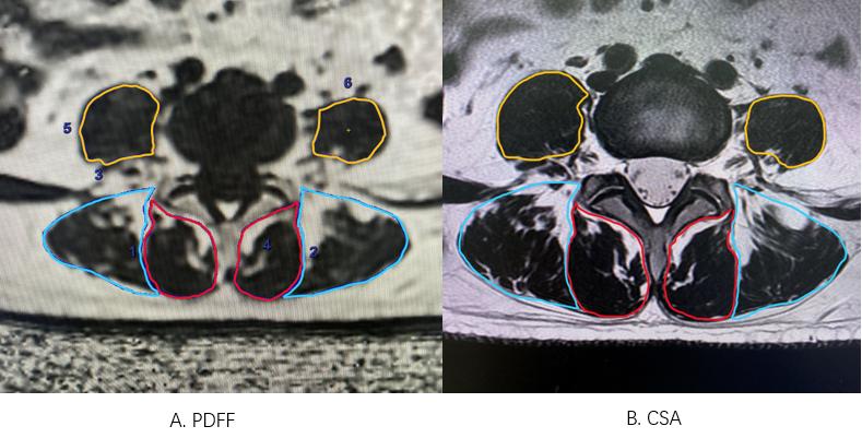
Patients:After IRB-approved written informed consent was obtained, 419 patients with low back pain were enrolled in this study. The discs’ condition were assessed using the modified Pfirrmann grading method (4). The patients were divided into three groups, with Pfirrmann grades 1-3 being low, grades 4-5 being mid and grades 6-8 being high. Fat infiltration of the L4-L5, L5-S1 paraspinal multifidus (MF), erector spinae (ES) and psoas major (PS) muscles was quantitatively assessed using the IDEAL-IQ sequence. The paraspinal muscle fat fraction (PDFF) and paraspinal muscle cross-sectional area (CSA) were compared with Pfirrmann's different classifications. We also analyzed the incidence of Modic changes in the different Pfirrmann classifications.Imaging acquisition and processing:TThe patients underwent MR examination (SIGNA Architect 3.0T, GE Healthcare, USA) with traditional T1 and T2 weighted imaging, and additional IDEAL-IQ sequence. The fat fraction map was generated with IDEAL-IQ raw images using the GE AW 4.7 workstation and the paraspinal muscle PDFF region of interest (ROI) was outlined on the axial fat fraction image, taking care to avoid the fatty tissue surrounding the muscle, as shown in Figure 1A. The area paraspinal muscle was measured with manually outlined region of interest on the axial T2 weighted image, again avoiding the fatty tissue surrounding the muscle, as shown in Figure 1B.
Statistical Analysis: All statistical analyses were performed using the IBM SPSS statistics for windows, v. 22.0 (IBM, Armonk, NY). One-way ANOVA test was used to compare the differences between patients' gender, age, BMI and paraspinal muscles PDFF and CSA. Correlations between Pfirrmann classification and paraspinal muscles PDFF and CSA were calculated using Spearman correlation analysis. The incidence of Modic changes in different Pfirrmann classifications was counted.
Results
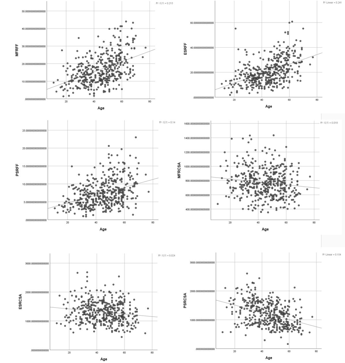
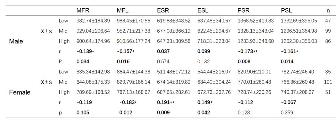
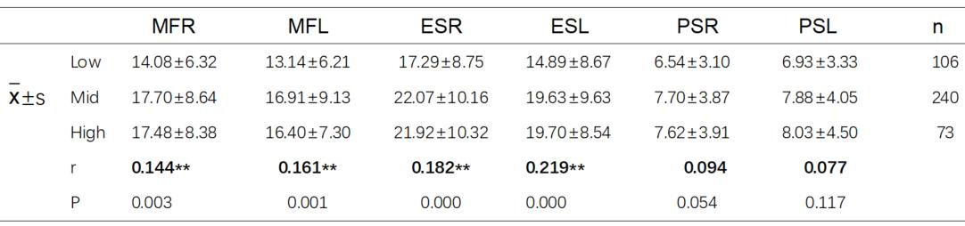
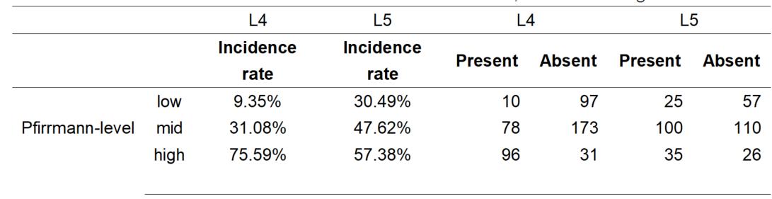
The mean age of the 419 patients was 47.43 (±12.50), with 232 males and 187 females. The statistical results showed significant differences in PDFF and CSA of paraspinal muscles between males and females, with the mean value of CSA of paraspinal muscles significantly greater in males than in females and the mean value of PDFF of paraspinal muscles significantly higher in females than in males. Age was statistically associated with PDFF of the paraspinal muscles, and the paraspinal muscle PDFF values increased with increasing age, as shown in Figure 2. In male subjects, CSA of PS and MF were negatively correlated with Pfirrmann grading; In female subjects, CSA of ES was positively correlated with Pfirrmann grading (Table 1). The PDFF of MF the ES were positively correlated with Pfirrmann grading, no significant correlation was found between Pfirrmann grading and PDFF for PS, as shown in Table 2. There are 9.35% of L4 and 30.49% of L5 Modic changes occurred in Pfirrmann-low group; 31.08% of L4 and 47.62% L5 Modic changes occurred in Pfirrmann-mid group; and 75.59% L4 and 57.38% L5 Modic change in Pfirrmann-high group (Table 3).Discussion and conclusion
As the grade of lumbar disc degeneration increases, the PDFF of the paraspinal muscles increases and so does the incidence of Modic changes. The fatty infiltration of the paraspinal muscles increases with age and the cross-sectional area of the paraspinal muscles decreases with age. This indicates that age and gender are important factors that need to be controlled in future study. Lee et, al (5) found The CSA of multifidus in men, but not in women, was smaller in the chronic LBP group than in those with improved LBP. Our results are similar except that instead of clinical symptom, we focused on Pfirrmann gradings. Teichtahl et, al (6) reported they found an association between fat infiltration in multifidus and high intensity low back pain and/ or disability. Our results are in agreement with theirs, except we also found association for erector spinae. In our study, no statistically significant correlation was found for psoas major. Another interesting finding is that, PDFF of multifidus and erector spinae increased markedly from Pfirrmann-Low to Mid group, but comparable between Mid and High groups. This may imply that paraspinal muscle fat infiltration is more important during early stage of disc degeneration. In conclusion, lumbar disc’s Pfirrmann grading is weakly correlated with cross-sectional area of multifidus, fat fraction of multifidus and erector spinae.Acknowledgements
No acknowledgement found.References
1. Ranger TA, Cicuttini FM, Jensen TS, Peiris WL, Hussain SM, Fairley J, et al. Are the size and composition of the paraspinal muscles associated with low back pain? A systematic review. Spine J. 2017;17(11):1729–48.
2. Kjaer P, Bendix T, Sorensen JS, Korsholm L, Leboeuf-Yde C. Are MRI-defined fat infiltrations in the multifidus muscles associated with low back pain? BMC Med. 2007 Jan 25;5(1):2.
3. Modic MT, Masaryk TJ, Ross JS, Carter JR. Imaging of degenerative disk disease. Radiology. 1988 Jul;168(1):177–86.
4. Griffith JF, Wang YXJ, Antonio GE, Choi KC, Yu A, Ahuja AT, et al. Modified Pfirrmann grading system for lumbar intervertebral disc degeneration. Spine. 2007;32(24):E708–12.
5. Lee HI, Lee ST, Kim M, Ryu JS. Sex differences in predicting chronicity of low-back pain after acute trauma using lumbar muscle area. Am J Phys Med Rehabil. 2015;94(2):123–30.
6. Teichtahl AJ, Urquhart DM, Wang Y, Wluka AE, Wijethilake P, O’Sullivan R, et al. Fat infiltration of paraspinal muscles is associated with low back pain, disability, and structural abnormalities in community-based adults. Spine J. 2015;15(7):1593–601.
Figures