2570
Age dependent changes of water exchange rate across the blood-brain barrier (BBB) in 115 subjects with age ranging from 8 to 87 years
Xingfeng Shao1, Qinyang Shou1, Kimberly Felix2, Brandon Ojogho1, Xuejuan Jiang2,3, Brian T. Gold4, Megan M. Herting2, and Danny JJ Wang1
1Laboratory of FMRI Technology (LOFT), Mark & Mary Stevens Neuroimaging and Informatics Institute, Keck School of Medicine, University of Southern California, Los Angeles, CA, United States, 2Department of Preventive Medicine, Keck School of Medicine, University of Southern California, Los Angeles, CA, United States, 3Department of Ophthalmology, Keck School of Medicine, University of Southern California, Los Angeles, CA, United States, 4Department of Neuroscience, College of Medicine, University of Kentucky, Lexington, KY, United States
1Laboratory of FMRI Technology (LOFT), Mark & Mary Stevens Neuroimaging and Informatics Institute, Keck School of Medicine, University of Southern California, Los Angeles, CA, United States, 2Department of Preventive Medicine, Keck School of Medicine, University of Southern California, Los Angeles, CA, United States, 3Department of Ophthalmology, Keck School of Medicine, University of Southern California, Los Angeles, CA, United States, 4Department of Neuroscience, College of Medicine, University of Kentucky, Lexington, KY, United States
Synopsis
Keywords: Arterial spin labelling, Permeability, BBB water exchange, permeability, ageing
We studied age-related BBB water exchange rate (kw) changes in a wide range of age groups with 115 subjects using diffusion prepared pseudo-continuous arterial spin labeling (DP-pCASL). We observed a significant trend of decreasing kw with age (slow when ≤50 years old and fast when >50 years old) and large spread of kw values in subjects >50 years, which may indicate compromised BBB function in some ageing populations. The inverted U-shaped correlations between kw versus age suggest a more complex age dependent changes in hippocampus and PHG regions.Background
Blood-brain-barrier (BBB) plays a critical role in the delivery of nutrients and oxygen in the brain. Previous studies have reported that BBB permeability to contrast agent (Ktrans) increases with age and may be related to microvascular diseases and dementia1. Recently, diffusion prepared pseudo-continuous arterial spin labeling (DP-pCASL) has been developed to assess the water exchange rate (kw) across the BBB without contrast2. The BBB mechanism underlying Ktrans and kw is likely to be different3. In this study, we analyzed DP-pCASL data collected on a wide range of age groups (8 to 87 years) to investigate age dependent changes of BBB water exchange rate.Method
The imaging parameters of DP-pCASL were: resolution = 3.5×3.5×8 mm3, TR = 4200ms, TE = 36.22ms, FOV=224 mm, 12 slices, labeling duration =1500 ms. Cerebral blood flow (CBF), arterial transit time (ATT) and kw were estimated by a two-stage approach with TGV regularized SPA modeling2. Total scan time was 10 mins. We studied a total of 115 subjects (54 Females/61 males) from five datasets:1) Pediatric subjects: N=9, age=13±2.9 (range 8-17) years, 6/3 males/females;
2) Young adults: N=17, age=26.9±5.8 (19-39) years, 9/8 males/females;
3) Elderly Latinx subjects: N=7, age=67.6±3.3 (64-72) years, 2/5 males/females;
4) Mid to Elderly African American subjects: N=39, age=63.9±10.2 (40-81) years, 24/15 males/females;
5) Elderly Caucasian subjects: N=43, age=73.0±6.0 (67-87) years, 21/22 males/females.
All subjects were cognitive normal and scanned on Siemens 3T Prisma scanners. kw values were extracted from whole brain and 11 brain regions including: Frontal lobe, temporal lobe, parietal lobe, hippocampus, para hippocampal gyrus (PGH), amygdala, caudata, ACC/PCC, precuneus and putamen. Multiple linear regressions were performed to study relationship between kw/CBF/ATT and age/gender. Quadratic regressions were also performed to study non-linear relationship between whole-brain and regional kw values versus age.
Results
Fig.1 shows the kw maps of seven representative subjects across the whole age range.Fig.2 shows the scatterplots of whole brain kw, CBF and ATT values versus age. Both CBF and kw significantly decreased with age, while ATT significantly increased with age (P<0.0001). Fig.2A dashed line shows the quadratic regression between kw and age, which indicate decrease of kw was slow with age ≤50 and became faster when age >50 years. kw from subjects >50 years old were 20% lower than the younger group (108.4 versus 126.5 min-1) with a larger spread (SD 22.9 versus 12.8 min-1). No significant difference between males and females was observed in whole-brain averaged kw (P=0.94) or ATT (P=0.30). Whole-brain averaged CBF was significantly higher in females (44.0ml/100g/min) than males (40.9ml/100g/min) (P=0.04).
Fig.3 shows scatter plots of 11 regional kw values versus age. Overall regional kw values decreased with age, while inverted U-shaped relationships with age were observed in the hippocampus and PHG regions.
Discussion and conclusion
We studied age-related kw changes in a wide range of age groups with 115 subjects using DP-pCASL. Water exchange rate across the BBB is related to the glymphatic function and clearance of brain waste. We observed a significant trend of decreasing kw with age and large spread kw values in subjects >50 years, which may indicate compromised BBB function in some ageing populations. Our finding is also consistent with a recent study that reported positive associations of kw with CSF amyloid-beta42 and episodic memory score in a cohort of aged subjects with normal cognition4. The inverted U-shaped correlations between kw versus age suggest a more complex age dependent changes in hippocampus and PHG regions5, which needs further evaluations.Acknowledgements
No acknowledgement found.References
1. Farrall, A. J., & Wardlaw, J. M. Neurobiology of aging, 2009, 30(3), 337-352.
2. Shao, X., et al., MRM, 2019, 81(5), 3065-3079.
3. Shao, X., et al., Frontiers in Neuroscience, 2020, 1236.
4. Gold, B.T., et al., Alzheimer's and Dementia, 2021, 17 (12), 2020-2029.
5. Satterthwaite, T. D., et al., PNAS, 2014, 111(23), 8643-8648.
Figures
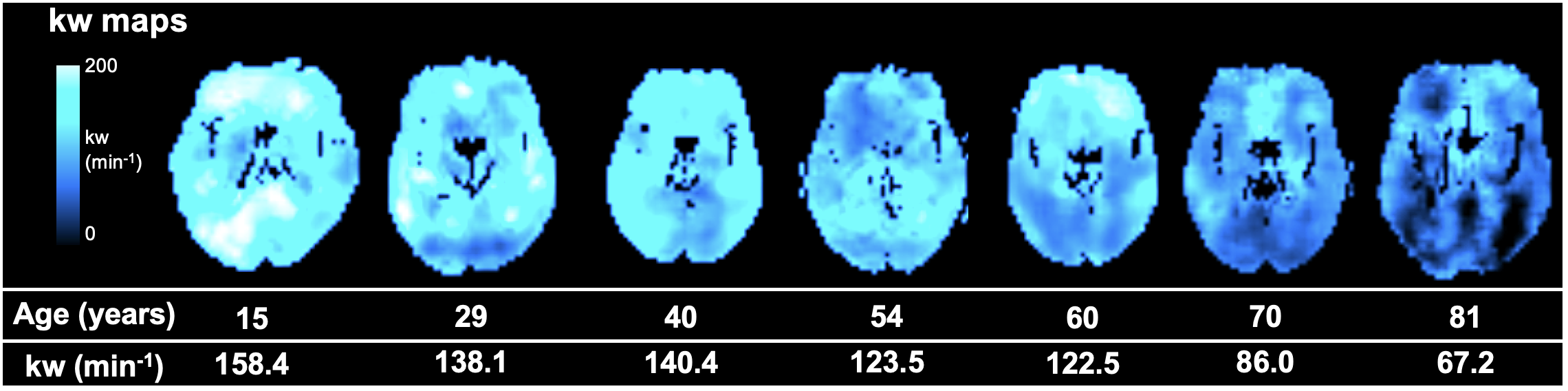
Figure 1. kw maps of 7 subjects across different ages.
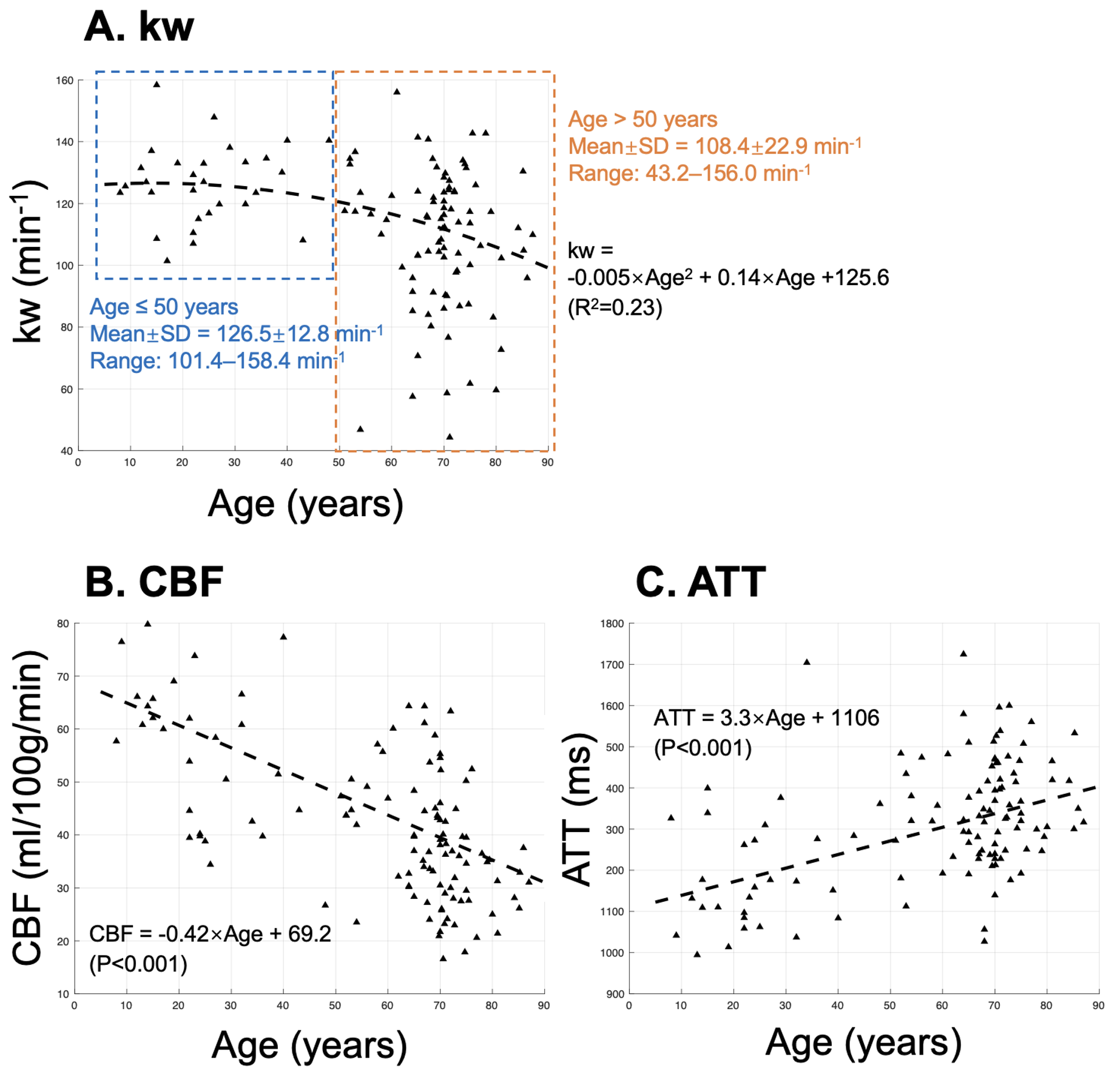
Figure 2. Scatter of whole-brain averaged kw (A), CBF (B) and ATT (C) versus age. The dashed black lines indicate quadratic regression results for kw and linear regression results for CBF/ATT. Blue and orange boxes show two clusters of kw values between subjects with age ≤50 and >50 years with slow and moderate rate of kw decrease. Significant CBF decrease was observed with ageing (P<0.001) while ATT significantly increased with age (P<0.001).
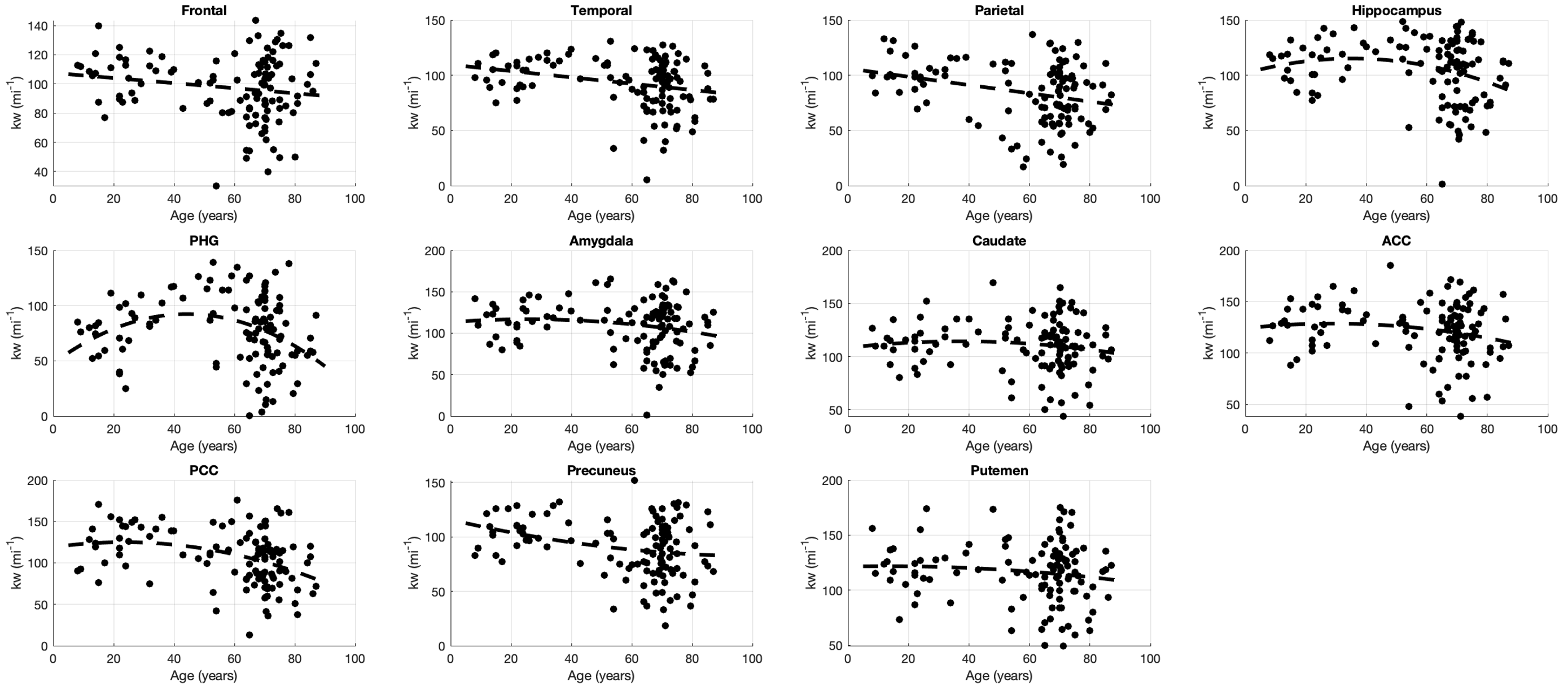
Figure 3. Scatter plot of regional kw values versus age. Inverted U-shape correlations can be observed between hippocampus/PHG and age.
DOI: https://doi.org/10.58530/2023/2570