2555
Comparison of ZOOMit DWI and Conventional DWI with the same geometric parameters at 3T in Patients with Breast Diseases1Xi'an International Medical Center Hospital, Xi'an, China, 2MR Scientific Marketing, Siemens Healthineers, Shanghai, China
Synopsis
Keywords: Breast, Diffusion/other diffusion imaging techniques
DWI performs an important role in the diagnosis of the benign and malignant breast lesions. This study compared the overall image quality, SNR,CNR and the diagnostic performance of ADC values between ZOOMit DWI and conventional DWI in breast cancer with same geometric parameters. The result showed that ZOOMit DWI provided significantly higher image quality and lesion conspicuity than C-DWI with no difference diagnostic performance for ADC values and with almost the same scanning time, which can help to improve the diagnosis efficiency in breast cancer.Introduction
Breast cancer in women has surpassed lung cancer as the most commonly diagnosed cancer in the world, according to a previous stduy[1]. Diffusion Weighted imaging(DWI) combined with Dynamic contrast enhanced(DCE) sequences can effectively distinguish the benign and malignant breast lesions[2,3],but the phase-encoding distortion artifacts and T2* blurring usually lead to a poor diagnostic performance of conventional DWI(C-DWI). ZOOMit DWI(Z-DWI)was designed to acquire data with high image quality[4].Previous Studies[5-7] which compared Z-DWI to C-DWI with different field of view(FOV)and matrix were not fair, because FOV and matrix could directly affect the quality of the images. The purpose of this study is to verify whether Z-DWI improves diagnosis compared to C-DWI with the same geometric parameters on breast MRI.Methods
81 patients with suspicious breast lesions confirmed by ultrasonography or mammography in our hospital from August 2020 to May 2021 were enrolled in this study, all the subjects were divided into two groups: group A :60 patients and group B: 21patients. All these patients performed Z-DWI and C-DWI scanning on a 3T MR scanner (MAGNETOM Prisma, Siemens Healthcare, Erlangen, Germany). Z-DWI and C-DWI had the same geometric parameters: TR/TE=6000/64ms; FOV=170×340mm; scan matrix=84×170; slice thickness=4mm; 35slices; b values=0,1000mm2/s; apparent diffusion coeffificients(ADC) noise level=20; parallel acceleration factor=2; phase oversampling=50%; acquisition time:3m12s(Z-DWI)/3m8s(C-DWI).The final clinical diagnosis were defined by pathology or long term imaging and clinical follow up within a year. For group A:a 5-point Likert scales(1=not diagnostic,5=excellent) image quality of were assessed by two senior doctors engaging on breast imaging diagnosis using Wilcoxon signed-rank test. Signal-to-noise ratio (SNR), lesion contrast-to-noise ratio (CNR) and ADC values of the lesions of were measured by two radiologists (with 12 years and 4 years of working experience) for comparison. The paired t test was used for the quantitative evaluation of SNR and CNR. Analysis of variance was performed for the ADC values comparison. Inter-reader agreement of quantitative measurements (SNR, CNR and ADC value) and qualitative image score were assessed by calculating respective intraclass correlation coefficients (ICC) and Cohen’s kappa, respectively. The receiver operating characteristic (ROC) curve analysis was performed to determine the cutoff values of ADC. For group B: a junior doctor assessed benign or malignant of lesions using the cutoff values of ADC counted from group A. The receiver operating characteristic (ROC) curve analysis of diagnostic performance was carried out.Results
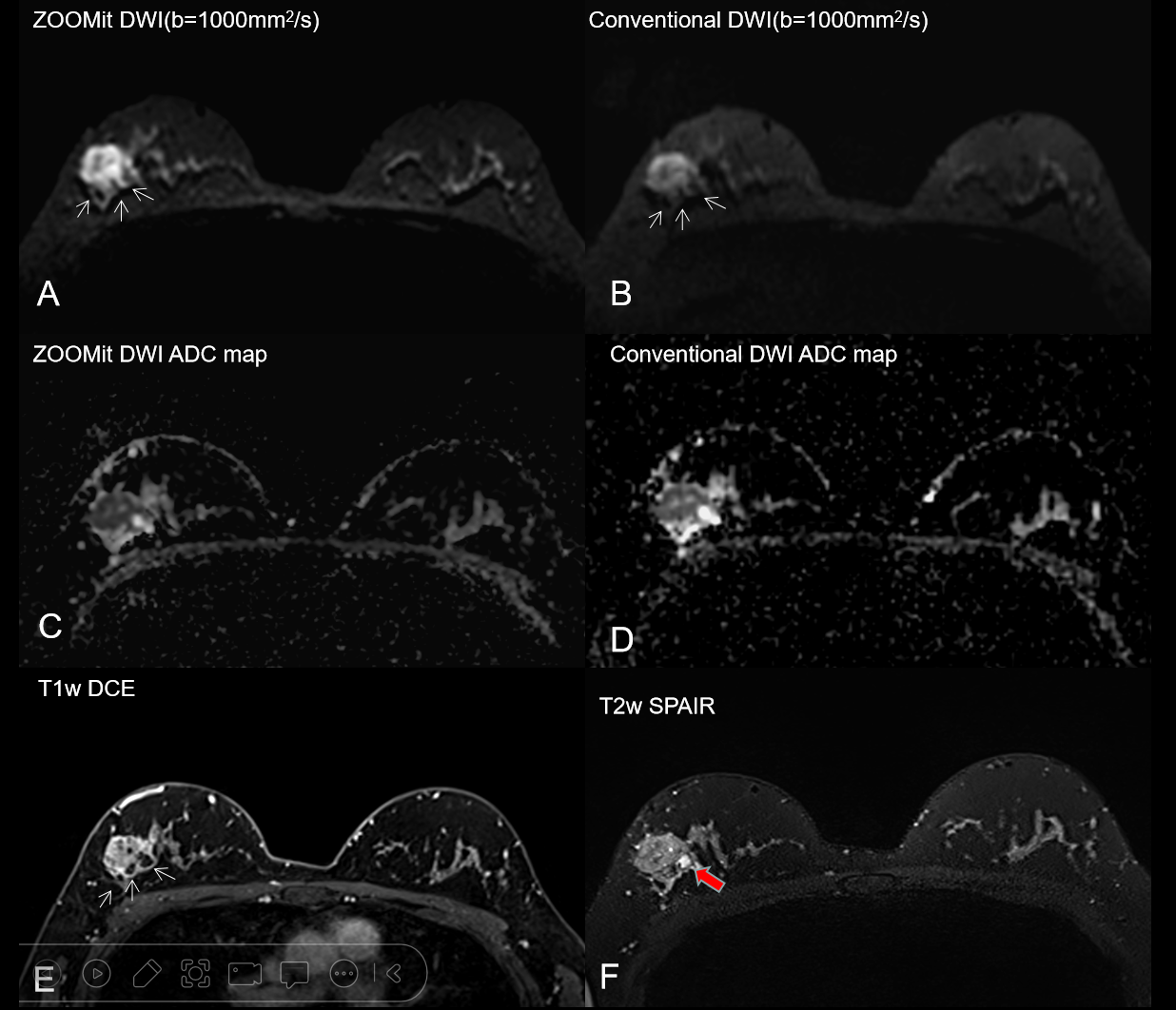
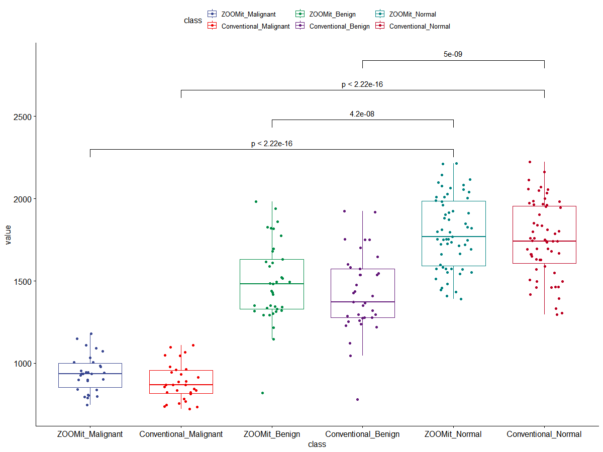
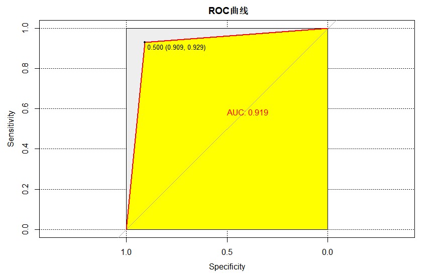
Group A:The inter-observer agreement was excellent (ICC=0.808) for the subjective assessments. The scores for Z-DWI images were considerably higher than those for C-DWI images in terms of overall picture quality(4.40±0.59 vs 3.09±0.56), sharpness of anatomical features(4.65±0.55 vs 3.80±0.48), fat suppression efficacy(4.57±0.56 vs 3.00±0.64), background noise(4.48±0.62 vs 3.02±0.62), and anatomic distortion(4.00±0.49 vs 3.17±0.67), (p <0.001);SNR and CNR in the Z-DWI series were both significantly higher than in the C-DWI sequence (77.72 ± 31.42 vs 8.59 ± 3.16, P <0.001) and (50.75 ± 26.43 vs 5.21 ± 3.04, P < 0.001);The mean ADC values of benign and malignant lesions in Z-DWI were higher than those in C-DWI (1.46×10⁻³ mm2/s vs1.37 ×10−3 mm2/s, p <0.001)and(0.87 × 10⁻³ mm2/s vs 0.83 × 10⁻³ mm2/s, p <0.001). The cutoff values of ADC for Z-DWI and C-DWI were 1.198×10⁻³ mm2/s and 1.114×10⁻³ mm2/s for benign and malignant lesions. Group B:The AUC was 0.919 in both Z-DWI and C-DWI when using the cutoff values of ADC counted from group A.Discussion
Z-DWI imaging enables reducing the region of interest while avoiding phase-warp artifacts. After the initial excitation pulse, refocusing pulse was rotated by an angle 90°, only the tissue signals in the area where the two pulses crossed can be acquired by scanner. Z-DWI obviating the need to encode a large extent in the phase-encode direction shortens the echo train. Thus, in our study, Z-DWI performs better than C-DWI in sharpness of anatomical and distortion. We can hardly find any background noise of the Z-DWI images, which was consistent with theory. So SNR and CNR of Z-DWI images were significantly higher. Fat suppression efficacy is higher in Z-DWI sequence due to the special excitation mode. The mean ADC values of benign lesions and malignant lesions in Z-DWI are higher than those in C-DWI, but which had no difference about diagnostic performance. These encouraging results demonstrated that Z-DWI can be a substitution for C-DWI with nearly the same acquisition time.Conclusion
ZOOMit DWI provides significantly higher image quality and lesion conspicuity than C-DWI with the same geometric parameters and nearly the same acquisition time. ZOOMit DWI can significantly improve the diagnostic efficiency of breast cancer.Acknowledgements
No acknowledgement found.References
[1]Sung, H, Ferlay, J, Siegel, RL, Laversanne, M, Soerjomataram, I, Jemal, A, Bray, F. Global cancer statistics 2020: GLOBOCAN estimates of incidence and mortality worldwide for 36 cancers in 185 countries. CA Cancer J Clin. 2021:71:209-249.
[2]Tezcan S, Ozturk FU, Uslu N, Akcay EY. The Role of Combined Diffusion-Weighted Imaging and Dynamic Contrast-Enhanced MRI for Differentiating Malignant From Benign Breast Lesions Presenting Washout Curve. Can Assoc Radiol J.2021Aug;72(3):460-469
[3]ang X, Dong M, Li S, Chai R, Zhang Z, Li N, Zhang L. Diffusion-weighted imaging or dynamic contrast-enhanced curve: a retrospective analysis of contrast-enhanced magnetic resonance imaging-based differential diagnoses of benign and malignant breast lesions. Eur Radiol. 2020 Sep;30(9):4795-4805.
[4]Banerjee S, Nishimura DG, Shankaranarayanan A, Saritas EU. Reduced field-of-view DWI with robust fat suppression and unrestricted slice coverage using tilted 2D RF excitation. Magn Reson Med. 2016 Dec;76(6):1668-1676.
[5]Sim KC, Park BJ, Han NY, Sung DJ, Kim MJ, Han YE. Efficacy of ZOOMit coronal diffusion-weighted imaging and MR texture analysis for differentiating between benign and malignant distal bile duct strictures. Abdom Radiol (NY). 2020 Aug;45(8):2418-2429.
[6]Yıldırım İO, Sağlık S, Çelik H. Conventional and ZOOMit DWI for Evaluation of Testis in Patients With Ipsilateral Varicocele. AJR Am J Roentgenol. 2017 May;208(5):1045-1050.
[7]Seeger A, Batra M, Süsskind D, Ernemann U, Hauser TK. Assessment of uveal melanomas using advanced diffusion-weighted imaging techniques: value of reduced field of view DWI ("zoomed DWI") and readout-segmented DWI (RESOLVE). Acta Radiol. 2019 Aug;60(8):977-984.
Figures