Joo Han1, Justin D. Sprick2,3, Lisa C. Krishnamurthy1,4, Serena Song1, and Venkatagiri Krishnamurthy1,5
1Center for Visual and Neurocognitive Rehabilitation, Atlanta VAMC, Decatur, GA, United States, 2Division of Renal Medicine, Department of Medicine, Emory University, Atlanta, GA, United States, 3University of North Texas Health Science Center, Denton, TX, United States, 4Department of Physics & Astronomy, Georgia State University, Atlanta, GA, United States, 5Division of Geriatrics and Gerontology, Department of Medicine, Emory University, Atlanta, GA, United States
Synopsis
Keywords: Signal Modeling, fMRI (task based)
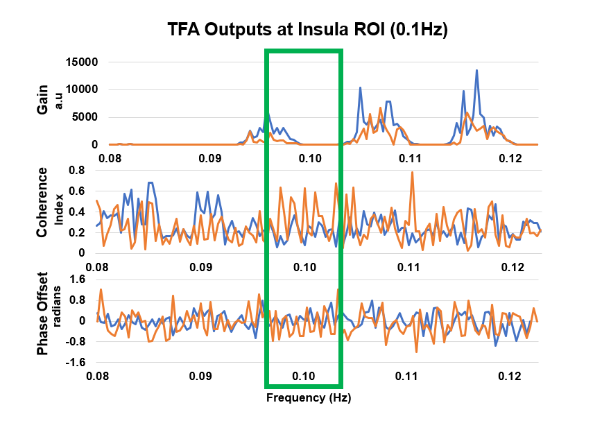
The limitation of estimating Dynamic Cerebral Autoregulation
(dCA) using Transcranial Doppler ultrasound is the lack of whole-brain estimation
capabilities. In this study, the data acquisition of whole-brain task-fMRI scan
during a novel Passive Cyclical Leg Raise (PCLR) task allows us to induce blood
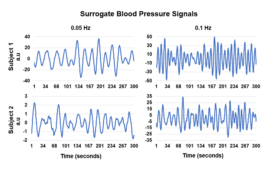
pressure (BP) changes. A surrogate BP signal was acquired from the brain stem
and depicts expected fluctuations based on the PCLR-task design. The whole-brain
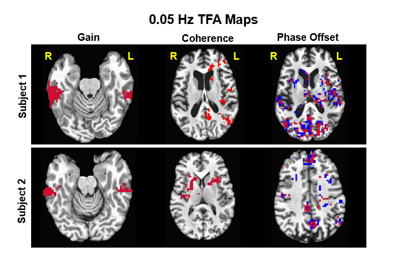
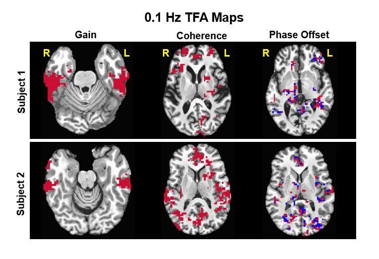
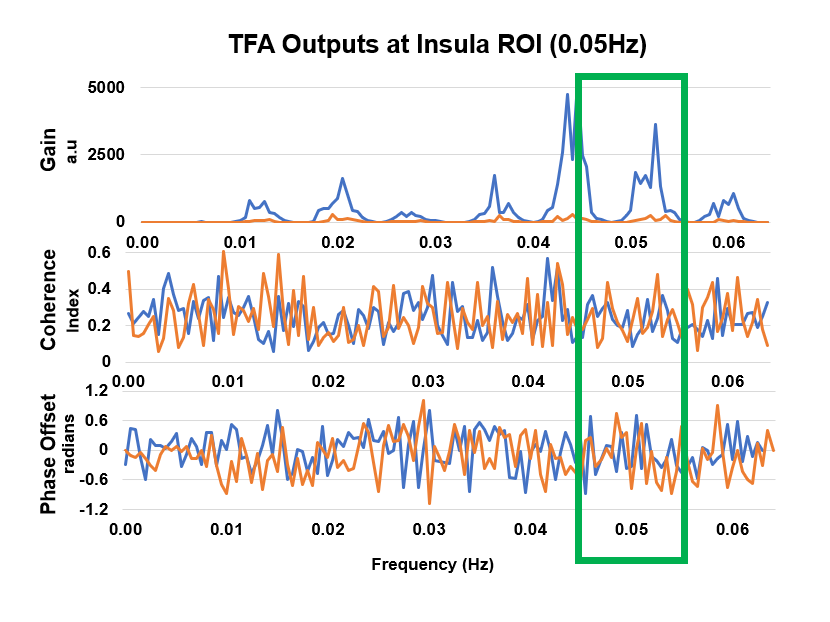
dCA was estimated using voxel-wise TFA to obtain the transfer gain, coherence,
and phase-offset. These results show similar significant brain regions but with
distinct values based on the participant’s varying level of constitution.
Introduction: Dynamic Cerebral
Autoregulation (dCA) is the brain’s inherent ability to modulate cerebral blood
flow (CBF) in response to changes in arterial blood pressure (BP). The dCA
modulatory signals have been previously discovered to be a frequency-dependent
phenomena1 and thus Transfer Function analysis (TFA) techniques are
most commonly utilized to estimate the modulation between the changes in CBF
following changes in the BP2). The most common approach of estimating
the CBF the has been through the continuous measurements of changes using a Transcranial
Doppler ultrasound (TCD) over the temples, and BP has been obtained through
arterial volume clamping of a finger artery2. However, the
limitation of this approach is that it not only cumbersome, but also the lack
of whole-brain estimation of dCA due to the limitations of TCD. In this study,
we employed a novel Passive Cyclical Leg Raise (PCLR) task to induce blood
pressure changes during a whole-brain rapid fMRI scan to acquire a surrogate BP
signal from the brain stem and then employ voxel-wise TFA to estimate whole-brain
dCA.
Methods: The task-fMRI
images were acquired from a healthy participant and a participant with
hypertension. The novel PCLR task occurred was time synced with a rapid fMRI
acquisition (TR=0.605sec) using an in-house apparatus to passively raise and
lower the participant legs at 0.05 and 0.1Hz. The apparatus was designed to
allow 60º elevation to induce cardiac load and thereby altering the BP. The 0.05Hz
oscillation occurred for five minutes before a five-minute washout and followed
up by five minutes of the 0.1Hz oscillation. The images were slice time corrected
before being separated into the 0.05Hz, 0.05Hz-washout, 0.1Hz, and 0.1Hz-washout
resulting in four images (each oscillation will be referred as conditions). The
0.1Hz-washout condition was removed from this analysis due to a sub five-minute
acquisition time2. The separated images were corrected for rigid body
motion followed by ICA denoising for various fMRI artifacts. The denoised fMRI
images were co-registered to the MPRAGE using FSL tools, temporally filtered
and then smoothed. The surrogate BP signal was estimated by extracting the BOLD
signal from the rostral Ventrolateral Medullary due to its relationship with
regulating blood pressure3. The signal was averaged within this ROI using
a five-millimeter sphere and then decomposed using a maximum overlap discrete
wavelet transform (MODWT) into six frequency bands4. The frequency
bands encompassing 0.05Hz and 0.1Hz was extracted as the surrogate BP signals
and used as the surrogate BP signal for its corresponding condition. The
temporal filtering steps were as follows: the 0.05Hz condition was low-pass
filtered at 0–0.06Hz, the washout low-pass filtered at 0-0.15Hz, and 0.1Hz
band-pass filtered at 0.08-0.12Hz. Considering that BOLD physiology inherently
involves CBF, and that BOLD offers improved temporal resolution and SNR, we
chose BOLD signal to index CBF changes. Finally, using in-house scripts for TFA
algorithm, the transfer gain, phase-offset, and coherence were estimated for
every voxel in the whole-brain image for every condition. The coherence images
were thresholded at a minimum of 0.34 to indicate a confidence value of 95% or
higher2, and the transfer gain and phase-offset were also
thresholded and clusterized to depict significant clusters of voxels. For a closer
inspection of TFA-based dCA functioning, we extracted the estimated gain, coherence,
and phase offset from left insula, as it has been shown to be in the baroreflex
control of sympathetic nerve activity3.
Results: Figure-1 depicts
the expected BP fluctuations at the designed PCLR frequency. Figure-2 and
3 depicts, the transfer gain, phase-offset, and coherence images for the 0.05Hz
and 0.1Hz condition where we observe that for both conditions, each participant
had similar significant brain regions in the transfer gain maps. Figure-4 shows
TFA outputs at 0.05Hz condition. The coherence index was greater in participant-2
at 0.015Hz and similar in other frequencies. The transfer gain was greater in
participant-1 but maintained peaks at similar frequencies. The phase-offset in participant-1
was more positive at 0.05Hz in comparison to participant-2, but otherwise,
shares similar trend. Figure-5 portrays TFA outputs at 0.1Hz condition. The
coherence index is greater in participant-2 at 0.1Hz. Further, the transfer
gain values are greater in participant-1, but the two participant’s transfer
gain values differ only by a small factor across all frequencies. The
phase-offset depicts larger shifts in participant-2 across all frequencies.
Discussion: Our preliminary
findings show results supporting that our novel PCLR approach combined with
advanced signal processing tools to estimate surrogate BP and dynamic CA are
promising and meaningful. While we note that the TFA outputs are represented in
similar brain regions between both participants, the values of said TFA outputs
are distinct between the participants. Participant-1 had larger and more
consistent values as compared to participant-2 with hypertension. Thus, the
capabilities of obtaining meaningful TFA outputs is also demonstrated in participants
with varying degree of associated health constitution.
Conclusion: Our novel MRI
compatible PCLR approach along with TFA modeling for dCA will be useful in
acute stroke and inpatient neurological settings to diagnose and regulate BP
fluctuations, and thereby minimizing secondary and/or severe damage from stroke
and other complications.Acknowledgements
We thank Dr. Keith McGregor for his assistance in data collection.References
1. R. Zhang, J. Zuckerman, C. Giller, and B. Levine. Transfer function analysis of dynamic cerebral autoregulation in humans. American Journal of Physiology-Heart and Circulatory Physiology, 274(1), pp. H233-H24. 1998. doi/10.1152/ajpheart.1998.274.1.H233.
2. Panerai RB, Brassard P, Burma JS, et al. Transfer function analysis of dynamic cerebral autoregulation: a CARNet white paper 2022 update. Journal of Cerebral Blood Flow & Metabolism, 0(0), 2022. doi:10.1177/0271678X221119760
3. V. Macefield, L. Henderson. Identification of the human sympathetic connectome involved in blood pressure regulation. NeuroImage, 202, 2019. doi.org/10.1016/j.neuroimage.2019.116119.
4. J. Whittaker, I. Driver, M. Venzi, M. Bright, and K. Murphy. Cerebral Autoregulation Evidenced by Synchronized Low Frequency Oscillations in Blood Pressure and Resting-State fMRI. Front. Neursci., 2019. doi.org/10.3389/fnins.2019.00433.