2527
Functional plasticity development of visual-motor cortex in neonatal brain is modulated by postnatal experience
Yifan Shuai1, Ruolin Li1, Yihan Wu1, Zhiyong Zhao1, and Dan Wu1
1Key Laboratory for Biomedical Engineering of Ministry of Education, Department of Biomedical Engineering, College of Biomedical Engineering & Instrument Science, Zhejiang University, Hangzhou, China
1Key Laboratory for Biomedical Engineering of Ministry of Education, Department of Biomedical Engineering, College of Biomedical Engineering & Instrument Science, Zhejiang University, Hangzhou, China
Synopsis
Keywords: Neonatal, Normal development, functional plasticity
The present study aimed to investigate how the brain entropy (BEN) changes with age during early development and whether it exists differences between term- and preterm-born neonates. The results found that the BEN in visual-motor cortex was positively correlated with PMA but had no significant correlation with GA, which showed an increase in preterm- than term-born neonates with matched PMA. Moreover, both age and preterm have a significant effect on the functional connectivity between visual-motor cortex and other regions. These findings suggest the functional plasticity development of visual-motor cortex is modulated by early postnatal experience in human neonates.Introduction
Brain entropy (BEN) is an effective model for evaluating brain complexity, which has been used to study brain functional development in adults. Studies have revealed that the BEN increases with age at 22-37 years which facilitates general brain functionality, whereas that at 65-95 years indicates progressive brain deteriorations 1,2. A recent study in individuals with ages ranging from 6 to 82 years found that the BEN change with age followed an inverted U-shaped trajectory with a peak point at age 40 3. However, the BEN analysis has not been utilized to study brain plasticity during early human brain development, although this method has been used in neonatal mice 4. In this study, we aimed to investigate the functional plasticity development of neonatal brain using BEN and compare the difference between term- and preterm-born neonates.Methods
Data acquisition: 280 neonatal resting-state fMRI (rs-fMRI) data (PMA at scan ranged from 37.5 to 44.5 weeks) were selected from the second release of Developing Human Connectome Project (dHCP), consisting of 244 term-born and 36 preterm-born infants. The rs-fMRI data were acquired using EPI sequence with the following parameters: TE/TR= 38/392ms, voxel size = 2.15 × 2.15 × 2.15 mm3, and 2300 volumes.Data processing: After the dHCP fMRI pipeline 5, the subsequent processing included slice time correction, head motion correction, normalization, nuisance signals regression, and spatial smoothing. Then, we calculated fALFF (which reflects the magnitude of spontaneous brain activity) using DPABI toolbox 6 and BEN maps after band-pass filtering (0.01-0.1Hz) using BEN mapping toolbox 7 for each neonate. The averaged maps of fALFF and BEN were computed at each week PMA.
Statistical analysis: Multiple regression was performed to detect the relationship between MRI measurements and age (244 term neonates for PMA and 155 term neonates with PMA-GA < 1 week for GA). Then two-sample t-tests were used to evaluate the difference between term and preterm birth (36 preterm and 36 term neonates matched) and between male and female (126 male and 108 female term neonates matched). A leave-one-out cross-validation (LOOCV) was applied to detect the effect of preterm birth, considering a relatively small sample size in preterm neonates. All P values were corrected by the FDR method (p < 0.05) to control the false positive discoveries.
Results
fALFF and BEN maps both showed a developmental change with age (Fig 1). We found that the BEN positively correlated with PMA in visual-motor cortical regions, including occipital lobe and precentral/postcentral gyrus (Fig. 2A), whereas the fALFF correlated with PMA in supplementary motor area, subcortical regions, and cerebellum (Fig. 2B) (adjusted p < 0.05). As the word cloud showed, the change with PMA for BEN was associated with sensorimotor and visual functions, and that for fALFF was associated with sensation and coordination functions. Moreover, the fALFF showed negative correlations with GA in cerebellum and subcortical regions, and positive correlations in occipital lobe and inferior parietal lobe (Fig. 2C) (adjusted p < 0.05). However, the BEN showed no significant correlation with GA (adjusted p > 0.05). In addition, compared with the term-born group, the preterm-born group displayed lower fALFF but higher BEN in the visual-motor cortical regions (Fig .3) (adjusted p < 0.05), which showed a high reproducibility by the LOOCV. There was no significant difference between sex in both fALFF and BEN (adjusted p > 0.05). We also constructed functional connectivity (FC) of the visual-motor cortical regions showing significant correlations between the PMA and the BEN but not fALFF. The results found that the FCs were significantly affected by age and preterm birth (Fig .4) (adjusted p < 0.05).Discussion and Conclusion
The present study applied BEN to investigate the development of brain functional plasticity in human neonates for the first time. We found that BEN in visual-motor cortical regions was significantly positively correlated with PMA, but had no significant association with GA. This demonstrated that the functional plasticity development of visual-motor cortex could be modulated by postnatal experience, which is consistent with recent reports that the development of visual-motor cortex in human neonates is selectively modified by postnatal experience 8,9. This finding was also supported by the between-group comparison that compared with term-born neonates, preterm-born neonates have weaker spontaneous brain activity (lower fALFF value) but higher plasticity (larger BEN) in the visual-motor cortex, which was in line with the previous finding that a high level of plasticity and capacity to recover from injuries associated with preterm birth through experiences 10. Thus, we speculate that the plasticity development may primarily occur in the functional connectivity between the regions but not the function of the brain region itself, which may be supported by a recent study 11 that found similar local activity but lower connectivity between regions and connectivity flexibility in neonates compared with adults. The subsequent FC analysis demonstrated that the FCs of visual-motor cortical regions showed significant correlations with infants’ age and significant differences between term- and preterm-born neonates. In summary, these findings revealed a significant effect of postnatal experience on functional plasticity development of visual-motor cortex in neonatal brain, and BEN analysis provides a novel insight toward the underlying mechanism of brain development.Acknowledgements
This work was supported by the Ministry of Science and Technology of the People’s Republic of China (2018YFE0114600, 2021ZD0200202), the National Natural Science Foundation of China (81971606, 82122032), and the Science and Technology Department of Zhejiang Province (202006140, 2022C03057).References
1. Wang, Z. The neurocognitive correlates of brain entropy estimated by resting state fMRI. Neuroimage. 2021; 232: 117893.2. Wang, Z., & Alzheimer’s Disease Neuroimaging Initiative. Brain entropy mapping in healthy aging and Alzheimer’s disease. Front Aging Neurosci. 2020; 12: 596122.
3. Niu, Y., Sun, J., Wang, B., Yang, Y., Wen, X., & Xiang, J. Trajectories of brain entropy across lifetime estimated by resting state functional magnetic resonance imaging. Hum Brain Mapp. 2022.
4. Fei, L., Chen, H., Ma, L., Weigao, E., Wang, R., Fang, X., ... & Guo, G. Systematic identification of cell fate regulatory programs using a single-cell atlas of mouse development. 2022.
5. Fitzgibbon, S. P., Harrison, S. J., Jenkinson, M., Baxter, L., Robinson, E. C., Bastiani, M., ... & Andersson, J. The developing Human Connectome Project (dHCP) automated resting-state functional processing framework for newborn infants. Neuroimage. 2020; 223: 117303.
6. Yan, C. G., Wang, X. D., Zuo, X. N., & Zang, Y. F. DPABI: data processing & analysis for (resting-state) brain imaging. Neuroinformatics. 2016; 14(3): 339-351.
7. Wang, Z., Li, Y., Childress, A. R., & Detre, J. A. Brain entropy mapping using fMRI. Plos One. 2014;9(3), e89948.
8. Li, M., Liu, T., Xu, X., Wen, Q., Zhao, Z., Dang, X., ... & Wu, D. Development of visual cortex in human neonates are selectively modified by postnatal experience. bioRxiv. 2022.
9. Natu, V. S., Rosenke, M., Wu, H., Querdasi, F. R., Kular, H., Lopez-Alvarez, N., ... & Grill-Spector, K. Infants’ cortex undergoes microstructural growth coupled with myelination during development. Comms Biol. 2021; 4(1):1-12.
10. Head, L. M., Baralt, M., & Mahoney, A. E. D. Bilingualism as a potential strategy to improve executive function in preterm infants: A review. J Pediatr Health Car. 2015; 29(2): 126-136.
11. Huang, Z., Wang, Q., Zhou, S., Tang, C., Yi, F., & Nie, J. Exploring functional brain activity in neonates: A resting-state fMRI study. Dev Cogn Neurosci. 2020; 45: 100850.
Figures
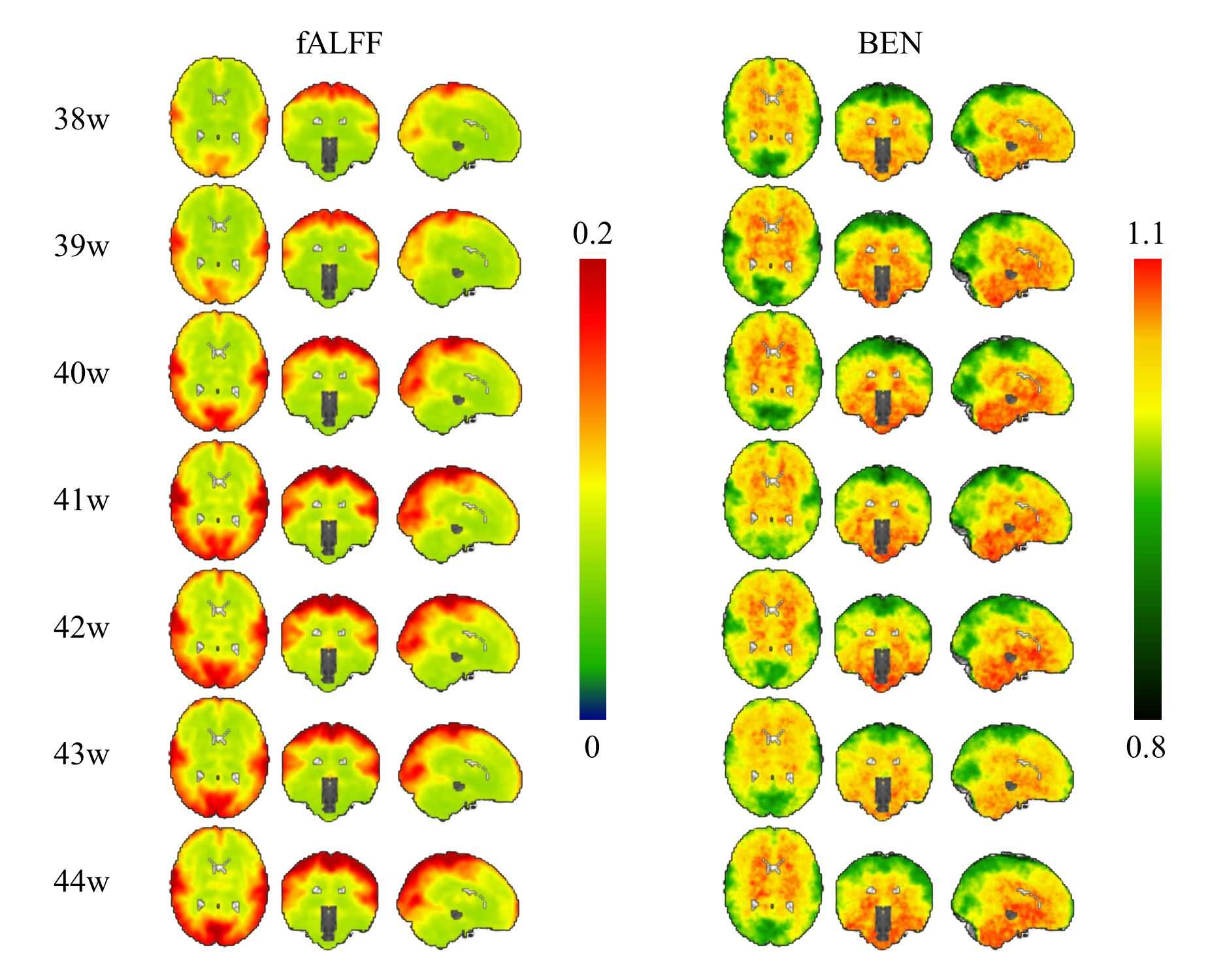
Figure 1. fALFF and BEN maps in term-born infants at each week PMA. Grouped-averaged maps of fALFF and BEN in term-born infants at 38-44 weeks PMA. fALFF and BEN both showed a trend of increase with age in cortical regions. Color bar represents the value of fALFF and brain entropy.
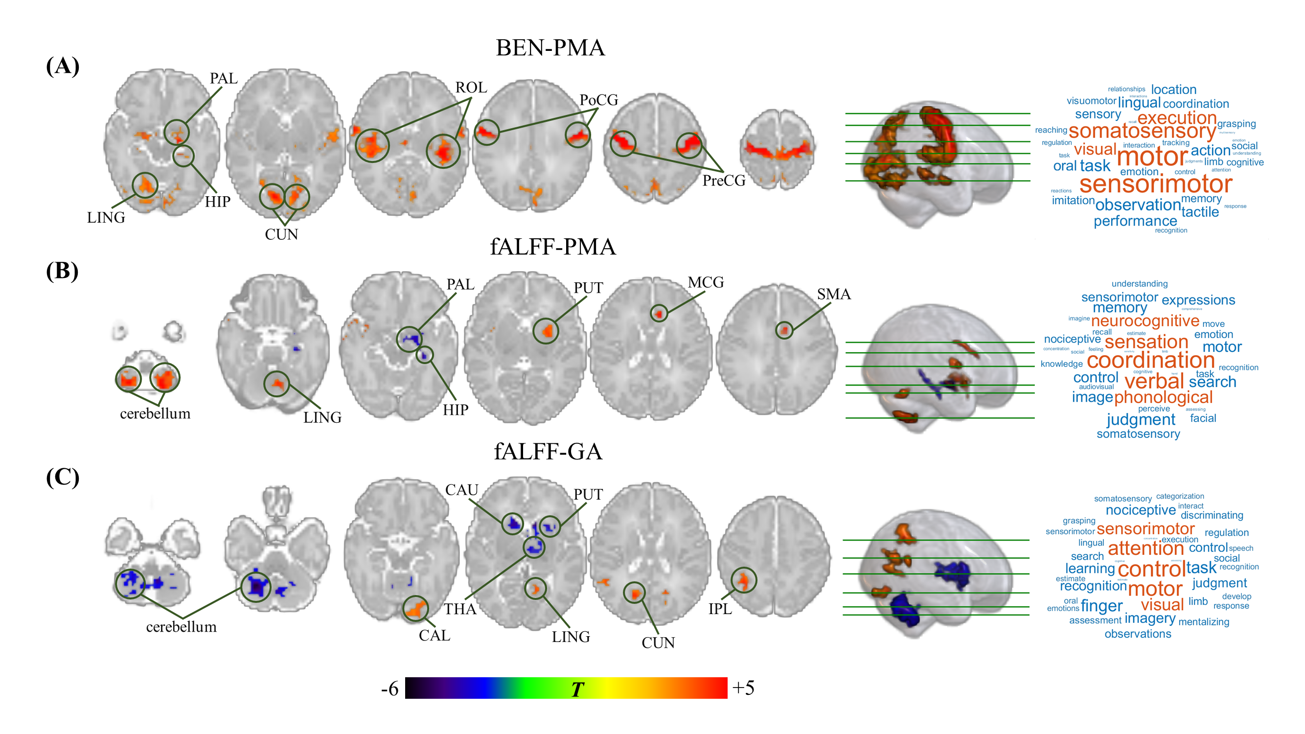
Figure 2. Relationship between BEN/fALFF and PMA/GA in term-born infants. Color bar represents T value. The brain regions with red and blue indicate positive and negative correlations, respectively. Word cloud represents the behavioral relevance with age, in which the larger the font, the higher the correlation. The BEN showed positive correlations with PMA in visual-motor regions but no correlation with GA, while the fALFF significantly correlated with PMA/GA in cerebellum, occipital lobe, and subcortical regions.
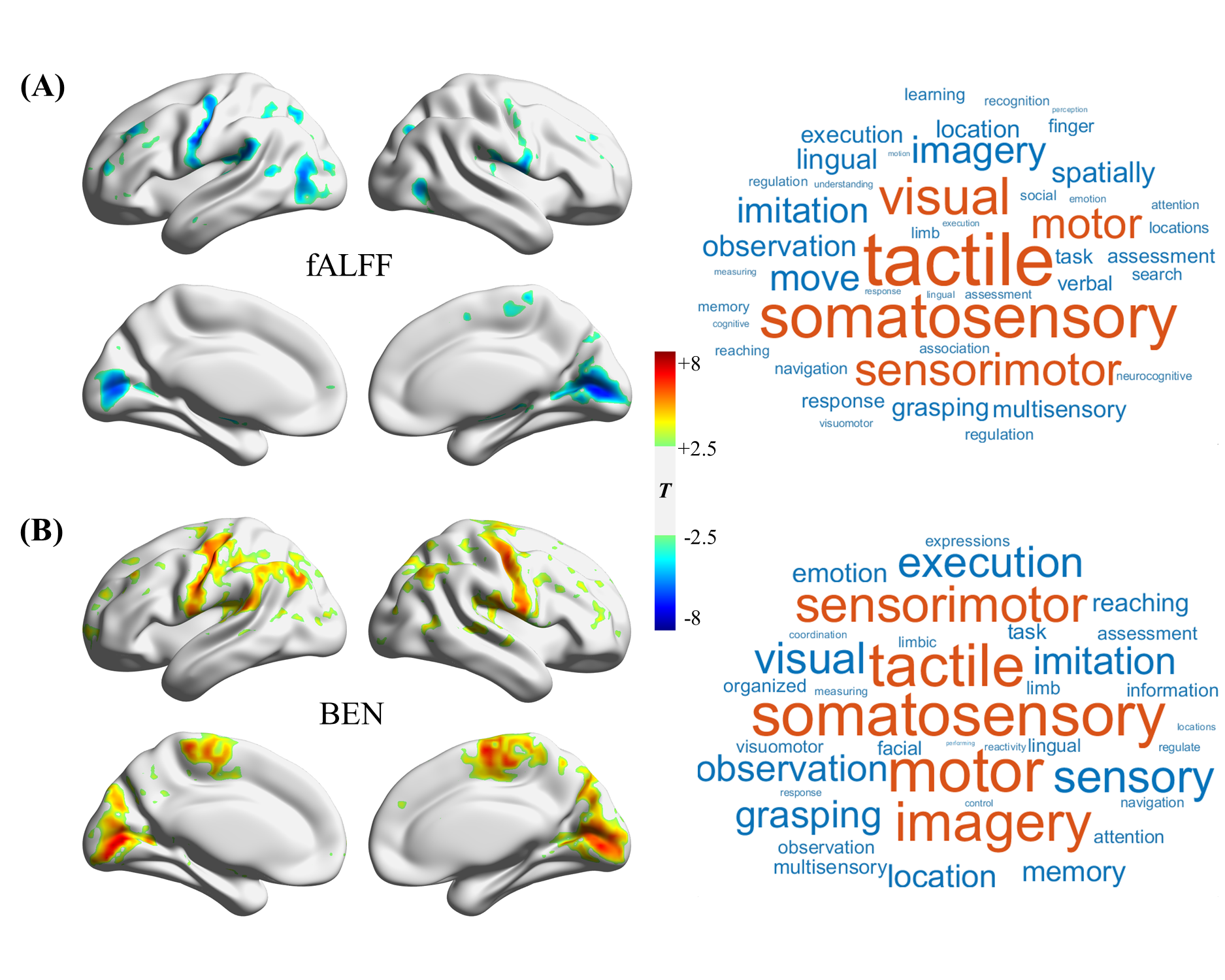
Figure 3. Effect of preterm birth on fALFF and BEN. The brain regions with red and blue indicate increased and decreased fALFF/BEN in preterm-born infants, respectively. Word cloud represents the behavioral relevance with the difference between the groups, in which the larger the font, the higher the correlation. The preterm-born group displayed lower fALFF but higher BEN in the visual-motor network, compared with the term-born group.
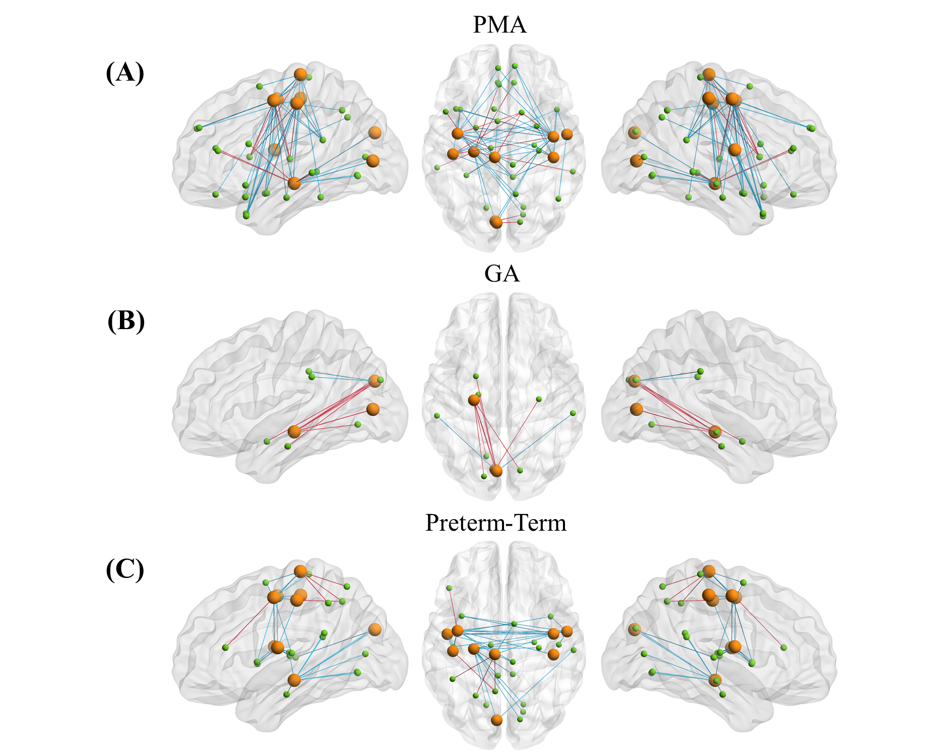
Figure 4. The effects of age and preterm functional connectivity in the visual-motor cortex. Yellow spheres represent visual-motor cortical regions showing significant correlations with PMA in BEN but not in fALFF. Green spheres represent other regions in the brain. The edges in red represent positive correlation or increased value in preterm, and those in blue represent negative correlation or decreased value in preterm. FCs in the visual-motor network showed significant correlations with age and significant differences between term- and preterm-born neonates.
DOI: https://doi.org/10.58530/2023/2527