2523
the value of ASL-based cerebral blood flow and network metrics in evaluating children with MR-negative epilepsy1Department of Radiology, The Affiliated Wuxi Children's Hospital of Nanjing Medical University, Wuxi, China, 2Department of Radiology, Wuxi People's Hospital Affiliated to Nanjing Medical University, Wuxi, China
Synopsis
Keywords: Neuro, Epilepsy
Epilepsy is the most common chronic neurological disorder with significant morbidity and mortality. Frontal lobe epilepsy is a common type of seizure after temporal lobe epilepsy, which makes the diagnosis of frontal lobe epilepsy and its treatment difficult due to its presence of not only motor symptoms but also other autonomic symptoms during seizures. Arterial spin labeling can be used to identify brain regions and brain network lesions associated with the propagation of epileptiform activity, and is of higher investigative value especially in children with negative MRI.
keyword: Epilepsy, brain network, arterial spin labeling, pediatrics
Introduction
The frontal lobe is located in the most anterior part of the cerebral hemisphere, accounting for the first third of the cerebral hemisphere, and its main functions are related to random movements and higher neuropsychiatric activities. Many afferent and efferent fibers in the frontal lobe area are connected to the limbic system respectively, so abnormal discharges in the frontal lobe can rapidly spread to other brain areas through these links, resulting in abnormal discharges and different functional clinical manifestations in these brain areas. This all causes great difficulties in clinical diagnosis and treatment, especially for children with negative MRI, where no obvious destruction of structural regions occurs, so the corresponding pathophysiological changes are more worthy of study and exploration.Arterial spin labeling (ASL) is a non-invasive MRI technique that uses magnetically labeled blood as a tracer to measure brain perfusion.ASL helps to localize the brain regions where epileptogenesis occurs and the corresponding brain networks. It provides information on altered brain function caused by epilepsy similar to PET, which is particularly relevant in patients with MR-negative epilepsy.
Methods
Seventeen children with seizures were enrolled while diagnosed with frontal lobe epilepsy after EEG examination. Twelve volunteers were also recruited with written consent from their guardians.MRI was performed using a Siemens Magnetom Aera 1.5T system. The 3D pulsed ASL (3D-pASL) sequence was set with the following parameters: axial acquisition, repetition time (TR)=2500ms, echo time (TE)=18ms, phase encoding=ap, 5mm thick, inversion time (TI)=1700ms, PLD=1000ms,For each subject, 120 images (60 label/control pairs) were acquired. A structural conventional MRI sequence acquired in the same session as 3D-pASL was also retrieved with the following parameters :MPRAGE; axial acquisition, TR=2200ms, TE=3.02ms, TI=900ms,voxel size acquisition and reconstructio=1x1x1 mm.
The magnetic resonance images were preprocessed using a matlab-based toolkit with processing steps including motion correction, background noise suppression, CBF numerical quantization, volume correction and MNI standard brain space alignment, and isotropic Gaussian kernel (FWHM=6 mm) for spatial smoothing.
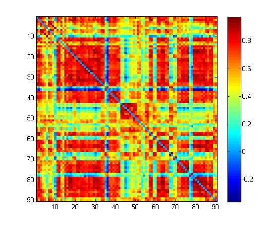
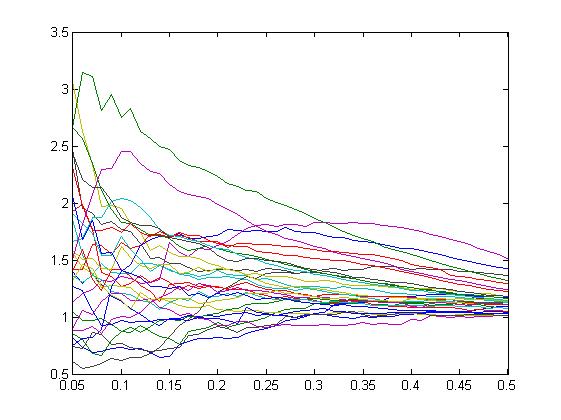
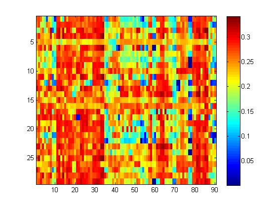
Network metrics were defined by using the Anatomical Automatic Labeling atlas with 90 regions(AAL90). Small-world network analysis was conducted on the individual level adjacency matrix. Network metrics were analyzed based on graph theory.
Results
CBF findings:Children in the case group had reduced CBF values in the Parietal_Sup_L,Parietal_Inf_L,Precentral_L,Frontal_Mid_L regions (voxel-level,P<0.001,uncorreted)Network metrics findings:Small-world characteristics of children in the case group increased relative to controls (measured in 1000 matched random networks).The nodal efficiency of children in the case group was reduced within the Frontal_Sup_Orb_L and Frontal_Sup_Orb_L brain regions (P < 0.05, FDR-corrected)
Conclusion
Arterial spin labeling is able to detect alterations in brain perfusion and network in children with magnetic resonance-negative epilepsy and is of potential clinical value in revealing the pathophysiological basis of epilepsy.Discussion
Brain perfusion can be abnormal in epileptic patients, and areas of altered perfusion provide important clues to the localization of epileptic lesions, especially in patients with negative MR. With some standardized image preprocessing, it is possible to pinpoint the lesion site to a specific brain region. The acquisition of ASL is more sensitive in children compared to adults because of the higher water content of the brain, higher blood flow and blood velocity in the carotid artery. we also observed extensive areas of perfusion changes in the frontal and parietal regions. This suggests that changes in the region of onset of epilepsy and even network connectivity may be altered. So ASL helps to better understand the extensive epileptic network. Our study uses the same time acquisition sequence in which CBF values and network metrics are extracted separately, maintaining the consistency of time scale and compensating for the disadvantage of low signal-to-noise ratio of ASL sequences as much as possible. In our later work, we will further optimize the exploration in terms of sample size, signal-to-noise ratio enhancement and network mapping selection.Acknowledgements
This work was supported by The Top Talents Project of Wuxi Taihu Lake Talent plan(2020THRC-GD-CHW),The Key Program of Wuxi health Committee(J202107), the General Project of Shaanxi Natural Science Basic Research Plan (2021JM-558).References
1.Haller S, Zaharchuk G, Thomas DL, Lovblad KO, Barkhof F, Golay X. Arterial Spin Labeling Perfusion of the Brain: Emerging Clinical Applications. Radiology. 2016;281(2):337-356. doi:10.1148/radiol.2016150789
2.Boscolo Galazzo I, Storti SF, Barnes A, et al. Arterial Spin Labeling Reveals Disrupted Brain Networks and Functional Connectivity in Drug-Resistant Temporal Epilepsy. Front Neuroinform. 2019;12:101. Published 2019 Mar 6. doi:10.3389/fninf.2018.00101
3.Pasca L, Sanvito F, Ballante E, et al. Arterial spin labelling qualitative assessment in paediatric patients with MRI-negative epilepsy. Clin Radiol. 2021;76(12):942.e15-942.e23. doi:10.1016/j.crad.2021.09.016
4.Kojan M, Gajdoš M, Říha P, Doležalová I, Řehák Z, Rektor I. Arterial Spin Labeling is a Useful MRI Method for Presurgical Evaluation in MRI-Negative Focal Epilepsy. Brain Topogr. 2021;34(4):504-510. doi:10.1007/s10548-021-00833-5
5.Liang X, Connelly A, Calamante F. Graph analysis of resting-state ASL perfusion MRI data: nonlinear correlations among CBF and network metrics. Neuroimage. 2014;87:265-275. doi:10.1016/j.neuroimage.2013.11.013
Figures