2494
Consistency between 2D and 3D amide proton weighted imaging on brain tumors1The First Affiliated Hospital of Dalian Medical University, DaLian, China, 2Philips Healthcare, BeiJing, China, 3The First Affiliated Hospital of Dalian Medical University, Dalian, China
Synopsis
Keywords: Tumors, Molecular Imaging
Amide Proton Transfer (APT) weighted imaging is a new type of magnetic resonance molecular imaging technology derived from Chemical Exchange Saturation Transfer (CEST) technology. APT weighted imaging can be carried out with 2D and 3D acquisition mode, so called as 2D- and 3D-APT respectively. There is no relevant report on the comparison of 2D and 3D-APT imaging on brain tumors. This study intends to compare and analyze the performance of 2D- and 3D-APT imaging on brain tumors through imaging and pathological data, and recommend a better APT imaging scheme to improve the diagnosis of brain tumors.Synopsis
Amide Proton Transfer (APT) weighted imaging is a new type of magnetic resonance molecular imaging technology derived from Chemical Exchange Saturation Transfer (CEST) technology. APT weighted imaging can be carried out with 2D and 3D acquisition mode, so called as 2D- and 3D-APT respectively. There is no relevant report on the comparison of 2D and 3D-APT imaging on brain tumors. This study intends to compare and analyze the performance of 2D- and 3D-APT imaging on brain tumors through imaging and pathological data, and recommend a better APT imaging scheme to improve the diagnosis of brain tumors.Summary of Main Findings
We compared the optimized 2D- and 3D-APT imaging in brain tumors. Results showed that the quantitative values measured by 2D and 3D-APT sequences (MTRasym (tumor), MTRasym(edema), MTRasym(CNAWM), △MTRasym(c-n), △MTRasym(d-n)) were not significantly different, indicating that the two imaging methods were highly consistent. The MTRasym (tumor) were significantly positively correlated with the pathological ki-67 index, showing that both 2D- and 3D-APT can reflect the elevated synthesis of protein and polypeptide resulted from the cell proliferation in tumor.Introduction
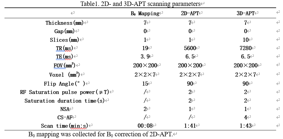
Amide proton transfer weighted imaging can be performed using the 3D volume acquisition (3D-APT) with high signal-to-noise ratio and less skull base artifacts, but the scan time can be long. The tolerance of patients with brain tumors tends to be poor, and the image quality of 3D-APT can be vulnerable to the interference from head motion. 2D-APT collects the image data on in slices in a 2D acquisition way, and the scan time can be short when less slices are collected. Besides, 2D APT can be implemented with flexible saturation schemes, which may provide the more accuracy fitting of the z-spectrum and thus the quantification of APT signals1~4. The B0 field correction for 2D- and 3D-APT imaging can also be different. This study aims to compare and analyze the performance of 2D- and 3D-APT imaging on brain tumors through imaging and pathological data, and recommend a better APT imaging scheme to improve the diagnosis of brain tumors.Materials and methods
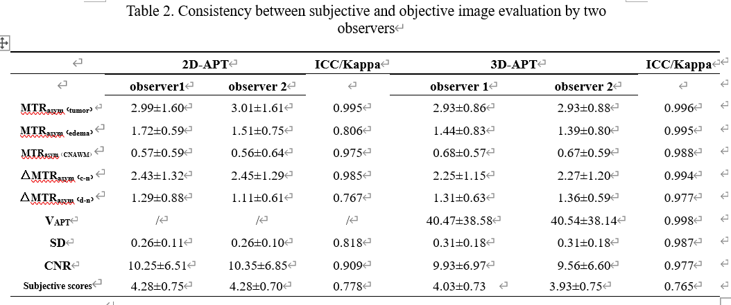
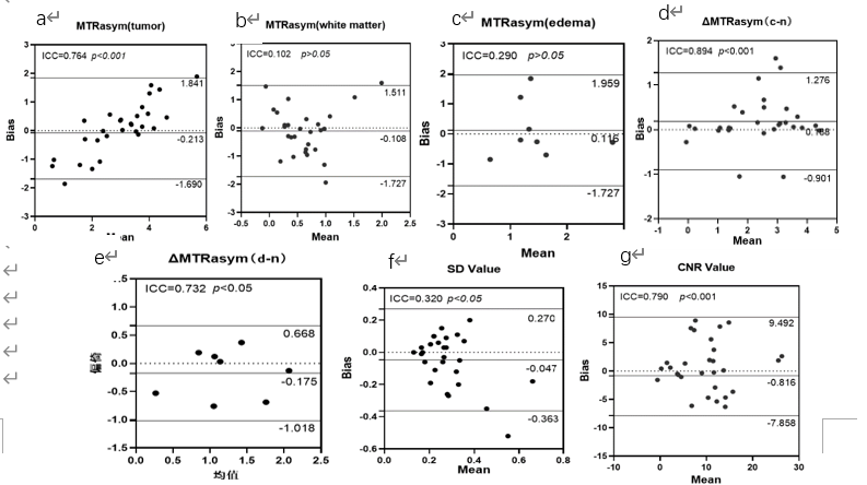
A total of 29 subjects (7 males, mean age: 54.90±13.11) were scanned using a 3.0 T MR scanner, the MR scans included 2D- and 3D-APT imaging as well as the conventional imaging sequences. APT images were subjectively evaluated by two radiologists with more than 3 years of diagnostic experience according to the Lipert Quintile, and MTRasym values were measured on 2D- and 3D-APT images, respectively, at the tumor core, peritumorous edema area, and the contralateral normal white matter area. The Intraclass Correlation Coefficient (ICC) and Kappa analysis were used to assess the consistency of measurement data and subjective scores, respectively, between the two observers. The difference of MTRasym(tumor), MTRasym(edema), MTRasym(CNAWM), △MTRasym(c-n), △MTRasym(d-n) and the contrast to noise ratio (CNR) measured from 2D- and 3D-APT images were compared by paired sample t test, and the subjective score of image quality was compared by Wilcoxon rank test. The correlation of MTRasym(tumor) with Ki-67 expression was analyzed by the Spearman correlation coefficient. Bland-Altman method was used to analyze the consistency of quantitative values between 2D- and 3D- APT imaging.Results
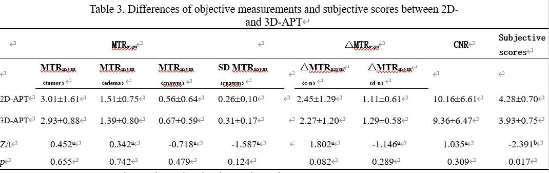
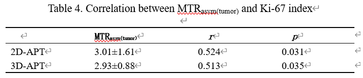
The quantitative measurements and the scores of image quality by the two observers were in good agreement (ICC: 0.767-0.998, Kappa: 0.765-0.778). The t-test of paired samples showed no significant difference of MTRasym(tumor), MTRasym(edema), MTRasym(CNAWM), △MTRasym(c-n), and △MTRasym(d-n) measured by 2D- and 3D-APT (p>0.05); the Wilcoxon rank test showed that the subjective scores of 2D-APT images was higher than those of 3D-APT (p<0.05); Spearman results showed that MTRasym(tumor) measured by 2D- and 3D-APT were both positively correlated with Ki-67 index (r=0.524, P=0.031; r=0.513, P=0.035). The Bland-Altman test showed good consistency of MTRasym(tumor), △MTRasym(c-n), and CNR values etc. for 2D- and 3D-APT images.Discussion and Conclusions
2D-APT may provide high image quality than 3D-APT for brain tumor imaging, while the measured MTRasym values for brain tumor by 2D- and 3D-APT show high consistency, which were both positively correlated with the Ki-67 index.Acknowledgements
No acknowledgement found.References
1 Park J E, Jung J E, Jung S C, et al. Amide proton transfer-weighted MRI can detect tissue acidosis and monitor recovery in a transient middle cerebral artery occlusion model compared with a permanent occlusion model in rats[J].EUR RADIOL. 2019,29(8):4096-4104.
2Zhao X, Wen Z, Zhang G, et al. Three-dimensional turbo-spin-echo amide proton transfer MR imaging at 3-Tesla and its application to high-grade human brain tumors[J]. MOL IMAGING BIOL. 2013,15(1):114-22.
3 Zhou J, Zhu H, Lim, M, et al. Three-dimensional amide proton transfer MR imaging of gliomas: Initial experience and comparison with gadolinium enhancement[J]. J MAGN RESON IMAGING. 2013,38(5):1119-28.
4 Zhou J, Zaiss M, Knutsson L, et al. Review and consensus recommendations on clinical APT-weighted imaging approaches at 3T: Application to brain tumors. MAGNET RESON MED,2022,88(2):546-574.
Figures