2479
Angiographic Comparison of Different Velocity-Selective Labeling Modules Directly on Cerebral Arteries1Kennedy Krieger Institute, Baltimore, MD, United States, 2The Russell H. Morgan Department of Radiology and Radiological Science, Johns Hopkins School of Medicine, Baltimore, MD, United States
Synopsis
Keywords: Blood vessels, Blood vessels, MR Angiography
Velocity-selective labeling has been utilized in both arterial spin labeling (VSASL) and angiography (VSMRA). This study evaluated the labeling characteristics directly on various cerebral arteries using VSMRA with different VS labeling modules, including double refocused hyperbolic tangent (DRHT), eight-segment B1-insensitive rotation (BIR-8), delay alternating with nutation for tailored excitation (DANTE), Fourier transform based velocity-selective saturation (FT-VSS) and inversion (FT-VSI). Their effects on both the cerebral arterial vasculature and static brain tissue were visualized angiographically and compared quantitatively. And the implication for VSASL and VSMRA are discussed.Introduction
Velocity-selective labeling has been utilized in both arterial spin labeling (VSASL)1–4 and angiography (VSMRA)5,6 for brain applications. This study evaluated the labeling characteristics directly on various cerebral arteries using VSMRA of different VS labeling modules, including double refocused hyperbolic tangent (DRHT), eight-segment B1-insensitive rotation (BIR-8), delay alternating with nutation for tailored excitation (DANTE), Fourier transform based velocity-selective saturation (FT-VSS) and inversion (FT-VSI).Methods
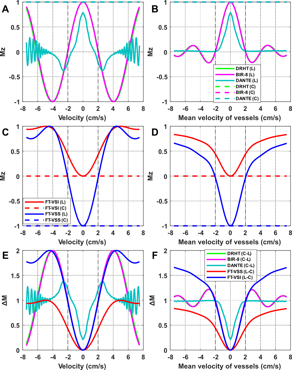
Five healthy volunteers (23-39yo, 3F) were recruited in this study. All experiments were performed on a Siemens Prisma 3T scanner with a 32-channel head coil for signal reception. All simulations and data processing were conducted with MATLAB 2022a.Five VS labeling modules were investigated, including DRHT (24ms, 5ms HT refocusing, 1.0ms gradient lobes with 0.3 ms ramp, 17mT/m), BIR-8 (24ms, 3ms HT refocusing, 1.0ms gradient lobes with 0.5 ms ramp, 30mT/m), DANTE (40ms, 1ms segment, 0.8ms gradient lobes with 0.4ms ramp, 59mT/m), FT-VSS and FT-VSI (64ms, 8ms segment, 1ms composite refocusing, 0.6ms gradient lobes with 0.3 ms ramp, 21mT/m). For DANTE, because of the existence of a banding signal pattern7, gradients were calculated so that the cycle of the banding is similar to the slice thickness to achieve a uniform background. Cutoff velocity was matched at 2cm/s, as shown in the simulation of Mz-velocity responses (Fig.1).
The VSMRA applied velocity-encoding gradients along foot-head direction. For all VS modules except FT-VSS, controls with gradient amplitude set to zero were acquired as well. A spatially selective inversion pulse was applied 900ms before the labeling modules to null venous signal6. Turbo-FLASH (TFL) readout with 180×180×104mm3 FOV and 0.7×0.7×1.0mm3 resolution was employed with 2-fold GRAPPA acceleration. The total time was 3.6min for a single label or control or 7.2min for the pair. A reference image without any labeling was acquired with the same acquisition and repetition time. A product time-of-flight (TOF) sequence was also acquired with an almost similar spatial coverage and a higher resolution of 0.5×0.5×1.0mm3, with a total scan time of 7min.
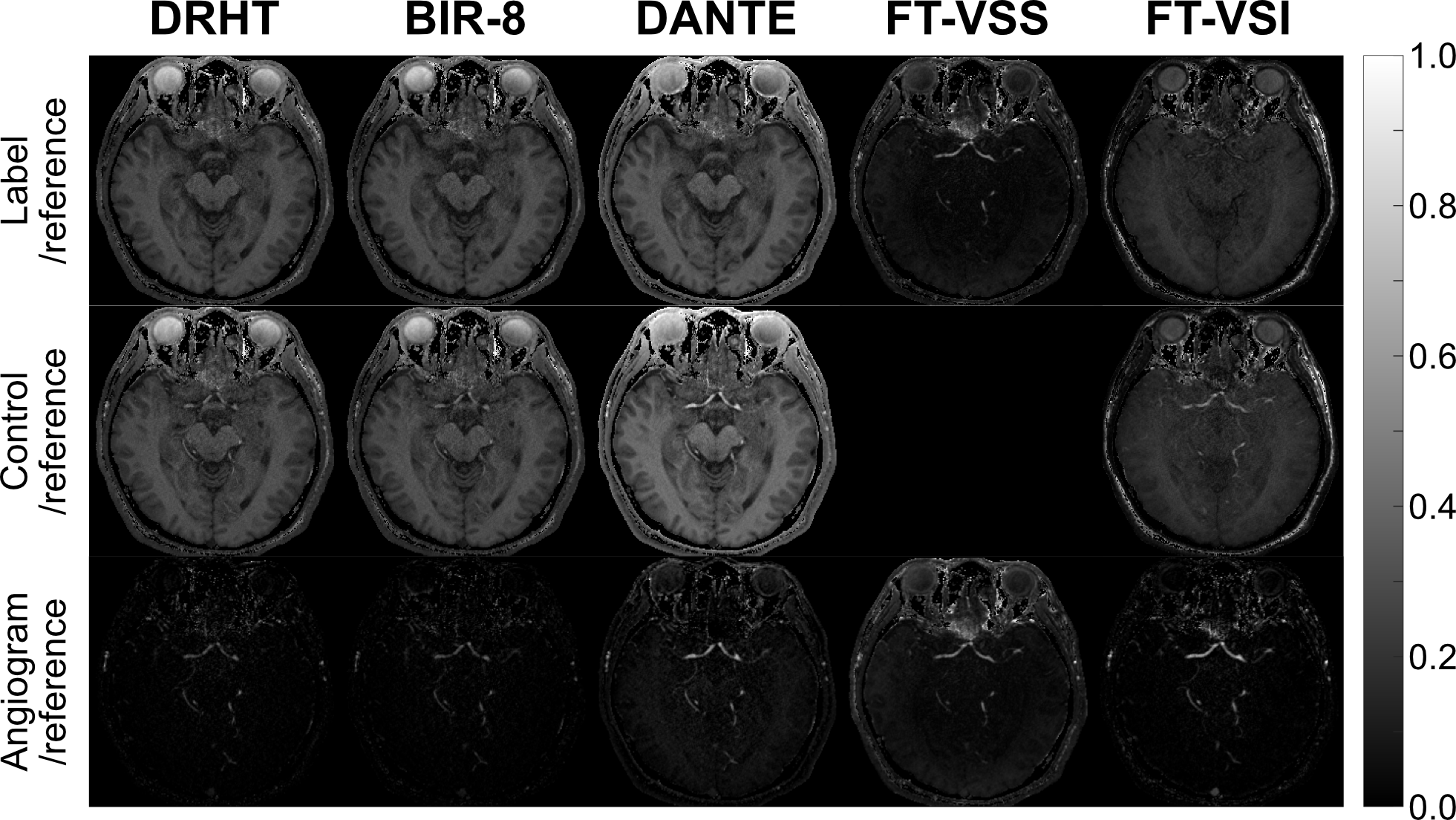
Arteriograms were obtained from subtraction of label and control for DRHT, BIR-8, DANTE, and FT-VSI, but not for FT-VSS, as its label alone yielded the angiogram. Coregistration was then applied to the angiogram images to correct head motion with SPM 12. Relative signal ratios between the label, control, angiographic images and the corresponding reference images were quantified (Figure 2). ROIs were drawn manually on arteriograms, including the first and second segments of ACA (A1 and A2), MCA (M1 and M2), PCA (P1 and P2), and tissue around them, respectively.
Results and Discussion
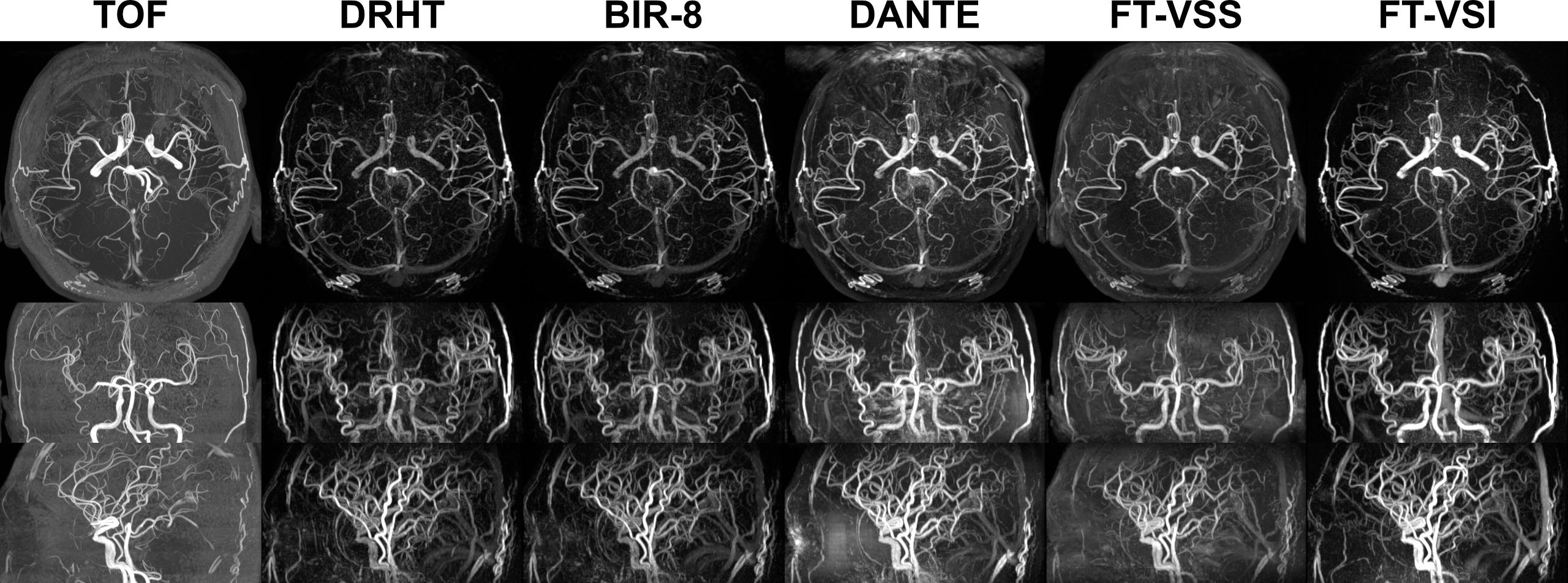
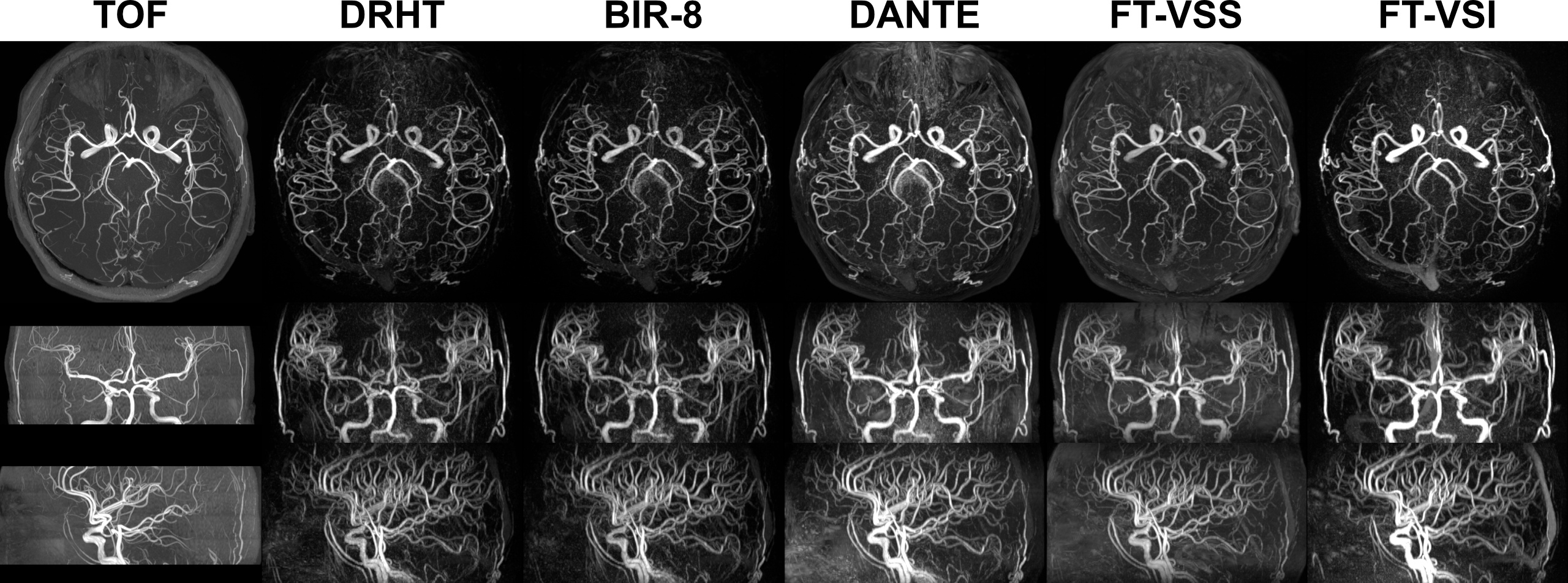
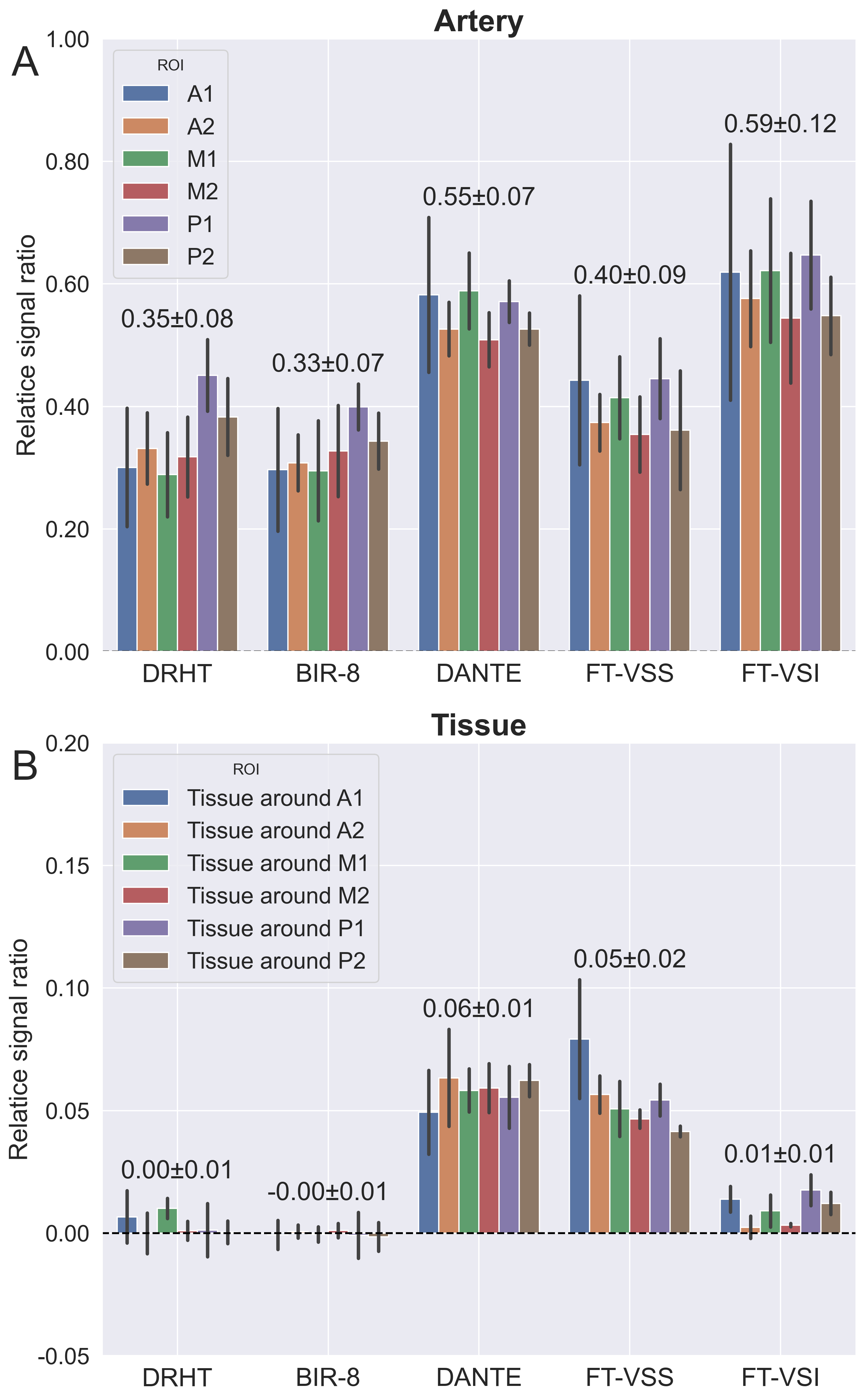
Representative VSMRA images of label, control, and the angiograms after their subtraction (label for FT-VSS) from a single slice of one subject are displayed in Fig.2. The orthogonal maximum-intensity-projection (MIP) images of arteriograms from two subjects are shown in Fig.3 and Fig.4, respectively. All VS modules depicted major arteries well. Compared to TOF and FT-VSS, the subtracted VSMRA (DRHT, BIR-8, DANTE, and FT-VSI) delivered more complete suppression of background tissue with better delineation of small arterial branches.Relative signal ratios of the major arterial segments and surrounding tissues for angiograms from five VS labeling modules are shown in Fig.5. DRHT and BIR-8 showed very similar labeling effect (Fig.2-4) without significant difference in comparison of relative signal ratio (Fig. 5). The residual tissue signal of FT-VSS is due to inhomogeneous B1 field. FT-VSI showed the highest relative signal ratio overall (Fig.5A), with p<0.05 compared to other methods except DANTE. The minor tissue signal observed in FT-VSI angiogram (~0.01 of reference image), was most likely due to the DC-bias, which could be improved by dynamic-phase cycling8.
Although DANTE yielded comparable relative signal ratio to FT-VSI, it also showed a net tissue signal after subtraction (Fig.5B), which were also observed in simulation (Fig.1E,F). This difference is caused by decrease of static signal in label only (Fig.1A,B). The gradients in DANTE labeling generate a spatially dependent phase between the RF pulses. For positions where this phase is zero or multiple of 2π, the label has similar signal as control. For any other position, the DANTE label will give a lower signal than control, yielding this net static signal after subtraction. Therefore, it may be challenging to use DANTE for ASL labeling. However, it still has the potential of application in MRA or flow suppression in ASL.
Conclusion
The effects of VSMRA with different velocity-selective modules on the cerebral arterial vasculature and static brain tissue were visualized angiographically and compared quantitatively. After subtraction, compared to DRHT and BIR-8, FT-VSI offered higher arterial signal in VSMRA, which correlates with the higher labeling efficiency for VSASL. DANTE also yielded high arterial signal along with residual tissue signal as subtraction error, which is a minor issue for VSMRA but poses a quantification challenge for VSASL. FT-VSS serves as an efficient non-subtractive VSMRA method with better delineation of small vessels than TOF.Acknowledgements
No acknowledgement found.References
1. Wong EC, Cronin M, Wu WC, Inglis B, Frank LR, Liu TT. Velocity-selective arterial spin labeling. Magnetic Resonance in Medicine. 2006;55(6):1334-1341. doi:10.1002/mrm.20906
2. Guo J, Meakin JA, Jezzard P, Wong EC. An optimized design to reduce eddy current sensitivity in velocity-selective arterial spin labeling using symmetric BIR-8 pulses. Magnetic Resonance in Medicine. 2015;73(3):1085-1094. doi:10.1002/mrm.25227
3. Qin Q, van Zijl PCM. Velocity-selective-inversion prepared arterial spin labeling. Magnetic Resonance in Medicine. 2016;76(4):1136-1148. doi:10.1002/mrm.26010
4. Matsuda T, Kimura H, Kabasawa H, Kanamoto M. Three-dimensional arterial spin labeling imaging with a DANTE preparation pulse. Magnetic Resonance Imaging. 2018;49:131-137. doi:10.1016/j.mri.2018.02.001
5. Qin Q, Shin T, Schär M, Guo H, Chen H, Qiao Y. Velocity-selective magnetization-prepared non-contrast-enhanced cerebral MR angiography at 3 Tesla: Improved immunity to B0/B1 inhomogeneity. Magnetic Resonance in Medicine. 2016;75(3):1232-1241. doi:10.1002/mrm.25764
6. Li W, Xu F, Schär M, et al. Whole-brain arteriography and venography: Using improved velocity-selective saturation pulse trains. Magnetic Resonance in Medicine. 2018;79(4):2014-2023. doi:10.1002/mrm.26864
7. Li L, Miller KL, Jezzard P. DANTE-prepared pulse trains: A novel approach to motion-sensitized and motion-suppressed quantitative magnetic resonance imaging: DANTE-Prepared Pulse Trains. Magn Reson Med. 2012;68(5):1423-1438. doi:10.1002/mrm.24142
8. Liu D, Li W, Xu F, Zhu D, Shin T, Qin Q. Ensuring both velocity and spatial responses robust to field inhomogeneities for velocity‐selective arterial spin labeling through dynamic phase‐cycling. Magn Reson Med. 2021;85(5):2723-2734. doi:10.1002/mrm.28622
Figures