2471
Correlation of Cerebral Blood Flow with Cognitive Impairment in Hemodialysis Patients Based on 3D pCASL1Department of Radiology, the First Affiliated Hospital of Dalian Medical University, Dalian, China, 2Clinical and Technical Support, Philips Healthcare, Beijing, China
Synopsis
Keywords: Blood vessels, Kidney, Cerebral Blood Flow、 Cognitive Impairment、 Hemodialysis Patients、 3D pCASL、 End-stage renal disease
This work aimed to use 3D pseudo-continuous arterial spin labeling (3D pCASL) imaging technology to compare the difference of cerebral blood flow between hemodialysis patients (HDs) and healthy controls (HCs), and to analyze the relationship between cerebral blood flow changes and cognitive impairment. Results showed that decreased cerebral blood flow in the left Fusiform (Fusiform-L) and the left Temporal Inferior of HDs compared with HCs. Mean CBF in Fusiform-L was positively correlated with recognition, and mean CBF in Fusiform-L was negatively correlated with psychomotor speed and visual attention in HDs.Introduction
End-stage renal disease (ESRD) is a disease with high morbidity and mortality but low awareness[1]. Hemodialysis is a common treatment for ESRD. Neurocognitive impairment is a common complication of hemodialysis, and the incidence of cognitive impairment increases with the prolongation of dialysis time[1,2]. Changes in cerebral hemodynamics, such as decreased local cerebral blood perfusion, may be a potential cause of cognitive decline[3]. Previous studies have found that during a single hemodialysis, the cerebral blood flow (CBF) value decreased after dialysis compared with before dialysis[4,5]. However, the long-term effects of hemodialysis on cerebral circulation are not consistent[2]. Compared with PET/CT and other imaging techniques, arterial spin labeling (ASL) MR imaging can non-invasively measure the brain CBF value of hemodialysis patients[6] without contrast agents. This work aimed to use 3D pseudo-continuous arterial spin labeling (3D pCASL) imaging technology to compare the difference of cerebral blood flow between hemodialysis patients (HDs) and healthy controls (HCs), and to analyze the relationship between cerebral blood flow changes and cognitive impairment.Materials and Methods
Thirty-seven HDs (mean age: 57.05 ± 10.02, range from 40 to 72 years; 16 males) and forty-eight HCs (mean age: 56.31 ± 8.32, range from 40 to 71 years; 28 males) were prospectively recruited and all were right-handed. Informed consent was acquired from each subject. All volunteers performed MR examination on a 3.0 T MR scanner (Ingenia CX, Philips Healthcare, Best, the Netherlands) with a 32-channel head coil. MR scans included 3D T1WI and 3D pCASL imaging. The original ASL data was automatically processed on the MR console to obtain the corresponding CBF maps. Then the CBF maps were masked with a whole brain gray matter template by using SPM12 software based on MATLAB. The gray matter CBF values were then compared between hemodialysis patients and normal controls, with age, gender, education level, diabetes, and hypertension included as covariates. Independent sample t test was used to compare the difference of cognitive related scores between HD and HC groups. Correlation analysis was used to explore the relationship between the average CBF value and cognitive function in the brain regions with significant difference in CBF between HD and HC groups.Results
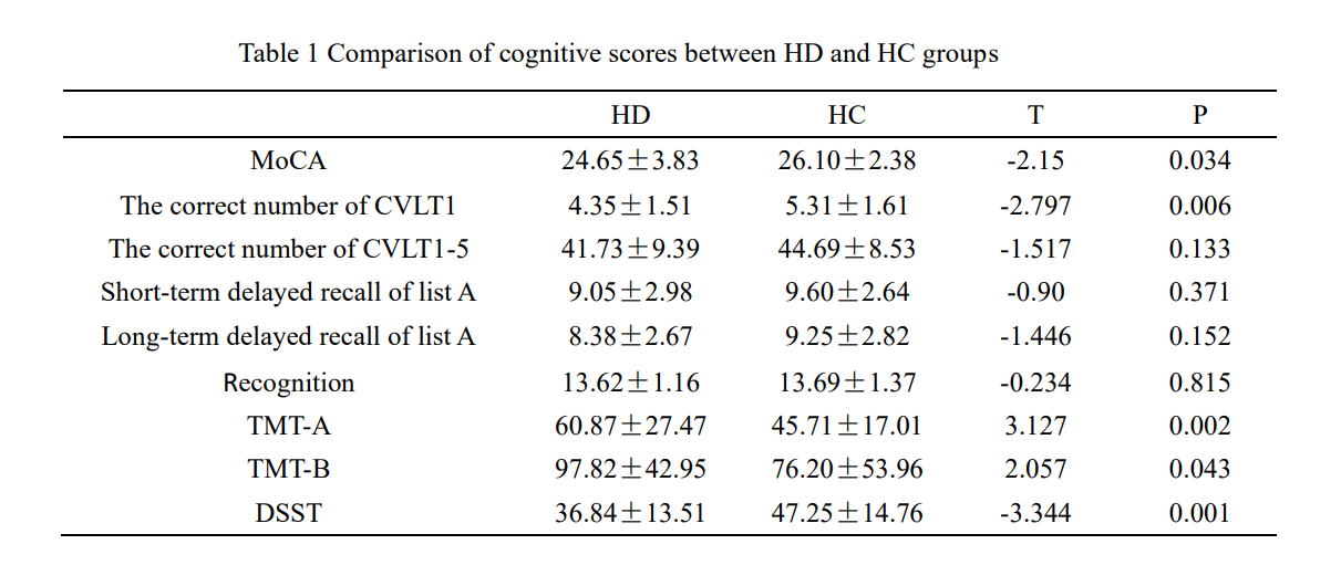
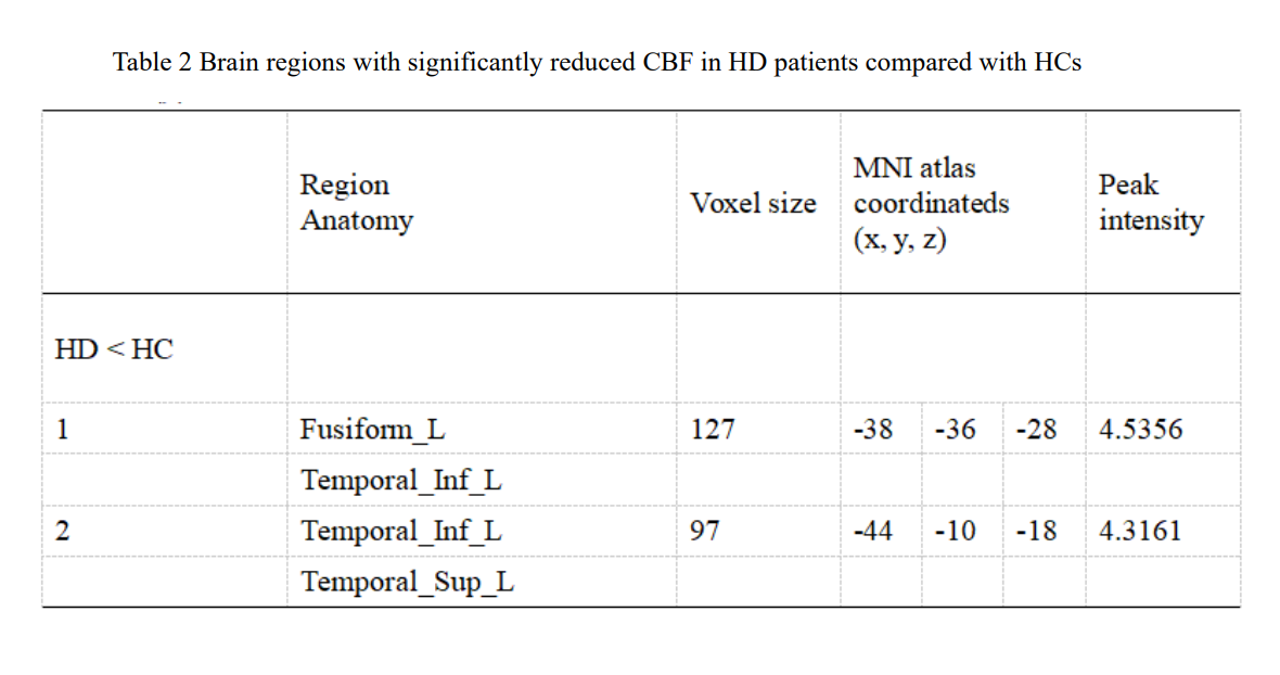
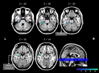
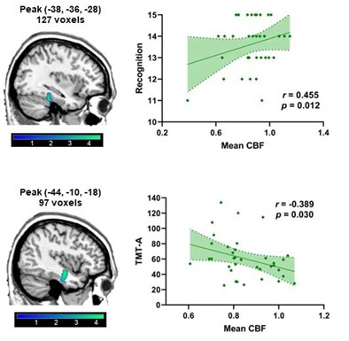
There were statistical differences in MoCA score, correct number of free recall in CVLT1 experiment, TMT-A, TMT-B and DSST between the two groups (p < 0.05) (Table 1). The CBF value was significantly reduced in brain regions of the left Fusiform (Fusiform-L) and the left Temporal Inferior (Temporal Inf – L) in the HD group compared with the HC group. The voxels, MNI coordinates, and peak intensity for these regions are shown in Table 2. Mean CBF in Fusiform -L was positively correlated with cognition (r = 0. 455, p = 0.012), and mean CBF in Temporal Inf - L was negatively correlated with TMT-A (r = -0.389, p = 0.030) (Figure 2).Discussion
Previous studies have shown that reduced regional cerebral blood flow is a potential factor in cognitive impairment [7]. In this study, the significantly reduced CBF values were observed in the HD group compared with the HC group were the brain regions of Fusiform -L and Temporal Inf -L. Moreover, the average CBF value of Fusiform -L was positively correlated with recognition, and the CBF value of Temporal Inf - L was negatively correlated with TMT-A. Temporal Inf - L plays an important role in cognitive learning, object memory and verbal fluency[8], while Fusiform -L is an important brain region involved in face recognition and object recognition[9]. The decrease of cerebral blood perfusion in Fusiform -L can lead to weakened cognitive ability; and the decrease of cerebral blood perfusion in Temporal Inf -L can result in prolonged time to complete the TMT-A experiment, thus reduced psychomotor speed and visual attention in subjects. Therefore, it is speculated that cognitive impairment in hemodialysis patients may be related to the decrease of CBF value in Fusiform -L and Temporal Inf - L.Conclusion
Decreased regional cerebral blood flow in hemodialysis patients is associated with cognitive impairment.Acknowledgements
Thanks to all my colleagues who participated in this studyReferences
[1]Lili Fu, Jun Liu, Jia Chen, et al. Effect of cognitive Dysfunction on Volume Overload in maintenance Hemodialysis Patients[J]. South China Journal of Defense Medicine, 2022, 36(09):702-707.
[2]Chai C, Zhang M, Wang H, et al. Increased cerebral blood flow is correlated with neurocognitive impairment in long-term hemodialysis patients: an arterial spin labeling MRI study. Brain Imaging Behav, 2021, 15(4):1828-1839.
[3]Tao Li, Zhensong Wang, Jianxian Liu, et al. A preliminary study of cerebral cortical blood flow in patients with type 2 diabetes mellitus by ASL[J]. Journal of Medical Imaging, 2022, 32(04):558-561.
[4]Polinder-Bos HA, García DV, Kuipers J, et al. Hemodialysis Induces an Acute Decline in Cerebral Blood Flow in Elderly Patients. J Am Soc Nephrol, 2018, 29(4):1317-1325.
[5]Findlay MD, Dawson J, Dickie DA, et al. Investigating the Relationship between Cerebral Blood Flow and Cognitive Function in Hemodialysis Patients. J Am Soc Nephrol, 2019, 30(1):147-158.
[6]Hernandez-Garcia L, Lahiri A, Schollenberger J. Recent progress in ASL. Neuroimage, 2019, 187:3-16.
[7]de Eulate RG, Goñi I, Galiano A, Vidorreta M, et al. Reduced Cerebral Blood Flow in Mild Cognitive Impairment Assessed Using Phase-Contrast MRI. J Alzheimers Dis, 2017, 58(2):585-595.
[8] Scheff SW, Price DA, Schmitt FA, et al. Synaptic loss in the inferior temporal gyrus in mild cognitive impairment and Alzheimer's disease. J Alzheimers Dis. 2011, 24(3):547-57.
[9]Cai S, Chong T, Zhang Y, et al. Altered Functional Connectivity of Fusiform Gyrus in Subjects with Amnestic Mild Cognitive Impairment: A Resting-State fMRI Study. Front Hum Neurosci, 2015, 27;9:471.
Figures